SlideShare una empresa de Scribd logo
1 de 52
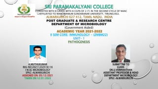
REACCREDITED WITH B GRADE WITH A CGPA OF 2.71 IN THE SECOND CYCLE OF NAAC
AFFILIATED TO MANOMANIUM SUNDARANAR UNIVERSITY, TIRUNELVELI.
ALWARKURICHI 627 412, TAMIL NADU, INDIA
POST GRADUATE & RESEARCH CENTRE
DEPARTMENT OF MICROBIOLOGY
(Government Aided)
ACADEMIC YEAR 2021-2022
II SEM CORE: IMMUNOLOGY - (ZMBM22)
UNIT- 1
PATHOGENESIS
A.MUTHUKUMAR
REG NO:20211232516116
I M.SC MICROBIOLOGY
SPKC-ALWARKURICHI
ASSIGNED ON: 05/12/2021
TAKEN ON:12/01/2022
SUBMITTED TO
GUIDE:
DR.S.VISWANATHAN
ASSISTANT PROFESSOR & HEAD
DEPARTMENT MICROBIOLOGY
SPKC-ALWARKURICHI
• INTRODUCTION
• GENERAL TERMS USED IN PATHOGENESIS
• VIRULENCE FACTORS
• EXAMPLES OF VIRULENCE FACTOR
• STAGES OF PATHOGENESIS
• HOW BACTERIAL PATHOGENS PENETRATE IN HOST DEFENSE
• TOXINS PRODUCED BY BACTERIA
• TRANSMISSION OF DISEASE
• REFERENCES
• SKILLACHIEVED BY SEMINAR
INTRODUCTION
• Pathogenesis is the process by which a disease or
disorder develops. It can include factors which contribute
not only to the onset of the disease or disorders, but also
to its progression and maintenance. The word comes from
Greek pathos means “suffering, disease” and genesis
means “creation”.
• The chain of events leading to that disease due to a series
of changes in the structure or function of the cell and
tissue and organ.
• Pathogenesis caused by a microbial ,chemical or physical
agent.
General Terms Used In Pathogenesis:
• A pathogen is a microorganism that is able to cause disease in a plant,
animal or insect.
• Pathogenicity is the ability to produce disease in a host organism.
Microbes express their pathogenicity by means of their virulence.
• Pathogenesis is a multi-factorial process which depends on the immune
status of the host, the nature of the species or strain (virulence factors) and
the number of organisms in the initial exposure.
• virulence, a term which refers to the degree of pathogenicity of the
microbe. Hence, the determinants of virulence of a pathogen are any of its
genetic or biochemical or structural features that enable it to produce
disease in a host.
Virulence factors :
 These are molecules expressed and secreted by pathogens (bacteria, viruses,
fungi and protozoa) that enable them to achieve the following:
 colonization of a niche in the host (this includes adhesion to cells)
 Immuno evasion, evasion of the host's immune response
 Immunosuppression, inhibition of the host's immune response
 entry into and exit out of cells (if the pathogen is an intracellular one)
 obtain nutrition from the host.
 Pathogens possess a wide array of virulence factors. Some are intrinsic to the
bacteria (e.g. capsules and endotoxin) whereas others are obtained from
plasmids (e.g. some toxins).
Examples Of Virulence
Factors:
• Examples of virulence factors for Staphylococcus aureus are hyaluronidase,
protease, coagulase, lipases, deoxyribonucleases and enterotoxins.
• Some examples of virulence factors for Streptococcus pyogenes are: M protein,
lipoteichoic acid, hyaluronic acid capsule,
• invasions such as streptokinase, hyaluronidase, and streptolysins, and exotoxins
• adhesion factors, extracellular enzymes, toxins and antiphagocytic factors.
Stages of Pathogenesis
To cause disease, a pathogen must successfully achieve
four steps or stages of pathogenesis: exposure (contact),
adhesion (colonization), invasion, and infection. The pathogen
must be able to gain entry to the host, travel to the location
where it can establish an infection, evade or overcome the
host’s immune response, and cause damage (i.e., disease) to the
host. In many cases, the cycle is completed when the pathogen
exits the host and is transmitted to a new host.
Exposure
An encounter with a potential pathogen is known as exposure or
contact. The food we eat and the objects we handle are all ways that
we can come into contact with potential pathogens. Yet, not all
contacts result in infection and disease. For a pathogen to cause
disease, it needs to be able to gain access into host tissue. An
anatomic site through which pathogens can pass into host tissue is
called a portal of entry. These are locations where the host cells are in
direct contact with the external environment. Major portals of entry
are include the skin, mucous membranes, and parenteral routes.
Portals Of Entry - Mucosal Surfaces
• Mucosal surfaces are the most important portals of entry for microbes; these include
the mucous membranes of the respiratory tract, the gastrointestinal tract, and the
genitourinary tract. Although most mucosal surfaces are in the interior of the body,
some are contiguous with the external skin at various body openings, including the
eyes, nose, mouth, urethra, and anus.
• Most pathogens are suited to a particular portal of entry. A pathogen’s portal
specificity is determined by the organism’s environmental adaptions and by the
enzymes and toxins they secrete. The respiratory and gastrointestinal tracts are
particularly vulnerable portals of entry because particles that include microorganisms
are constantly inhaled or ingested, respectively.
Portals Of Entry - Parenteral Route
• Pathogens can also enter through a breach in the protective barriers of the skin and mucous membranes.
Pathogens that enter the body in this way are said to enter by the parenteral route. For example, the skin is
a good natural barrier to pathogens, but breaks in the skin (e.g., wounds, insect bites, animal bites, needle
pricks) can provide a parenteral portal of entry for microorganisms.
• In pregnant women, the placenta normally prevents microorganisms from passing from the mother to the
fetus. However, a few pathogens are capable of crossing the blood-placental barrier. The gram-positive
bacterium Listeria monocytogenes, which causes the foodborne disease listeriosis, is one example that
poses a serious risk to the fetus and can sometimes lead to spontaneous abortion. Other pathogens that
can pass the placental barrier to infect the fetus are known collectively by the acronym TORCH .
• Transmission of infectious diseases from mother to baby is also a concern at the time of birth when the
baby passes through the birth canal. Babies whose mothers have active chlamydia or gonorrhea infections
may be exposed to the causative pathogens in the vagina, which can result in eye infections that lead to
blindness. To prevent this, it is standard practice to administer antibiotic drops to infants’ eyes shortly after
birth.
Adhesion
• Following the initial exposure, the pathogen adheres at the portal of entry. The term
adhesion refers to the capability of pathogenic microbes to attach to the cells of the body
using adhesion factors, and different pathogens use various mechanisms to adhere to the
cells of host tissues.
• Molecules (either proteins or carbohydrates) called adhesins are found on the surface of
certain pathogens and bind to specific receptors (glycoproteins) on host cells. Adhesins
are present on the fimbriae and flagella of bacteria, the cilia of protozoa, and the capsids
or membranes of viruses. Protozoans can also use hooks and barbs for adhesion; spike
proteins on viruses also enhance viral adhesion. The production of glycocalyces (slime
layers and capsules), with their high sugar and protein content, can also allow certain
bacterial pathogens to attach to cells.
Biofilm growth can also act as an adhesion factor. A biofilm is a community of bacteria
that produce a glycocalyx, which contributes to the extrapolymeric substances (EPS) that
allows the biofilm to attach to a surface. Persistent Pseudomonas aeruginosa infections are
common in patients suffering from cystic fibrosis, burn wounds, and middle-ear infections
(otitis media) because P. aeruginosa produces a biofilm. The EPS allows the microbe to
adhere to the host cells and makes it harder for the host to physically remove the
pathogen. The EPS not only allows for attachment but provides protection against the
immune system and antibiotic or antimicrobial treatments, preventing the medications
from reaching the cells within the biofilm. In addition, not all bacteria in a biofilm are
rapidly growing; some are in stationary phase. Since antibiotics are most effective against
rapidly growing bacteria, portions of bacteria in a biofilm are protected against antibiotics.
Biofilm - Adhesion Factor
Invasion
Once adhesion is successful, invasion can proceed. Invasion involves the dissemination of a
pathogen throughout local tissues or the body. Pathogens may produce exoenzymes or toxins,
which serve as virulence factors that allow them to colonize and damage host tissues as they
spread deeper into the body. Pathogens may also produce virulence factors that protect them
against immune system defenses. A pathogen’s specific virulence factors determine the degree
of tissue damage that occurs.
Intracellular pathogens achieve invasion by entering the host’s cells and reproducing. Some are
obligate intracellular pathogens (meaning they can only reproduce inside of host cells) and
others are facultative intracellular pathogens (meaning they can reproduce either inside or
outside of host cells). By entering the host cells, intracellular pathogens are able to evade
some mechanisms of the immune system while also exploiting the nutrients in the host cell.
• Entry to a cell can occur by endocytosis. For most kinds of host cells, pathogens use one of two
different mechanisms for endocytosis and entry.
• One mechanism relies on effector proteins secreted by the pathogen; these effector proteins
trigger entry into the host cell. This is the method that Salmonella and Shigella use when
invading intestinal epithelial cells. When these pathogens come in contact with epithelial cells in
the intestine, they secrete effector molecules that cause protrusions of membrane ruffles that
bring the bacterial cell in. This process is called membrane ruffling.
• The second mechanism relies on surface proteins expressed on the pathogen that bind to
receptors on the host cell, resulting in entry. For example, Yersinia pseudotuberculosis produces
a surface protein known as invasin that binds to beta-1 integrins expressed on the surface of
host cells.
Invasion -Endocytosis
• Some host cells, such as white blood cells and other phagocytes of the immune system, actively
endocytose pathogens in a process called phagocytosis.
• Although phagocytosis allows the pathogen to gain entry to the host cell, in most cases, the host
cell kills and degrades the pathogen by using digestive enzymes. Normally, when a pathogen is
ingested by a phagocyte, it is enclosed within a phagosome in the cytoplasm; the phagosome fuses
with a lysosome to form a phagolysosome, where digestive enzymes kill the pathogen.
• However, some intracellular pathogens have the ability to survive and multiply within phagocytes.
Examples include Listeria monocytogenes and Shigella; these bacteria produce proteins that lyse
the phagosome before it fuses with the lysosome, allowing the bacteria to escape into the
phagocyte’s cytoplasm where they can multiply.
• Bacteria such as Mycobacterium tuberculosis, Legionella pneumophila, and Salmonella species use
a slightly different mechanism to evade being digested by the phagocyte. These bacteria prevent
the fusion of the phagosome with the lysosome, thus remaining alive and dividing within the
phagosome.
Invasion - Phagocytosis
Infection
Following invasion, successful multiplication
of the pathogen leads to infection.
Infections can be described as local, focal, or
systemic, depending on the extent of the
infection.
Local Infection
A local infection is confined to a small area of the body,
typically near the portal of entry. For example, a hair follicle
infected by Staphylococcus aureus infection may result in a boil
around the site of infection, but the bacterium is largely contained
to this small location. Other examples of local infections that
involve more extensive tissue involvement include urinary tract
infections confined to the bladder or pneumonia confined to the
lungs.
Focal Infection
In a focal infection, a localized pathogen, or the toxins it
produces, can spread to a secondary location. For example, a dental
hygienist nicking the gum with a sharp tool can lead to a local
infection in the gum by Streptococcus bacteria of the normal oral
microbiota. These Streptococcus spp. May then gain access to the
bloodstream and make their way to other locations in the body,
resulting in a secondary infection.
Systemic Infection
When an infection becomes disseminated throughout the body, we
call it a systemic infection. For example, infection by the varicella-zoster
virus typically gains entry through a mucous membrane of the upper
respiratory system. It then spreads throughout the body, resulting in
the classic red skin lesions associated with chickenpox. Since these
lesions are not sites of initial infection, they are signs of a systemic
infection.
Sometimes a primary infection, the initial infection caused by one pathogen,
can lead to a secondary infection by another pathogen. For example, the immune
system of a patient with a primary infection by HIV becomes compromised,
making the patient more susceptible to secondary diseases like oral thrush and
others caused by opportunistic pathogens. Similarly, a primary infection by
Influenzavirus damages and decreases the defense mechanisms of the lungs,
making patients more susceptible to a secondary pneumonia by a bacterial
pathogen like Haemophilus influenzae or Streptococcus pneumoniae. Some
secondary infections can even develop as a result of treatment for a primary
infection. Antibiotic therapy targeting the primary pathogen can cause collateral
damage to the normal microbiota, creating an opening for opportunistic
Transmission Of Disease
For a pathogen to persist, it must put itself in a position to be transmitted to a
new host, leaving the infected host through a portal of exit . As with portals of
entry, many pathogens are adapted to use a particular portal of exit. Similar to
portals of entry, the most common portals of exit include the skin and the
respiratory, urogenital, and gastrointestinal tracts. Coughing and sneezing can expel
pathogens from the respiratory tract. A single sneeze can send thousands of virus
particles into the air. Secretions and excretions can transport pathogens out of
other portals of exit. Feces, urine, semen, vaginal secretions, tears, sweat, and shed
skin cells can all serve as vehicles for a pathogen to leave the body. Pathogens that
rely on insect vectors for transmission exit the body in the blood extracted by a
biting insect. Similarly, some pathogens exit the body in blood extracted by
needles.
• F. Savino et al. “Pain Assessment in Children Undergoing Venipuncture: The Wong–Baker Faces Scale Versus Skin
Conductance Fluctuations.” PeerJ 1 (2013):e37; https://peerj.com/articles/37/
• J.G. Kusters et al. Pathogenesis of Helicobacter pylori Infection. Clinical Microbiology Reviews 19 no. 3
(2006):449–490.
• N.R. Salama et al. “Life in the Human Stomach: Persistence Strategies of the Bacterial Pathogen Helicobacter
pylori.” Nature Reviews Microbiology 11 (2013):385–399.
• C. Owens. “P. aeruginosa survives in sinks 10 years after hospital outbreak.” 2015. www.healio.com/infectious-
dis...pital-outbreak
• Food and Drug Administration. “Bad Bug Book, Foodborne Pathogenic Microorganisms and Natural Toxins.” 2nd
ed. Silver Spring, MD: US Food and Drug Administration; 2012.
• M. Otto. “Staphylococcus epidermidis—The ‘Accidental’ Pathogen.” Nature Reviews Microbiology 7 no. 8
(2009):555–567.
• The O in TORCH stands for “other.”
• D. Davies. “Understanding Biofilm Resistance to Antibacterial Agents.” Nature Reviews Drug Discovery 2
(2003):114–122.
• Comunication skills
• Confidence
• Gained subject knowledge
better
• Presentation skills
• Motivations
PATHOGENESIS

Más contenido relacionado

La actualidad más candente

La actualidad más candente (20)

VIRULENCE FACTORS (1).pptx
VIRULENCE FACTORS (1).pptxVIRULENCE FACTORS (1).pptx
VIRULENCE FACTORS (1).pptx
 
Human microbial flora
Human microbial floraHuman microbial flora
Human microbial flora
 
Antigen
AntigenAntigen
Antigen
 
Staphylococci
StaphylococciStaphylococci
Staphylococci
 
Bacterial physiology ppt
Bacterial physiology pptBacterial physiology ppt
Bacterial physiology ppt
 
Types of infection
Types of infectionTypes of infection
Types of infection
 
Types of immunity
Types of immunityTypes of immunity
Types of immunity
 
Infection ppt
Infection pptInfection ppt
Infection ppt
 
Normal Microflora of Human body
Normal Microflora of Human bodyNormal Microflora of Human body
Normal Microflora of Human body
 
Antibodies
AntibodiesAntibodies
Antibodies
 
1.1 Classsification Of Microorganisms
1.1 Classsification  Of  Microorganisms1.1 Classsification  Of  Microorganisms
1.1 Classsification Of Microorganisms
 
Introduction to microbiology
Introduction to microbiologyIntroduction to microbiology
Introduction to microbiology
 
Bacterial cell wall
Bacterial cell wallBacterial cell wall
Bacterial cell wall
 
Classification of microorganisms lecture 2
Classification of microorganisms lecture 2Classification of microorganisms lecture 2
Classification of microorganisms lecture 2
 
Bacterial Pathogenesis
Bacterial PathogenesisBacterial Pathogenesis
Bacterial Pathogenesis
 
Normal Flora
Normal FloraNormal Flora
Normal Flora
 
Bacterial genetics
Bacterial geneticsBacterial genetics
Bacterial genetics
 
Factors affecting growth of bacteria
Factors affecting growth of bacteriaFactors affecting growth of bacteria
Factors affecting growth of bacteria
 
Properties of viruses
Properties of virusesProperties of viruses
Properties of viruses
 
Microbial pathogenicity
Microbial pathogenicityMicrobial pathogenicity
Microbial pathogenicity
 

Similar a PATHOGENESIS

pathogenicity_of_microbes (1).pptx
pathogenicity_of_microbes (1).pptxpathogenicity_of_microbes (1).pptx
pathogenicity_of_microbes (1).pptxChrisTina960888
 
15. principles of infectious disease 1
15. principles of infectious disease 115. principles of infectious disease 1
15. principles of infectious disease 1Ahmad Hamadi
 
STAINSStains and dyes are frequently used in histology, in cytology, and in t...
STAINSStains and dyes are frequently used in histology, in cytology, and in t...STAINSStains and dyes are frequently used in histology, in cytology, and in t...
STAINSStains and dyes are frequently used in histology, in cytology, and in t...AyushiSharma843565
 
BACTERIAL PATHOGENICITY AND VIRULENCE FACTORS.pptx
BACTERIAL PATHOGENICITY AND VIRULENCE FACTORS.pptxBACTERIAL PATHOGENICITY AND VIRULENCE FACTORS.pptx
BACTERIAL PATHOGENICITY AND VIRULENCE FACTORS.pptxanshulsharma905846
 
host pathogen interaction, Mechanism of pathogenesis
host pathogen interaction, Mechanism of pathogenesis host pathogen interaction, Mechanism of pathogenesis
host pathogen interaction, Mechanism of pathogenesis rashmi816961
 
Mechanism of bacterial pathogenesis
Mechanism of bacterial pathogenesisMechanism of bacterial pathogenesis
Mechanism of bacterial pathogenesisjigisha pancholi
 
Microbial_Pathogenicity.pdf
Microbial_Pathogenicity.pdfMicrobial_Pathogenicity.pdf
Microbial_Pathogenicity.pdfMidhatSarfraz
 
Innate Immunity 2022.pptx
Innate Immunity 2022.pptxInnate Immunity 2022.pptx
Innate Immunity 2022.pptxMercyDaka3
 
Microbes, Man and Environment (Microbial pathogenicity) .pptx
Microbes, Man and Environment (Microbial pathogenicity) .pptxMicrobes, Man and Environment (Microbial pathogenicity) .pptx
Microbes, Man and Environment (Microbial pathogenicity) .pptxMidhatSarfraz
 
Mechanism of pathogenicity-Exotoxin and endotoxin
Mechanism of pathogenicity-Exotoxin and endotoxinMechanism of pathogenicity-Exotoxin and endotoxin
Mechanism of pathogenicity-Exotoxin and endotoxinaiswarya thomas
 
BASIC NURSING,FIRST AID AND STERILIZATION.pptx
BASIC NURSING,FIRST AID AND STERILIZATION.pptxBASIC NURSING,FIRST AID AND STERILIZATION.pptx
BASIC NURSING,FIRST AID AND STERILIZATION.pptxFrancisKazoba
 
An Overview of Immunity to Viruses, Bacteria, Fungi and Ptozoans
An Overview of Immunity to Viruses, Bacteria, Fungi and PtozoansAn Overview of Immunity to Viruses, Bacteria, Fungi and Ptozoans
An Overview of Immunity to Viruses, Bacteria, Fungi and PtozoansAtifa Ambreen
 
Host pathogen interactions
 Host pathogen interactions Host pathogen interactions
Host pathogen interactionsPALANIANANTH.S
 
Introduction of medical microbiology unit 1
Introduction of medical microbiology unit 1Introduction of medical microbiology unit 1
Introduction of medical microbiology unit 1meghashridhar
 
MCB 814-Principles of Bacterial Pathogenesis.pptx
MCB 814-Principles of Bacterial Pathogenesis.pptxMCB 814-Principles of Bacterial Pathogenesis.pptx
MCB 814-Principles of Bacterial Pathogenesis.pptxjohnojonugwaprecious
 
Bacterial Virulence Factors in Periodontology
Bacterial Virulence Factors in PeriodontologyBacterial Virulence Factors in Periodontology
Bacterial Virulence Factors in PeriodontologyDr Ripunjay Tripathi
 
Bacterial virulence
Bacterial virulenceBacterial virulence
Bacterial virulenceSaritaSahu18
 

Similar a PATHOGENESIS (20)

pathogenicity_of_microbes (1).pptx
pathogenicity_of_microbes (1).pptxpathogenicity_of_microbes (1).pptx
pathogenicity_of_microbes (1).pptx
 
15. principles of infectious disease 1
15. principles of infectious disease 115. principles of infectious disease 1
15. principles of infectious disease 1
 
STAINSStains and dyes are frequently used in histology, in cytology, and in t...
STAINSStains and dyes are frequently used in histology, in cytology, and in t...STAINSStains and dyes are frequently used in histology, in cytology, and in t...
STAINSStains and dyes are frequently used in histology, in cytology, and in t...
 
BACTERIAL PATHOGENICITY AND VIRULENCE FACTORS.pptx
BACTERIAL PATHOGENICITY AND VIRULENCE FACTORS.pptxBACTERIAL PATHOGENICITY AND VIRULENCE FACTORS.pptx
BACTERIAL PATHOGENICITY AND VIRULENCE FACTORS.pptx
 
host pathogen interaction, Mechanism of pathogenesis
host pathogen interaction, Mechanism of pathogenesis host pathogen interaction, Mechanism of pathogenesis
host pathogen interaction, Mechanism of pathogenesis
 
Mechanism of bacterial pathogenesis
Mechanism of bacterial pathogenesisMechanism of bacterial pathogenesis
Mechanism of bacterial pathogenesis
 
Microbial_Pathogenicity.pdf
Microbial_Pathogenicity.pdfMicrobial_Pathogenicity.pdf
Microbial_Pathogenicity.pdf
 
Innate Immunity 2022.pptx
Innate Immunity 2022.pptxInnate Immunity 2022.pptx
Innate Immunity 2022.pptx
 
Paraprotex
ParaprotexParaprotex
Paraprotex
 
Microbes, Man and Environment (Microbial pathogenicity) .pptx
Microbes, Man and Environment (Microbial pathogenicity) .pptxMicrobes, Man and Environment (Microbial pathogenicity) .pptx
Microbes, Man and Environment (Microbial pathogenicity) .pptx
 
Mechanism of pathogenicity-Exotoxin and endotoxin
Mechanism of pathogenicity-Exotoxin and endotoxinMechanism of pathogenicity-Exotoxin and endotoxin
Mechanism of pathogenicity-Exotoxin and endotoxin
 
BASIC NURSING,FIRST AID AND STERILIZATION.pptx
BASIC NURSING,FIRST AID AND STERILIZATION.pptxBASIC NURSING,FIRST AID AND STERILIZATION.pptx
BASIC NURSING,FIRST AID AND STERILIZATION.pptx
 
An Overview of Immunity to Viruses, Bacteria, Fungi and Ptozoans
An Overview of Immunity to Viruses, Bacteria, Fungi and PtozoansAn Overview of Immunity to Viruses, Bacteria, Fungi and Ptozoans
An Overview of Immunity to Viruses, Bacteria, Fungi and Ptozoans
 
Host pathogen interactions
 Host pathogen interactions Host pathogen interactions
Host pathogen interactions
 
host parasite relationship
host parasite relationshiphost parasite relationship
host parasite relationship
 
Introduction of medical microbiology unit 1
Introduction of medical microbiology unit 1Introduction of medical microbiology unit 1
Introduction of medical microbiology unit 1
 
MCB 814-Principles of Bacterial Pathogenesis.pptx
MCB 814-Principles of Bacterial Pathogenesis.pptxMCB 814-Principles of Bacterial Pathogenesis.pptx
MCB 814-Principles of Bacterial Pathogenesis.pptx
 
Bacterial Virulence Factors in Periodontology
Bacterial Virulence Factors in PeriodontologyBacterial Virulence Factors in Periodontology
Bacterial Virulence Factors in Periodontology
 
Bacterial virulence
Bacterial virulenceBacterial virulence
Bacterial virulence
 
Pharm 314
Pharm 314Pharm 314
Pharm 314
 

Más de akshyhari

FED BATCH CULTURE-2.pptx
FED BATCH CULTURE-2.pptxFED BATCH CULTURE-2.pptx
FED BATCH CULTURE-2.pptxakshyhari
 
INNATE IMMUNITY
INNATE IMMUNITYINNATE IMMUNITY
INNATE IMMUNITYakshyhari
 
IMMUNOFLOURESCENCE.pptx
IMMUNOFLOURESCENCE.pptxIMMUNOFLOURESCENCE.pptx
IMMUNOFLOURESCENCE.pptxakshyhari
 
COMPLEMENT FIXATION.pptx
COMPLEMENT FIXATION.pptxCOMPLEMENT FIXATION.pptx
COMPLEMENT FIXATION.pptxakshyhari
 
Cytokine, lympokine,interferon.pptx
Cytokine, lympokine,interferon.pptxCytokine, lympokine,interferon.pptx
Cytokine, lympokine,interferon.pptxakshyhari
 
Infection.pptx
Infection.pptxInfection.pptx
Infection.pptxakshyhari
 
HISTORY AND DEVELOPMENT OF IMMUNOLOGY.pptx
HISTORY AND DEVELOPMENT OF IMMUNOLOGY.pptxHISTORY AND DEVELOPMENT OF IMMUNOLOGY.pptx
HISTORY AND DEVELOPMENT OF IMMUNOLOGY.pptxakshyhari
 
ACQUIRED IMMUNITY.pdf
ACQUIRED IMMUNITY.pdfACQUIRED IMMUNITY.pdf
ACQUIRED IMMUNITY.pdfakshyhari
 
Secondary lymphoid organ.pptx
Secondary lymphoid organ.pptxSecondary lymphoid organ.pptx
Secondary lymphoid organ.pptxakshyhari
 
LYMPHOID TIASUES.pdf
LYMPHOID TIASUES.pdfLYMPHOID TIASUES.pdf
LYMPHOID TIASUES.pdfakshyhari
 
NUTRIENT TRANSPORT MECHANISM.pdf
NUTRIENT TRANSPORT MECHANISM.pdfNUTRIENT TRANSPORT MECHANISM.pdf
NUTRIENT TRANSPORT MECHANISM.pdfakshyhari
 
Flow cytometry.pptx
Flow cytometry.pptxFlow cytometry.pptx
Flow cytometry.pptxakshyhari
 
immunology-immunization schedule.pdf
immunology-immunization schedule.pdfimmunology-immunization schedule.pdf
immunology-immunization schedule.pdfakshyhari
 
EMP,ED,GLYOXYLATE PATHWAY
EMP,ED,GLYOXYLATE PATHWAYEMP,ED,GLYOXYLATE PATHWAY
EMP,ED,GLYOXYLATE PATHWAYakshyhari
 
INFLAMMATION.pptx
INFLAMMATION.pptxINFLAMMATION.pptx
INFLAMMATION.pptxakshyhari
 
PASTEUR EFFECT.pptx
PASTEUR EFFECT.pptxPASTEUR EFFECT.pptx
PASTEUR EFFECT.pptxakshyhari
 
RADIOIMMUNOASSAY.pptx
RADIOIMMUNOASSAY.pptxRADIOIMMUNOASSAY.pptx
RADIOIMMUNOASSAY.pptxakshyhari
 
HETEROCYST.pdf
HETEROCYST.pdfHETEROCYST.pdf
HETEROCYST.pdfakshyhari
 
green sulfur bacteria .pptx
green sulfur bacteria .pptxgreen sulfur bacteria .pptx
green sulfur bacteria .pptxakshyhari
 

Más de akshyhari (20)

FED BATCH CULTURE-2.pptx
FED BATCH CULTURE-2.pptxFED BATCH CULTURE-2.pptx
FED BATCH CULTURE-2.pptx
 
INNATE IMMUNITY
INNATE IMMUNITYINNATE IMMUNITY
INNATE IMMUNITY
 
IMMUNOFLOURESCENCE.pptx
IMMUNOFLOURESCENCE.pptxIMMUNOFLOURESCENCE.pptx
IMMUNOFLOURESCENCE.pptx
 
COMPLEMENT FIXATION.pptx
COMPLEMENT FIXATION.pptxCOMPLEMENT FIXATION.pptx
COMPLEMENT FIXATION.pptx
 
Cytokine, lympokine,interferon.pptx
Cytokine, lympokine,interferon.pptxCytokine, lympokine,interferon.pptx
Cytokine, lympokine,interferon.pptx
 
Infection.pptx
Infection.pptxInfection.pptx
Infection.pptx
 
HISTORY AND DEVELOPMENT OF IMMUNOLOGY.pptx
HISTORY AND DEVELOPMENT OF IMMUNOLOGY.pptxHISTORY AND DEVELOPMENT OF IMMUNOLOGY.pptx
HISTORY AND DEVELOPMENT OF IMMUNOLOGY.pptx
 
ACQUIRED IMMUNITY.pdf
ACQUIRED IMMUNITY.pdfACQUIRED IMMUNITY.pdf
ACQUIRED IMMUNITY.pdf
 
Secondary lymphoid organ.pptx
Secondary lymphoid organ.pptxSecondary lymphoid organ.pptx
Secondary lymphoid organ.pptx
 
LYMPHOID TIASUES.pdf
LYMPHOID TIASUES.pdfLYMPHOID TIASUES.pdf
LYMPHOID TIASUES.pdf
 
NUTRIENT TRANSPORT MECHANISM.pdf
NUTRIENT TRANSPORT MECHANISM.pdfNUTRIENT TRANSPORT MECHANISM.pdf
NUTRIENT TRANSPORT MECHANISM.pdf
 
TCA CYCLE
TCA CYCLETCA CYCLE
TCA CYCLE
 
Flow cytometry.pptx
Flow cytometry.pptxFlow cytometry.pptx
Flow cytometry.pptx
 
immunology-immunization schedule.pdf
immunology-immunization schedule.pdfimmunology-immunization schedule.pdf
immunology-immunization schedule.pdf
 
EMP,ED,GLYOXYLATE PATHWAY
EMP,ED,GLYOXYLATE PATHWAYEMP,ED,GLYOXYLATE PATHWAY
EMP,ED,GLYOXYLATE PATHWAY
 
INFLAMMATION.pptx
INFLAMMATION.pptxINFLAMMATION.pptx
INFLAMMATION.pptx
 
PASTEUR EFFECT.pptx
PASTEUR EFFECT.pptxPASTEUR EFFECT.pptx
PASTEUR EFFECT.pptx
 
RADIOIMMUNOASSAY.pptx
RADIOIMMUNOASSAY.pptxRADIOIMMUNOASSAY.pptx
RADIOIMMUNOASSAY.pptx
 
HETEROCYST.pdf
HETEROCYST.pdfHETEROCYST.pdf
HETEROCYST.pdf
 
green sulfur bacteria .pptx
green sulfur bacteria .pptxgreen sulfur bacteria .pptx
green sulfur bacteria .pptx
 

Último

Formation of low mass protostars and their circumstellar disks
Formation of low mass protostars and their circumstellar disksFormation of low mass protostars and their circumstellar disks
Formation of low mass protostars and their circumstellar disksSérgio Sacani
 
Biopesticide (2).pptx .This slides helps to know the different types of biop...
Biopesticide (2).pptx  .This slides helps to know the different types of biop...Biopesticide (2).pptx  .This slides helps to know the different types of biop...
Biopesticide (2).pptx .This slides helps to know the different types of biop...RohitNehra6
 
Nightside clouds and disequilibrium chemistry on the hot Jupiter WASP-43b
Nightside clouds and disequilibrium chemistry on the hot Jupiter WASP-43bNightside clouds and disequilibrium chemistry on the hot Jupiter WASP-43b
Nightside clouds and disequilibrium chemistry on the hot Jupiter WASP-43bSérgio Sacani
 
Pests of mustard_Identification_Management_Dr.UPR.pdf
Pests of mustard_Identification_Management_Dr.UPR.pdfPests of mustard_Identification_Management_Dr.UPR.pdf
Pests of mustard_Identification_Management_Dr.UPR.pdfPirithiRaju
 
GBSN - Microbiology (Unit 1)
GBSN - Microbiology (Unit 1)GBSN - Microbiology (Unit 1)
GBSN - Microbiology (Unit 1)Areesha Ahmad
 
VIRUSES structure and classification ppt by Dr.Prince C P
VIRUSES structure and classification ppt by Dr.Prince C PVIRUSES structure and classification ppt by Dr.Prince C P
VIRUSES structure and classification ppt by Dr.Prince C PPRINCE C P
 
Isotopic evidence of long-lived volcanism on Io
Isotopic evidence of long-lived volcanism on IoIsotopic evidence of long-lived volcanism on Io
Isotopic evidence of long-lived volcanism on IoSérgio Sacani
 
Discovery of an Accretion Streamer and a Slow Wide-angle Outflow around FUOri...
Discovery of an Accretion Streamer and a Slow Wide-angle Outflow around FUOri...Discovery of an Accretion Streamer and a Slow Wide-angle Outflow around FUOri...
Discovery of an Accretion Streamer and a Slow Wide-angle Outflow around FUOri...Sérgio Sacani
 
❤Jammu Kashmir Call Girls 8617697112 Personal Whatsapp Number 💦✅.
❤Jammu Kashmir Call Girls 8617697112 Personal Whatsapp Number 💦✅.❤Jammu Kashmir Call Girls 8617697112 Personal Whatsapp Number 💦✅.
❤Jammu Kashmir Call Girls 8617697112 Personal Whatsapp Number 💦✅.Nitya salvi
 
Nanoparticles synthesis and characterization​ ​
Nanoparticles synthesis and characterization​  ​Nanoparticles synthesis and characterization​  ​
Nanoparticles synthesis and characterization​ ​kaibalyasahoo82800
 
SAMASTIPUR CALL GIRL 7857803690 LOW PRICE ESCORT SERVICE
SAMASTIPUR CALL GIRL 7857803690  LOW PRICE  ESCORT SERVICESAMASTIPUR CALL GIRL 7857803690  LOW PRICE  ESCORT SERVICE
SAMASTIPUR CALL GIRL 7857803690 LOW PRICE ESCORT SERVICEayushi9330
 
Pulmonary drug delivery system M.pharm -2nd sem P'ceutics
Pulmonary drug delivery system M.pharm -2nd sem P'ceuticsPulmonary drug delivery system M.pharm -2nd sem P'ceutics
Pulmonary drug delivery system M.pharm -2nd sem P'ceuticssakshisoni2385
 
Animal Communication- Auditory and Visual.pptx
Animal Communication- Auditory and Visual.pptxAnimal Communication- Auditory and Visual.pptx
Animal Communication- Auditory and Visual.pptxUmerFayaz5
 
GUIDELINES ON SIMILAR BIOLOGICS Regulatory Requirements for Marketing Authori...
GUIDELINES ON SIMILAR BIOLOGICS Regulatory Requirements for Marketing Authori...GUIDELINES ON SIMILAR BIOLOGICS Regulatory Requirements for Marketing Authori...
GUIDELINES ON SIMILAR BIOLOGICS Regulatory Requirements for Marketing Authori...Lokesh Kothari
 
Botany 4th semester series (krishna).pdf
Botany 4th semester series (krishna).pdfBotany 4th semester series (krishna).pdf
Botany 4th semester series (krishna).pdfSumit Kumar yadav
 
Hubble Asteroid Hunter III. Physical properties of newly found asteroids
Hubble Asteroid Hunter III. Physical properties of newly found asteroidsHubble Asteroid Hunter III. Physical properties of newly found asteroids
Hubble Asteroid Hunter III. Physical properties of newly found asteroidsSérgio Sacani
 
Green chemistry and Sustainable development.pptx
Green chemistry  and Sustainable development.pptxGreen chemistry  and Sustainable development.pptx
Green chemistry and Sustainable development.pptxRajatChauhan518211
 
High Class Escorts in Hyderabad ₹7.5k Pick Up & Drop With Cash Payment 969456...
High Class Escorts in Hyderabad ₹7.5k Pick Up & Drop With Cash Payment 969456...High Class Escorts in Hyderabad ₹7.5k Pick Up & Drop With Cash Payment 969456...
High Class Escorts in Hyderabad ₹7.5k Pick Up & Drop With Cash Payment 969456...chandars293
 
GBSN - Biochemistry (Unit 1)
GBSN - Biochemistry (Unit 1)GBSN - Biochemistry (Unit 1)
GBSN - Biochemistry (Unit 1)Areesha Ahmad
 

Último (20)

Formation of low mass protostars and their circumstellar disks
Formation of low mass protostars and their circumstellar disksFormation of low mass protostars and their circumstellar disks
Formation of low mass protostars and their circumstellar disks
 
Biopesticide (2).pptx .This slides helps to know the different types of biop...
Biopesticide (2).pptx  .This slides helps to know the different types of biop...Biopesticide (2).pptx  .This slides helps to know the different types of biop...
Biopesticide (2).pptx .This slides helps to know the different types of biop...
 
Nightside clouds and disequilibrium chemistry on the hot Jupiter WASP-43b
Nightside clouds and disequilibrium chemistry on the hot Jupiter WASP-43bNightside clouds and disequilibrium chemistry on the hot Jupiter WASP-43b
Nightside clouds and disequilibrium chemistry on the hot Jupiter WASP-43b
 
Pests of mustard_Identification_Management_Dr.UPR.pdf
Pests of mustard_Identification_Management_Dr.UPR.pdfPests of mustard_Identification_Management_Dr.UPR.pdf
Pests of mustard_Identification_Management_Dr.UPR.pdf
 
GBSN - Microbiology (Unit 1)
GBSN - Microbiology (Unit 1)GBSN - Microbiology (Unit 1)
GBSN - Microbiology (Unit 1)
 
VIRUSES structure and classification ppt by Dr.Prince C P
VIRUSES structure and classification ppt by Dr.Prince C PVIRUSES structure and classification ppt by Dr.Prince C P
VIRUSES structure and classification ppt by Dr.Prince C P
 
Isotopic evidence of long-lived volcanism on Io
Isotopic evidence of long-lived volcanism on IoIsotopic evidence of long-lived volcanism on Io
Isotopic evidence of long-lived volcanism on Io
 
Discovery of an Accretion Streamer and a Slow Wide-angle Outflow around FUOri...
Discovery of an Accretion Streamer and a Slow Wide-angle Outflow around FUOri...Discovery of an Accretion Streamer and a Slow Wide-angle Outflow around FUOri...
Discovery of an Accretion Streamer and a Slow Wide-angle Outflow around FUOri...
 
❤Jammu Kashmir Call Girls 8617697112 Personal Whatsapp Number 💦✅.
❤Jammu Kashmir Call Girls 8617697112 Personal Whatsapp Number 💦✅.❤Jammu Kashmir Call Girls 8617697112 Personal Whatsapp Number 💦✅.
❤Jammu Kashmir Call Girls 8617697112 Personal Whatsapp Number 💦✅.
 
CELL -Structural and Functional unit of life.pdf
CELL -Structural and Functional unit of life.pdfCELL -Structural and Functional unit of life.pdf
CELL -Structural and Functional unit of life.pdf
 
Nanoparticles synthesis and characterization​ ​
Nanoparticles synthesis and characterization​  ​Nanoparticles synthesis and characterization​  ​
Nanoparticles synthesis and characterization​ ​
 
SAMASTIPUR CALL GIRL 7857803690 LOW PRICE ESCORT SERVICE
SAMASTIPUR CALL GIRL 7857803690  LOW PRICE  ESCORT SERVICESAMASTIPUR CALL GIRL 7857803690  LOW PRICE  ESCORT SERVICE
SAMASTIPUR CALL GIRL 7857803690 LOW PRICE ESCORT SERVICE
 
Pulmonary drug delivery system M.pharm -2nd sem P'ceutics
Pulmonary drug delivery system M.pharm -2nd sem P'ceuticsPulmonary drug delivery system M.pharm -2nd sem P'ceutics
Pulmonary drug delivery system M.pharm -2nd sem P'ceutics
 
Animal Communication- Auditory and Visual.pptx
Animal Communication- Auditory and Visual.pptxAnimal Communication- Auditory and Visual.pptx
Animal Communication- Auditory and Visual.pptx
 
GUIDELINES ON SIMILAR BIOLOGICS Regulatory Requirements for Marketing Authori...
GUIDELINES ON SIMILAR BIOLOGICS Regulatory Requirements for Marketing Authori...GUIDELINES ON SIMILAR BIOLOGICS Regulatory Requirements for Marketing Authori...
GUIDELINES ON SIMILAR BIOLOGICS Regulatory Requirements for Marketing Authori...
 
Botany 4th semester series (krishna).pdf
Botany 4th semester series (krishna).pdfBotany 4th semester series (krishna).pdf
Botany 4th semester series (krishna).pdf
 
Hubble Asteroid Hunter III. Physical properties of newly found asteroids
Hubble Asteroid Hunter III. Physical properties of newly found asteroidsHubble Asteroid Hunter III. Physical properties of newly found asteroids
Hubble Asteroid Hunter III. Physical properties of newly found asteroids
 
Green chemistry and Sustainable development.pptx
Green chemistry  and Sustainable development.pptxGreen chemistry  and Sustainable development.pptx
Green chemistry and Sustainable development.pptx
 
High Class Escorts in Hyderabad ₹7.5k Pick Up & Drop With Cash Payment 969456...
High Class Escorts in Hyderabad ₹7.5k Pick Up & Drop With Cash Payment 969456...High Class Escorts in Hyderabad ₹7.5k Pick Up & Drop With Cash Payment 969456...
High Class Escorts in Hyderabad ₹7.5k Pick Up & Drop With Cash Payment 969456...
 
GBSN - Biochemistry (Unit 1)
GBSN - Biochemistry (Unit 1)GBSN - Biochemistry (Unit 1)
GBSN - Biochemistry (Unit 1)
 

PATHOGENESIS

  • 1. REACCREDITED WITH B GRADE WITH A CGPA OF 2.71 IN THE SECOND CYCLE OF NAAC AFFILIATED TO MANOMANIUM SUNDARANAR UNIVERSITY, TIRUNELVELI. ALWARKURICHI 627 412, TAMIL NADU, INDIA POST GRADUATE & RESEARCH CENTRE DEPARTMENT OF MICROBIOLOGY (Government Aided) ACADEMIC YEAR 2021-2022 II SEM CORE: IMMUNOLOGY - (ZMBM22) UNIT- 1 PATHOGENESIS A.MUTHUKUMAR REG NO:20211232516116 I M.SC MICROBIOLOGY SPKC-ALWARKURICHI ASSIGNED ON: 05/12/2021 TAKEN ON:12/01/2022 SUBMITTED TO GUIDE: DR.S.VISWANATHAN ASSISTANT PROFESSOR & HEAD DEPARTMENT MICROBIOLOGY SPKC-ALWARKURICHI
  • 2. • INTRODUCTION • GENERAL TERMS USED IN PATHOGENESIS • VIRULENCE FACTORS • EXAMPLES OF VIRULENCE FACTOR • STAGES OF PATHOGENESIS • HOW BACTERIAL PATHOGENS PENETRATE IN HOST DEFENSE • TOXINS PRODUCED BY BACTERIA • TRANSMISSION OF DISEASE • REFERENCES • SKILLACHIEVED BY SEMINAR
  • 3. INTRODUCTION • Pathogenesis is the process by which a disease or disorder develops. It can include factors which contribute not only to the onset of the disease or disorders, but also to its progression and maintenance. The word comes from Greek pathos means “suffering, disease” and genesis means “creation”. • The chain of events leading to that disease due to a series of changes in the structure or function of the cell and tissue and organ. • Pathogenesis caused by a microbial ,chemical or physical agent.
  • 4. General Terms Used In Pathogenesis: • A pathogen is a microorganism that is able to cause disease in a plant, animal or insect. • Pathogenicity is the ability to produce disease in a host organism. Microbes express their pathogenicity by means of their virulence. • Pathogenesis is a multi-factorial process which depends on the immune status of the host, the nature of the species or strain (virulence factors) and the number of organisms in the initial exposure. • virulence, a term which refers to the degree of pathogenicity of the microbe. Hence, the determinants of virulence of a pathogen are any of its genetic or biochemical or structural features that enable it to produce disease in a host.
  • 5. Virulence factors :  These are molecules expressed and secreted by pathogens (bacteria, viruses, fungi and protozoa) that enable them to achieve the following:  colonization of a niche in the host (this includes adhesion to cells)  Immuno evasion, evasion of the host's immune response  Immunosuppression, inhibition of the host's immune response  entry into and exit out of cells (if the pathogen is an intracellular one)  obtain nutrition from the host.  Pathogens possess a wide array of virulence factors. Some are intrinsic to the bacteria (e.g. capsules and endotoxin) whereas others are obtained from plasmids (e.g. some toxins).
  • 6. Examples Of Virulence Factors: • Examples of virulence factors for Staphylococcus aureus are hyaluronidase, protease, coagulase, lipases, deoxyribonucleases and enterotoxins. • Some examples of virulence factors for Streptococcus pyogenes are: M protein, lipoteichoic acid, hyaluronic acid capsule, • invasions such as streptokinase, hyaluronidase, and streptolysins, and exotoxins • adhesion factors, extracellular enzymes, toxins and antiphagocytic factors.
  • 7. Stages of Pathogenesis To cause disease, a pathogen must successfully achieve four steps or stages of pathogenesis: exposure (contact), adhesion (colonization), invasion, and infection. The pathogen must be able to gain entry to the host, travel to the location where it can establish an infection, evade or overcome the host’s immune response, and cause damage (i.e., disease) to the host. In many cases, the cycle is completed when the pathogen exits the host and is transmitted to a new host.
  • 8. Exposure An encounter with a potential pathogen is known as exposure or contact. The food we eat and the objects we handle are all ways that we can come into contact with potential pathogens. Yet, not all contacts result in infection and disease. For a pathogen to cause disease, it needs to be able to gain access into host tissue. An anatomic site through which pathogens can pass into host tissue is called a portal of entry. These are locations where the host cells are in direct contact with the external environment. Major portals of entry are include the skin, mucous membranes, and parenteral routes.
  • 9. Portals Of Entry - Mucosal Surfaces • Mucosal surfaces are the most important portals of entry for microbes; these include the mucous membranes of the respiratory tract, the gastrointestinal tract, and the genitourinary tract. Although most mucosal surfaces are in the interior of the body, some are contiguous with the external skin at various body openings, including the eyes, nose, mouth, urethra, and anus. • Most pathogens are suited to a particular portal of entry. A pathogen’s portal specificity is determined by the organism’s environmental adaptions and by the enzymes and toxins they secrete. The respiratory and gastrointestinal tracts are particularly vulnerable portals of entry because particles that include microorganisms are constantly inhaled or ingested, respectively.
  • 10. Portals Of Entry - Parenteral Route • Pathogens can also enter through a breach in the protective barriers of the skin and mucous membranes. Pathogens that enter the body in this way are said to enter by the parenteral route. For example, the skin is a good natural barrier to pathogens, but breaks in the skin (e.g., wounds, insect bites, animal bites, needle pricks) can provide a parenteral portal of entry for microorganisms. • In pregnant women, the placenta normally prevents microorganisms from passing from the mother to the fetus. However, a few pathogens are capable of crossing the blood-placental barrier. The gram-positive bacterium Listeria monocytogenes, which causes the foodborne disease listeriosis, is one example that poses a serious risk to the fetus and can sometimes lead to spontaneous abortion. Other pathogens that can pass the placental barrier to infect the fetus are known collectively by the acronym TORCH . • Transmission of infectious diseases from mother to baby is also a concern at the time of birth when the baby passes through the birth canal. Babies whose mothers have active chlamydia or gonorrhea infections may be exposed to the causative pathogens in the vagina, which can result in eye infections that lead to blindness. To prevent this, it is standard practice to administer antibiotic drops to infants’ eyes shortly after birth.
  • 11.
  • 12. Adhesion • Following the initial exposure, the pathogen adheres at the portal of entry. The term adhesion refers to the capability of pathogenic microbes to attach to the cells of the body using adhesion factors, and different pathogens use various mechanisms to adhere to the cells of host tissues. • Molecules (either proteins or carbohydrates) called adhesins are found on the surface of certain pathogens and bind to specific receptors (glycoproteins) on host cells. Adhesins are present on the fimbriae and flagella of bacteria, the cilia of protozoa, and the capsids or membranes of viruses. Protozoans can also use hooks and barbs for adhesion; spike proteins on viruses also enhance viral adhesion. The production of glycocalyces (slime layers and capsules), with their high sugar and protein content, can also allow certain bacterial pathogens to attach to cells.
  • 13. Biofilm growth can also act as an adhesion factor. A biofilm is a community of bacteria that produce a glycocalyx, which contributes to the extrapolymeric substances (EPS) that allows the biofilm to attach to a surface. Persistent Pseudomonas aeruginosa infections are common in patients suffering from cystic fibrosis, burn wounds, and middle-ear infections (otitis media) because P. aeruginosa produces a biofilm. The EPS allows the microbe to adhere to the host cells and makes it harder for the host to physically remove the pathogen. The EPS not only allows for attachment but provides protection against the immune system and antibiotic or antimicrobial treatments, preventing the medications from reaching the cells within the biofilm. In addition, not all bacteria in a biofilm are rapidly growing; some are in stationary phase. Since antibiotics are most effective against rapidly growing bacteria, portions of bacteria in a biofilm are protected against antibiotics. Biofilm - Adhesion Factor
  • 14.
  • 15.
  • 16. Invasion Once adhesion is successful, invasion can proceed. Invasion involves the dissemination of a pathogen throughout local tissues or the body. Pathogens may produce exoenzymes or toxins, which serve as virulence factors that allow them to colonize and damage host tissues as they spread deeper into the body. Pathogens may also produce virulence factors that protect them against immune system defenses. A pathogen’s specific virulence factors determine the degree of tissue damage that occurs. Intracellular pathogens achieve invasion by entering the host’s cells and reproducing. Some are obligate intracellular pathogens (meaning they can only reproduce inside of host cells) and others are facultative intracellular pathogens (meaning they can reproduce either inside or outside of host cells). By entering the host cells, intracellular pathogens are able to evade some mechanisms of the immune system while also exploiting the nutrients in the host cell.
  • 17. • Entry to a cell can occur by endocytosis. For most kinds of host cells, pathogens use one of two different mechanisms for endocytosis and entry. • One mechanism relies on effector proteins secreted by the pathogen; these effector proteins trigger entry into the host cell. This is the method that Salmonella and Shigella use when invading intestinal epithelial cells. When these pathogens come in contact with epithelial cells in the intestine, they secrete effector molecules that cause protrusions of membrane ruffles that bring the bacterial cell in. This process is called membrane ruffling. • The second mechanism relies on surface proteins expressed on the pathogen that bind to receptors on the host cell, resulting in entry. For example, Yersinia pseudotuberculosis produces a surface protein known as invasin that binds to beta-1 integrins expressed on the surface of host cells. Invasion -Endocytosis
  • 18. • Some host cells, such as white blood cells and other phagocytes of the immune system, actively endocytose pathogens in a process called phagocytosis. • Although phagocytosis allows the pathogen to gain entry to the host cell, in most cases, the host cell kills and degrades the pathogen by using digestive enzymes. Normally, when a pathogen is ingested by a phagocyte, it is enclosed within a phagosome in the cytoplasm; the phagosome fuses with a lysosome to form a phagolysosome, where digestive enzymes kill the pathogen. • However, some intracellular pathogens have the ability to survive and multiply within phagocytes. Examples include Listeria monocytogenes and Shigella; these bacteria produce proteins that lyse the phagosome before it fuses with the lysosome, allowing the bacteria to escape into the phagocyte’s cytoplasm where they can multiply. • Bacteria such as Mycobacterium tuberculosis, Legionella pneumophila, and Salmonella species use a slightly different mechanism to evade being digested by the phagocyte. These bacteria prevent the fusion of the phagosome with the lysosome, thus remaining alive and dividing within the phagosome. Invasion - Phagocytosis
  • 19.
  • 20.
  • 21.
  • 22.
  • 23.
  • 24.
  • 25.
  • 26.
  • 27.
  • 28.
  • 29.
  • 30.
  • 31.
  • 32.
  • 33.
  • 34.
  • 35.
  • 36.
  • 37.
  • 38.
  • 39.
  • 40. Infection Following invasion, successful multiplication of the pathogen leads to infection. Infections can be described as local, focal, or systemic, depending on the extent of the infection.
  • 41. Local Infection A local infection is confined to a small area of the body, typically near the portal of entry. For example, a hair follicle infected by Staphylococcus aureus infection may result in a boil around the site of infection, but the bacterium is largely contained to this small location. Other examples of local infections that involve more extensive tissue involvement include urinary tract infections confined to the bladder or pneumonia confined to the lungs.
  • 42. Focal Infection In a focal infection, a localized pathogen, or the toxins it produces, can spread to a secondary location. For example, a dental hygienist nicking the gum with a sharp tool can lead to a local infection in the gum by Streptococcus bacteria of the normal oral microbiota. These Streptococcus spp. May then gain access to the bloodstream and make their way to other locations in the body, resulting in a secondary infection.
  • 43. Systemic Infection When an infection becomes disseminated throughout the body, we call it a systemic infection. For example, infection by the varicella-zoster virus typically gains entry through a mucous membrane of the upper respiratory system. It then spreads throughout the body, resulting in the classic red skin lesions associated with chickenpox. Since these lesions are not sites of initial infection, they are signs of a systemic infection.
  • 44. Sometimes a primary infection, the initial infection caused by one pathogen, can lead to a secondary infection by another pathogen. For example, the immune system of a patient with a primary infection by HIV becomes compromised, making the patient more susceptible to secondary diseases like oral thrush and others caused by opportunistic pathogens. Similarly, a primary infection by Influenzavirus damages and decreases the defense mechanisms of the lungs, making patients more susceptible to a secondary pneumonia by a bacterial pathogen like Haemophilus influenzae or Streptococcus pneumoniae. Some secondary infections can even develop as a result of treatment for a primary infection. Antibiotic therapy targeting the primary pathogen can cause collateral damage to the normal microbiota, creating an opening for opportunistic
  • 45. Transmission Of Disease For a pathogen to persist, it must put itself in a position to be transmitted to a new host, leaving the infected host through a portal of exit . As with portals of entry, many pathogens are adapted to use a particular portal of exit. Similar to portals of entry, the most common portals of exit include the skin and the respiratory, urogenital, and gastrointestinal tracts. Coughing and sneezing can expel pathogens from the respiratory tract. A single sneeze can send thousands of virus particles into the air. Secretions and excretions can transport pathogens out of other portals of exit. Feces, urine, semen, vaginal secretions, tears, sweat, and shed skin cells can all serve as vehicles for a pathogen to leave the body. Pathogens that rely on insect vectors for transmission exit the body in the blood extracted by a biting insect. Similarly, some pathogens exit the body in blood extracted by needles.
  • 46.
  • 47. • F. Savino et al. “Pain Assessment in Children Undergoing Venipuncture: The Wong–Baker Faces Scale Versus Skin Conductance Fluctuations.” PeerJ 1 (2013):e37; https://peerj.com/articles/37/ • J.G. Kusters et al. Pathogenesis of Helicobacter pylori Infection. Clinical Microbiology Reviews 19 no. 3 (2006):449–490. • N.R. Salama et al. “Life in the Human Stomach: Persistence Strategies of the Bacterial Pathogen Helicobacter pylori.” Nature Reviews Microbiology 11 (2013):385–399. • C. Owens. “P. aeruginosa survives in sinks 10 years after hospital outbreak.” 2015. www.healio.com/infectious- dis...pital-outbreak • Food and Drug Administration. “Bad Bug Book, Foodborne Pathogenic Microorganisms and Natural Toxins.” 2nd ed. Silver Spring, MD: US Food and Drug Administration; 2012. • M. Otto. “Staphylococcus epidermidis—The ‘Accidental’ Pathogen.” Nature Reviews Microbiology 7 no. 8 (2009):555–567. • The O in TORCH stands for “other.” • D. Davies. “Understanding Biofilm Resistance to Antibacterial Agents.” Nature Reviews Drug Discovery 2 (2003):114–122.
  • 48.
  • 49.
  • 50.
  • 51. • Comunication skills • Confidence • Gained subject knowledge better • Presentation skills • Motivations