Suturing techniques involved in dental surgery
•Descargar como PPTX, PDF•
321 recomendaciones•122,552 vistas

This was my presentation during Oral Surgery rotation at the University of Detroit Mercy School of Dentistry (UDMSD)

Recomendados
Más contenido relacionado
La actualidad más candente
La actualidad más candente (20)
Destacado
Destacado (15)
Similar a Suturing techniques involved in dental surgery
Similar a Suturing techniques involved in dental surgery (20)
Último
Último (20)
Suturing techniques involved in dental surgery
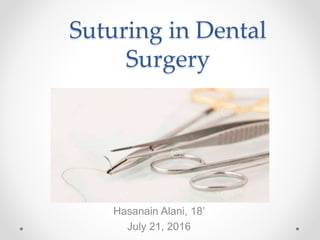
- 1. Suturing in Dental Surgery Hasanain Alani, 18’ July 21, 2016
- 2. Overview • Objectives • Review of suturing materials • Techniques used in Oral Surgery
- 3. What is Suturing? • The primary objective is to position and secure surgical flaps to promote optimal healing. • When performed properly, healing by primary intention occurs. • Performed intra- and extra-orally o Achieve functional and esthetic results o Decreasing the potential for postoperative infections occurance
- 4. Suture Armamentarium • Needle Holder Suture scissors Adson forceps Suture Needle
- 5. Suture Materials: Needle A surgical needle has 3 parts: the needle point, the needle body, and the swaged (press-fit) end The most commonly used are the 3/8 and ½ circle needles. The common shapes: • ROUND o Less traumatic than the other two, requires more force • REVERSE CUTTING: o The sharp TIP is DOWNWARD. o More safe when working in delicate tissue. • Cutting o Sharp TIP is UPWARD. o Extra sharp tip in is more likely to tear the tissue.
- 6. Suture Materials: Thread • Properties o Tensile strength o Biocompatibility o Ease of tying o Least tissue irritation and reaction o Diameter and size o Coefficient of friction • Classification o Origin o Structure o Duration
- 7. Durability • Resorbable o Natural • Plain gut • Chromic gut o Synthetic • Polyglycolic acid (PGA) • Poliglecaprone 25 How do sutures resorb? o Antigenic Reaction o Acidic Environment Nonresorbable Silk Polyester Monofilament type ‘nylon’ Polytetrafluoroethylen e (PTFE)
- 8. Silk Sutures • This is the most universally used material in dentistry • Advantages: o Inexpensive o Easy to handle and tie • Disadvantages: o It must be removed o It is multifilament • When Should we avoid using silk? And what are the alternatives?
- 9. Example • Patient diagnosed with bulimia presented to OS clinic for #30 extraction, and it was determined that the flap edges need to be positioned by sutures. • What is the minimum coaptation time for tissue flaps? • Synthetic vs Organic thread? • Fast Absorbing Polyglycolic Acid (PGA-FA)
- 10. Diameter • Thread materials range in diameter from 1 to 10, and the higher number corresponds to the thinner, more delicate thread. • periodontal plastic surgery: 5–0 for soft tissue grafts, 4-0 mucoperiosteal grafts and implants surgery.
- 11. Knots • Art of suturing! • An appropriate type of know should be used for the specific suture material • Slip knot: used with silk, chromic or plain gut suture • Surgeon’s knot: used with synthetic resorbable and other nonresorbable synthetic suture materials to prevent untimely knot untying.
- 12. Techniques • Interrupted Suture • Simple Continuous Suture • External Horizontal Mattress Suture • External Vertical Mattress Suture • Figure-of-eight Suture • Criss-cross Suture
- 13. Interrupted Suture • Do the pass technique, two loops around the needle holder, then grab the tail and do the knot. • Indications: Single tooth extraction, third molar extraction flap, biopsies, implants, ..etc. • Advantages: It is the most commonly used technique, preferred in urgent situations and it is easy to remove. Failure of one is inconsequential of the others. • Disadvantages: It does not bring all surfaces into contact and less supportive for healing of the flap margins.
- 14. Simple Continuous Suture • Start it with simple interrupted suture • Then you cut the tail off and leave that last piece loose then you can do your loops. • Indications: Bone graft, removal of mandibular tori, tuberosity reduction and where esthetics are not important • Advantages: It is very easy to produce and offers a more water tight closure • Disadvantages: if you cut one part of it, you lost all of it.
- 15. Horizontal Mattress Suture • The strongest type of sutures, very far away (8 mm from the edge) • Indications: large distances between tissues, bone grafts and implants, and closure of extraction socket. • Advantages: Good for hemostasis, less prominent scarring. • Disadvantages: Leave a gap between flaps and it is difficult to remove.
- 16. Vertical Mattress Suture • The far far, near near technique. • Indications: where the wound edges tend to evert • Advantages: greater closure strength and better distribution of wound tension • Disadvantages: Scar formation and the formation of edge necrosis.
- 17. Figure of 8 sutures • Pattern goes 1-2-3-4-1 • Indication: Extraction socket closure, adaptation of ginigival papilla around the tooth, and bone graft placement in socket • Advantages: Rapid closure • Disadvantages: Due to its orientation, it is difficult to remove and it leaves a significant amount of suture threads inside the socket.
- 18. General Principles • 1- Grasp the needle 2/3 front, and 1/3 behind the needle driver. • 2- The needle should pass perpendicular to the tissue • 3- The needle should pass at an equal depth and distance on both sides of the wound • 4- Pass from the thinner to the thicker tissue • 5- The suture should never be closed under tension (no blanch). • 6- The knot should be placed at 2-3 mm from the incision • 7- Suture should pass over the dental papilla, not the empty socket.
- 19. Conclusion • Due to the daily surgical procedures carried by dentists, a greater knowledge of suturing armamentarium and materials and is needed. • The success of technique-sensitive surgeries depends on the clinician’s knowledge and skills to close the wound and achieve optimal healing • The innovations in suturing materials decrease the potential for postoperative infections.
- 20. Refrences • 1- Silverstein, Lee H., Gregori M. Kurtzman, and Peter C. Shatz. "Suturing for optimal soft-tissue management." Journal of Oral Implantology 35.2 (2009): 82-90. • 2- Chu, Chih-Chang, J. Anthony Von Fraunhofer, and Howard P. Greisler, eds.Wound closure biomaterials and devices. CRC Press, 1996. • 3- Int J Periodontics Restorative Dent. 1998 Oct;18(5):474-87. Oral tissue reactions to suture materials.Selvig KA(1), Biagiotti GR, Leknes KN, Wikesjö UM.
Notas del editor
- I will discuss the rationale of specific suturing techniques and suture materials and how soft tissues react to them, to help the clinician obtain optimal wound closure.
- When used properly, surgical sutures should hold flap edges in apposition until the wound has healed enough to withstand normal functional stresses. When the proper suture technique is used with the appropriate thread type and diameter, tension is placed on the wound margins so primary intention healing occurs.
- Needle Holder to drive the needle Suture scissors to cut the suture Adson forceps = tissue pick Suture needle with suture material attached to it.
- The most commonly used suture needles in dentistry are the 3/8 and 1/2 circle needles3,4 (Figure 2). The 3/8 needle allows the clinician to pass from the buccal surface to the lingual surface in one motion by rotating the needle on a central axis. In contrast, the 1/2 circle needle is traditionally used in more restricted areas, for instance, in the buccal of the maxillary molars and the facial aspect of the maxillary and mandibular incisors. In addition, the 1/2 circle needle is routinely used for periosteal and mucogingival surgery Round: the rest of the needle is dull and blunt so it needs lots of pressure. , and it is used for subcutaneous tissues and blood vessels. Reverse cutting: the most common one, safer than next type. Cutting: It is the least safe type, it could tear up the tissue because it is too sharp. BOTH the Reverse Cutting and the Cutting have a triangular shape with 3 cutting edges. But note the location of the cutting edges is different. Generally in dentistry, the 3/8 reverse cutting needle with a 3–0 or 4–0 thread diameter and the 1/2 reverse cutting needle with the thinner and more delicate 5–0 or 6–0 thread diameter are the most commonly used needle-and-thread combinations, in the author's experienc
- Properties: 1- Tensile Strength: clinical should use a resorbable suture that loses its tensile strength at the same rate as the tissue gains strength. 2- Biocompatibility: It is a two-way relationship, the extent of tissue reactions to suture depends largely on the chemical structure and their degradation products if they are absorbable. Sutures of natural origin like silk usually provoke more tissue reactions than synthetic ones, and this reactions is due to the availability of enzymes to react with natural biopolymers. 3- Ease of tying to prevent slipping of the knot Threads are classified based on either their origin (Synthetic or organic), or Duration (resorbable or non-resorbable), or structure (mono vs multifilament)… monofilament is less traumatic and has less chance of infection because there are no spaces between filaments. Here it is important to mention that the physical factors come into play, not only chemical… stiff suture materials in monofilament sutures with stiff projection ends in a know where cut, and that could cause irritation to the surrounding tissue, this problem is not commonly found in multiflament threads.
- First, sutures of biological origin, such as surgical gut (eg, plain and chromic gut), are gradually digested by intraoral enzymes.2 This suture material is made from an animal protein and can potentially induce an antigenic reaction. When used intraorally, this material loses most of its tensile strength in 24 to 48 hours, unless it is coated with a chromic compound that extends absorption up to 7 to 10 days and extends loss of tensile strength for up to 5 days.5 Second, surgical gut sutures may break too rapidly if used in patients with a very low intraoral pH. A decrease in intraoral pH may be caused by a plethora of physiological events, such as metabolic disorders (eg, epigastric reflux, hiatal hernia, bulimia). Autoimmunity caused by Sjögren's syndrome, chemotherapy, radiation therapy, and some medications (eg, maximum acid output inhibitors, angiotensin-converting inhibitors, antipsychotics, diuretics, antihypertensive agents, antipsoriasis medications, and steroid inhalers) can also result in dry mouth and a low intraoral pH.2,6
- Classically, silk has been the most universally used material in dentistry and many other surgical disciplines.8 Silk is easy to handle, ties with a slip knot, and is relatively inexpensive compared with other nonabsorbable suture materials currently available. However, silk has distinct disadvantages. First, it is nonabsorbable, so it must be removed, usually a week or so later when the patient is not numb. Second, silk specifically is a multifilament that “wicks” or pulls bacteria and fluids into the wound site.9 Therefore, silk is not the suture material of choice when any sterile materials are placed under a mucoperiosteal flap (eg, dental implant, bone graft, or regenerative barrier) or when there is clinical evidence of an infection at the surgical site. Instead of silk, other nonabsorbable sutures that can be used in these situations, such as nylon, polyester, polyethylene, polypropylene, or expanded polytetrafluoroethylene (e-PTFE).
- The minimum coaptation time for tissue flaps is approximately 5 days.5 Therefore, clinicians should select a fast-absorbing PGA suture for indications in which there is a low intraoral pH, when surgical gut sutures are contraindicated. The PGA-FA suture material is manufactured from synthetic polymers and is principally broken down by hydrolysis in tissue fluids in approximately 7 to 10 days; it is not affected by a low intraoral pH.1,2 The PGA-FA suture also has a higher tensile strength than surgical gut suture material; however, it absorbs at a rate comparable to that of surgical gut sutures under normal intraoral physiologic conditions.1,2
- Surgical knot tying is an important component to the art of suturing. For knot security and to prevent untimely knot untying, it is essential that the appropriate surgical knot be used for the specific suture material being secured. For instance, when using silk or chromic gut, or plain gut suture material, a slip surgical knot should be used. SLIP KNOT involves a tie in one direction, and a second tie in the same direction, and then a third tie in the opposite direction so square the knot and hold it permenantely. However, with synthetic both resorbable and nonabsorbable suture materials, a surgeon's knot must be used to prevent untimely knot untying4 (Figures 4a through 4c). SURGEON’S KNOT INVOLVES placing the leader and the tippet side by side. Use both lines to form a loop with enough overlap to tie a double overhand knot. Pull both ends through the loop and then through a second time. Lubricate the knot and pull it tight. Trim the ends.
- GOOD STRESS DISTRIBUTION It is strong and can be used in areas of stress Loosening of one suture does not cause the lossening of the others. In case of infection or hematoma, removing some of the sutures may offer a satisfactory treatment.
- 8 mm from the edge, while it is usually 3-5 mm.
- The far-far near near suture placement passes 4 to 8 mm from the wound edge, fairly deep in the wound below the dermis. Then The near-near placement occurs at a shallow depth (about 1 mm) and should be in the upper dermis. The near-near placement should be within 1 to 2 mm of the wound edge. Following the near-near passage of the needle, both ends of the suture thread should be tied on one side of the wound.
- The needle is first inserted into the outer surface of the buccal flap and then either through the outer epithelialized surface under the surface. </li></ul><ul><li>The needle is then returned through the embrasure and tied buccally.
- Blanching causes necrosis or scarring