Electrophoresis by Anubhav Singh, M.pharm
•Descargar como PPT, PDF•
12 recomendaciones•7,193 vistas

Easy Presentation on Electrophoresis

Recomendados
Más contenido relacionado
La actualidad más candente
La actualidad más candente (20)
Destacado
Destacado (20)
Similar a Electrophoresis by Anubhav Singh, M.pharm
Similar a Electrophoresis by Anubhav Singh, M.pharm (20)
Más de Anubhav Singh
Más de Anubhav Singh (6)
Último
Último (20)
Electrophoresis by Anubhav Singh, M.pharm
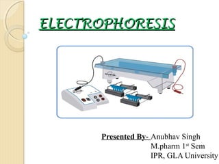
- 1. ELECTROPHORESIS Presented By- Anubhav Singh M.pharm 1st Sem IPR, GLA University
- 2. Introduction• When a potential difference is applied between the two electrodes in a colloidal solution, It has been observed that the colloidal particles are carried to either the positive or negative electrode. • In other words , they behave as if they are electrically charged w.r.t. the dispersion medium. This phenomenon is known as electrophoresis. • Many important biological molecules, such as amino acids, peptides, proteins, nucleotides and nucleic acids, possess ionisable groups and, therefore, at any given pH, exist in solution as electrically charged species either as cations or anions. • Under the influence of an electric field these charged particles will migrate either to the cathode or to the anode, depending on the nature of their net charge. • The common support is a polymer of agarose and acrylamide.
- 3. Why to Use Electrophoresis? • Electrophoresis is a technique used for the separation of biological molecules based on their movement due to the influence of a direct electric current. • The technique was pioneered in 1937 by the Swedish chemist Arne Tiselius for the separation of proteins. • It has now been extended to the separation of many other different classes of biomolecules including nucleic acids, carbohydrates and amino acids. • Electrophoresis has become increasingly important in the laboratory for basic research, biomedical research and in clinical settings for the diagnosis of disease. • However, it is valuable as an analytical technique for detecting and quantifying minute traces of many biomolecules in a mixture. Cont.
- 4. • It is also useful for determining certain physical properties such as molecular mass, isoelectric point, and biological activity. • It is useful to be able to separate the pieces (for recovering particular pieces of DNA, for forensic work or for sequencing).
- 5. Types of ElectrophoresisThere are quite a number of types of electrophoresis commonly used. It is not possible to go through them all in any detail here, but a brief description of a few of the most common types follows: A. SDS Electrophoresis- One of the most common means of analyzing proteins by electrophoresis is by using Sodium Dodecyl Sulfate Polyacrylamide Gel Electrophoresis. SDS is a detergent which denatures proteins by binding to the hydrophobic regions and essentially coating the linear protein sequence with a set of SDS molecules. The SDS is negatively charged and thus becomes the dominant charge of the complex. The number of SDS molecules that bind is simply proportional to the size of the protein. Therefore the charge to mass ratio should not change with size. In solution (water), in principle all different sized proteins covered with SDS would run at about the same mobility. However, the proteins are not run through water. Instead they are run through an inert polymer, polyacrylamide. The density and pore size of this polymer can be varied by just how you make it (concentration of monomer and of cross-linking agent). Thus, the size of molecules that can pass through the matrix can be varied. This determines in what molecular weight range the gel will have the highest resolving power.
- 6. B. Native Gels- It is also possible to run protein gels without the SDS. These are called native gels in that one does not purposely denature the protein. Here, the native charge on the protein (divided by its mass) determines how fast the protein will travel and in what direction. C. Electrofocusing Gels- Another variation of gel electrophoresis is to pour a gel that purposely has a pH gradient from one end to the other. As the protein travels through this pH gradient, its various ionizable groups with either pick up or lose protons. Eventually, it will find a pH where its charge is zero and it will get stuck (focused) at that point. E. DNA Agarose Gels- A simple way of separating fairly large fragments of DNA from one another by size is to use an agarose gel. Agarose is another type of matrix used for many purposes (such as the support for the growth of bacteria on plates). DNA does not need a detergent, since it already has a large under of negative phosphate groups evenly spaced. Thus, as with SDSPAGE, the charge to mass ratio is constant. Also like SDS-PAGE, the separation results from the matrix itself. The range of size sensitivity can be varied by changing the density of the agarose.
- 7. F. Capillary electrophoresis- It has become popular to separate molecules electrophoretically by running them into and through a capillary tube. This is fast and accurate, but does not allow much sample to be loaded on the gel at once.
- 9. Electrophoresis Equipment's And ProcessA. SAMPLE DELIVERY INSTRUMENTSa. Variable Automatic Micropipettes: An automatic micropipette is used to deliver accurate, reproducible volumes of sample. • For the electrophoresis of dyes, load the well with 35-38 microliters of sample. • Use a clean micropipette tip for loading each sample.
- 10. b. Transfer Pipets: •Disposable plastic transfer pipets can be used, but they are not precise. Because their volumes cannot be accurately controlled, their use can result in significant sample waste.
- 11. c. Fixed Volume Micropipettes: •Accurate sample delivery can also be achieved using fixed volume Micropipettes. These types of Micropipettes are pre-set to a specific volume. Although the volume can not be changed, these types of Micropipettes operate similarly to the variable automatic Micropipettes. Most fixed volume pipets do not have ejector buttons, so the tips must be removed manually.
- 12. B.
- 13. C. PREPARING THE GEL BED1. Close off the open ends of a clean and dry gel bed (casting tray) by using rubber dams or tape. A. Using Rubber dams: • Place a rubber dam on each end of the bed. Make sure the rubber dam fits firmly in contact with the sides and bottom of the bed. B. Taping with labeling or masking tape: • With 3/4 inch wide tape, extend the tape over the sides and bottom edge of the bed. • Fold the extended edges of the tape back onto the sides and bottom. Press contact points firmly to form a good seal.
- 14. 2. Place a well-former template (comb) in the middle set of notches. Make sure the comb sits firmly and evenly across the bed. CASTING AGAROSE GELS 3. Use a 250 ml flask to prepare the gel solution. Add the following components to the flask as specified for your experiment •Buffer concentrate •Distilled water •Agarose powder
- 15. 4. Swirl the mixture to disperse clumps of agarose powder. 5. With a marking pen, indicate the level of the solution volume on the outside of the flask. 6. Heat the mixture to dissolve the agarose powder. The final solution should appear clear (like water) without any undissolved particles. i.. Microwave method: Cover the flask with plastic wrap to minimize evaporation. •Heat the mixture on High for 1 minute. •Swirl the mixture and heat on High in bursts of 25 seconds until all the agarose is completely dissolved. ii. Hot plate method: •Cover the flask with aluminum foil to prevent excess evaporation. •Heat the mixture to boiling over a burner with occasional swirling. Boil until all the agarose is completely dissolved.
- 16. 7. Cool the agarose solution to 55°C with careful swirling to promote even dissipation of heat. If detectable evaporation has occurred, add distilled water to bring the solution up to the original volume as marked on the flask in step 5. 8. Seal the interface of the gel bed and tape to prevent the agarose solution from leaking. •Use a transfer pipet to deposit a small amount of cooled agarose to both inside ends of the bed. •Wait approximately 1 minute for the agarose to solidify. 9. Pour the cooled agarose solution into the bed. Make sure the bed is on a level surface. 10. Allow the gel to completely solidify. It will become firm and cool to the touch after approximately 20 minutes.
- 17. D. Prepare Gel for Electrophoresis-
- 19. F. Loading of Sample. G. Running of gel. H. Result formation•A variety of documentation methods can be used, including drawing a picture of the gel, taking a photograph, or scanning an image of the gel on a flatbed scanner.
- 22. Digital Camera/Gel Documentation System
- 23. Tube Gel Units
- 24. Slab Gel Units
- 25. Capillary Electrophoresis • Capillary electrophoresis has grown to become a collection of a range of separation techniques which involve the application of high voltages across buffer filled capillaries to achieve separations . • Capillary electrophoresis, then, is the technique of performing electrophoresis in buffer-filled, narrow-bore capillaries, normally from 25 to 100 pm in internal diameter (ID). • A high voltage (typically 10-30 kV) is applied across a narrow bore (25-100 mm) capillary. • The capillary is filled with electrolyte solution which conducts current through the inside of the capillary. The ends of the capillary are dipped into reservoirs filled with the electrolyte. • Electrodes (platinum) are inserted into the electrolyte reservoirs to complete the electrical circuit
- 26. • A small volume of sample is moved into one end of the capillary. The capillary passes through a detector, usually a UV absorbance detector, at the opposite end of the capillary. • Application of a voltage causes movement of sample ions towards their appropriate electrode usually passing through the detector • A plot of detector response with time is generated which is termed an electropherogram.
- 27. Two-Dimensional Gel Electrophoresis • Two-dimensional gel electrophoresis is widely used to separate complex mixtures of proteins into many more components than is possible in conventional one-dimensional electrophoresis. • Each dimension separates proteins according to different properties.
- 28. Applications Of Electrophoresis • DNA Sequencing • Blotting (DNA Hybridization /Southern Blot) • Medical Research • Protein research/purification • Agricultural testing • Many others
- 29. References • Sharma B.K; “ Instrumental Methods of Chemical Analysis”, pp- C-269 – C281. • Electrophoresis journals: Journal of Separation Science (JSS), Electrophoresis. • Societies: The American Electrophoresis Society (www.aesociety.org). • Principles and Practice of Agarose Gel Electrophoresis. The Biotechnology Education Company, EDVO-Kit- 101. • http://wiki.answers.com • http://www.public.asu.edu/~laserweb/woodbury/classes/chm467/bioanalytical/Ele