
Recomendados
Recomendados
Más contenido relacionado
La actualidad más candente
La actualidad más candente (20)
Similar a Anatomy of pharynx
Similar a Anatomy of pharynx (20)
Más de anwaradil4
Último
Último (20)
Anatomy of pharynx
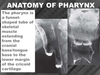
- 1. ANATOMY OF PHARYNX The pharynx is a funnel- shaped tube of skeletal muscle extending from the cranial base/tongue base to the lower margin of the cricoid cartilage
- 2. ANATOMY OF PHARYNX The pharynx lies anterior to the vertebral bodies of the cervical spine, prevertebral muscles, and loose connective tissue of the retropharyngeal space. The pharynx is confined laterally by the muscles of the neck, lateral portions of the hyoid bone and thyroid cartilage, and carotid sheath
- 3. ANATOMY OF PHARYNX The pharynx and larynx are intimately related, in embryologic origin and anatomically. The epiglottis and remainder of the supraglottis are of pharyngeal, not laryngeal origin.
- 4. ANATOMY OF PHARYNX 1. Superior surface of the tongue. 2. Tonsillar fossa. 3. Valleculae. 4. Lateral wall of the piriform sinus. 5. Piriform sinus 6. The median glossoepiglottic fold. 7. Surface of the base of the tongue
- 5. ANATOMY OF PHARYNX 1.Soft palate. 2.Base of the tongue. 3.Epiglottis. 4.Vallecula. 5.Pyriform fossa/sinus. 6.Posterior pharyngeal wall. 7.Pharyngo- esophageal region.
- 6. ANATOMY OF PHARYNX 1.Salpingopharyngeal folds. 2.Eustachian tube orifice. 3.Palatopharyngeal fold. 4.Soft palate. 5.Adenoidal lymphoid tissue. A C-shaped prominence is seen radiographically near the torus tubarius. The salpingopharyngeal fold overlying salpingopharyngeal muscle courses inferiorly from the torus along the lateral pharyngeal wall to the level of the soft palate. The posterior nasopharyngeal wall has a variably nodular surface because of underlying adenoidal tissue.
- 7. ANATOMY OF PHARYNX The vertical (pharyngeal) surface of the base of the tongue is variably nodular because of underlying lymphoid tissue of the lingual tonsil
- 8. ANATOMY OF PHARYNX 1.Median glossoepiglottic fold. 2.Vallecula. 3.Pharyngoepiglottic fold. 4.Epiglottic tip. 5.Aryepiglottic fold. 6.Interarytenoid notch. The median glossoepiglottic fold overlies the glossoepiglottic ligament, which courses from the base of the tongue to the epiglottis. The median glossoepiglottic fold divides the space between the tongue and the epiglottis into two sacs, the valleculae.
- 9. ANATOMY OF PHARYNX 1.Uvular tip. 2.Paired pharyngoepiglottic folds. 3.Valleculae. 4.Anterior walls of pyriform fossae. 5.Muscular processes of aretenoid cartilages. The lateral glossoepiglottic folds form the lateral walls of the valleculae. The pharyngoepiglottic folds course from the posterolateral portion of the valleculae into the lateral pharyngeal wall
- 10. ANATOMY OF PHARYNX The tonsillar fossa forms part of the lateral oropharyngeal wall. Each tonsillar fossa is bounded anteriorly by a palatoglossal fold (anterior tonsillar pillar; and posteriorly by a palatopharyngeal fold (posterior tonsillar pillar) overlying the palatopharyngeal muscle
- 11. ANATOMY OF PHARYNX During swallowing, redundant mucosa along the anterior wall of the pharyngoesophageal segment ust posterior to the cricoid cartilage may create an undulating or plaquelike contour. To rule out a subtle stricture, web, or infiltrating lesion, the radiologist must be certain that this mucosal nodularity changes size and shape and flattens during swallowing.