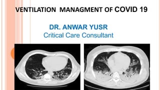
Covid 19 mechanical ventilation managment
•Descargar como PPTX, PDF•
8 recomendaciones•244 vistas

critical care

Recomendados
Recomendados
Más contenido relacionado
La actualidad más candente
La actualidad más candente (20)
Similar a Covid 19 mechanical ventilation managment
Similar a Covid 19 mechanical ventilation managment (20)
Más de Anwar Yusr
Más de Anwar Yusr (10)
Último
Último (20)
Covid 19 mechanical ventilation managment
- 1. VENTILATION MANAGMENT OF COVID 19 DR. ANWAR YUSR Critical Care Consultant
- 2. The interaction between three factors: 1) The severity of the infection, the host response, physiological reserve and comorbidities; 2) The ventilatory responsiveness of the patient to hypoxemia; 3) The time elapsed between the onset of the disease and the observation in the hospital. The interaction between these factors leads to the development of a time-related disease spectrum within two primary “phenotypes”: Type L characterized by Low elastance (i.e., high compliance), Low ventilation-to- perfusion ratio, Low lung weight and Low recruitability. Type H characterized by High elastance, High right-to left shunt, High lung weight and High recruitability. COVID-19 Pneumonia Phenotypes
- 3. COVID-19 pneumonia, Type L Low elastance. Low ventilation-to-perfusion (VA/Q) ratio. Since the gas volume is nearly normal, hypoxemia may be best explained by the loss of regulation of perfusion and by loss of hypoxic vasoconstriction. Accordingly, at this stage, the pulmonary artery pressure should be near normal. Low lung weight. Only ground-glass densities are present on CT scan, primarily located sub-Pleurally and along the lung fissures. Consequently, lung weight is only moderately increased. Low lung Recruitability. Therefore, severe hypoxemia is primarily due to ventilation/perfusion (VA/Q) mismatch. High PEEP and prone positioning do not improve oxygenation through recruitment of collapsed areas.
- 4. COVID-19 pneumonia, Type H High Elastance. The decrease in gas volume due to increased edema accounts for the increased lung elastance. High right-to-left shunt. This is due to the fraction of cardiac output perfusing the non-aerated tissue which develops in the dependent lung regions due to the increased edema and superimposed pressure. High lung weight. Quantitative analysis of the CT scan shows a remarkable increase in lung weight (> 1.5 kg), on the order of magnitude of severe ARDS. High lung Recruitability. The increased amount of non-aerated tissue is associated, as in severe ARDS, with increased recruitability. The Type H pattern, 20–30% of patients in our series, fully fits the severe ARDS criteria: hypoxemia, bilateral infiltrates, decreased the respiratory system compliance, increased lung weight and potential for recruitment.
- 6. Type L lung weight (1192 g), gas volume (2774 ml), percentage of non-aerated tissue (8.4%), venous admixture (56%), P/F (68), and respiratory system compliance (80 ml/cmH2O). Type H lung weight (1441 g), gas volume (1640 ml), percentage of non-aerated tissue (39%), venous admixture (49%), P/F (61), and respiratory system compliance (43 ml/cmH2O). Type L Type H
- 7. Type L ( Type 1) patients: PEEP levels should be kept lower in patients with high pulmonary compliance. Tidal volume thresholds should not be limited at 6 ml/kg. Respiratory rate should not exceed 20 breaths/min. Patients should be left “quiet”; avoiding doing too much is of higher benefit than intervening at any cost. Type H (Type 2) patients: Standard treatment for severe ARDS should be applied (lower tidal volume, prone positioning, and relatively high PEEP).
- 8. Respiratory management Goals of Oxygen therapy: 1. Target saturation SpO2 92%-96%. 2. Maintain stable work of breathing: Goal respiratory rate < 24. Target normal respiratory effort (no signs of accessory muscle use or obvious increased respiratory work). Supplemental oxygen support: 1. Initial oxygen delivery should be humidified nasal cannula (NC) titrated from 1 to 6 LPM to meet goals of therapy. 2. If goals of therapy are not met at 6 LPM NC then advance to either: Oxymizer mustache: Initiate at 6 LPM. Titrate to maximum of 12 LPM to meet goals of therapy. Venturi mask Initiate at 12 LPM and FiO2 40%. Titrate to maximum of FiO2 60% to meet goals of therapy.
- 9. Respiratory management Start by oxygen mask 5 L/min. Escalate to face mask with bag 10-15 L/min. Consider awake prone positioning with non-rebreathing mask. Criteria of failure of oxygen therapy with face mask. ▪ Respiratory rate > 30 /min and SPo2 < 90% or PaO2 < 60 mmHg. Escalate oxygen therapy to high flow nasal oxygen (if available): Start by FIO2 100%. Flow rate start @ 40 L/min to minimize aerosol spread and escalate to 60 L/min. Consider prone position with HFNC. Monitor for 1 to 2 hours. if reached the target continue on HFNC. if failed or HFNC is not available short trial of non-invasive ventilation.
- 10. Apply airborne precautions. Pt in isolation room. Pt wear surgical mask or N95 during HFNC. Consider using high-flow oxygen systems if patient is: awake, cooperative. with normal haemodynamics. and without urgent need for intubation. (PaCO2 < 45 mmHg). Safe when compared with NIV in patients with ARDS: may be associated with less mortality. nearly 40% of patients still require intubation. If high flow tried and unsuccessful DO NOT delay High Flow Oxygen Systems intubation.
- 11. Non-Invasive Ventilation NIPPV: Initiation of NIPPV (bilevel positive airway pressure [BiPAP]/ continuous positive airway pressure [CPAP]) requires attending approval; strongly recommended to avoid NIPPV (BiPAP/CPAP) in persons under investigation and confirmed COVID-19 cases. Rare exceptions are No intubation for those with acute indications for NIV or HFNC. Patients who use NIV chronically or are currently stable or improving on NIV or HFNC. Exacerbations that are expected to have a rapid reversal such as congestive heart failure. Extubation failure or high risk for reintubation. Equipment shortages in which milder disease could be managed to save invasive ventilation devices.
- 12. Non-Invasive Ventilation Use of non-invasive ventilation is controversial. Because the atelectasis is the main cause of hypoxemia in these patients, CPAP is preferred over BiPAP. Initial settings for high level of CPAP 13-15 cmH2O. Titrate FIO2 against oxygen saturation. Falling FIO2 indicate successful therapy. Predictors of failure (the patient should be evaluated within 30-60min after starting NIV) High Tidal volume exceeding 9.5 ml/kg predicts excess work of breathing Progressive worsening of blood gases. Increase work of breathing.
- 13. Non-Invasive Ventilation NIV is continuous positive airway pressure (CPAP) or bi-level positive airway pressure delivered via a tight- fitting mask. Not generally recommended for treatment of patients with ARDS: may preclude achieving low tidal volumes and adequate PEEP level. complications: facial skin breakdown, poor nutrition, failure to rest respiratory muscles. If used, apply airborne precautions.It can be difficult to achieve a tight-fit with face masks in children and infants. EMERGENCIES|
- 14. Some experts use NIV in carefully selected patients with mild ARDS: cooperative, stable haemodynamics, few secretions, without urgent need for intubation. Can be used as a temporizing measure until IMV is initiated. If NIV tried and unsuccessful, do not delay intubation: i.e. inability to reverse gas exchange dysfunction within 2–4 hours. Non-Invasive Ventilation |
- 15. GAP: Escalation to invasive ventilation G: Gas exchange abnormality: COVID-19 respiratory failure is usually hypoxemic, not hypercarbic. Worsening oxygenation: PaO2/FIO2 or SpO2/FiO2 <150. NIV with FIO2 >0.6 and can’t maintain SpO2 >90%. Oxygenation unresponsive to HFNC therapy. Hypercapnia with acidosis, pH <7.3. Increased work of breathing suggests deterioration of respiratory function. A: Airway protection: Altered mental status attributed to respiratory failure. Neurological dysfunction. P: Pulmonary toilet: Increased airway secretions.
- 16. Indication of intubation: Worsening hypercarbia. Acidemia. Altered mental status. Fatigue. Hemodynamic instability. Indications of mechanical ventilation: Respiratory exhaustion. Refractory severe hypoxemia (PaO2 <50-60 mmHg) on maximal oxygen therapy. Progressive CO2 retention with academia. Failure of NIV. Non-respiratory indications especially refractory hemodynamic compromise and Deep coma.
- 17. Intubation protocol Apply disposable mask, goggles, footwear, gown and gloves. Consider adopting the double glove technique. Designate the most experienced anesthesia professional available to perform intubation. Avoid awake fiberoptic intubation unless specifically indicated. Use filters between the bag valve & ET tube, and on the ventilator circuit. Ensure the absence of leaks on patient circuit. Having vasopressor for bolus or infusion immediately available for managing hypotension. Ensure the placement of a high quality HMEF. Place nasogastric tube after tracheal intubation. Re-sheath the laryngoscope immediately post intubation (double glove technique).
- 20. Ventilatory management protocol: 1. Calculate Ideal Body Weight Set Initial respiratory rate Typical starting rates will be 16-24 titrated to goal minute ventilation of 5-8 L/min. Consider starting rates of 24-28 titrated to goal minute ventilation of 8-12 L/min in setting of acidosis (pH < 7.25) pre-intubation.
- 21. Initial settings Ventilator settings: Lung protective ventilation Initial mode of ventilation: Assist control/ PRVC Tidal volume: 6 mL/kg PBW (calculate this from height and gender) Male patients: 50 + 2.3 [height (inches) – 60] Female patients: 45.5 + 2.3 [height (inches) – 60] PEEP 10 cm H2O: Monitor hemodynamics with increasing PEEP. Respiratory rate: 20-25 Consider patients’ preintubation respiratory rate. Goal: Limit overdistention of alveoli and ensure adequate oxygenation and ventilation. Overdistention causes inflammation, organ dysfunction, decreased venous return, and worsens ARDS.
- 22. Maintenance: Goals of therapy Oxygenation PaO2 > 60 / SpO2 88-98% FIO2 to maintain a SpO2 of 88-98% FIO2 <0.6 Try to avoid 100% oxygen, which favors de-nitrogen atelectasis. Lower FIO2 of 0.7-0.9 may not drastically change oxygenation due to high levels of shunt. Ventilation Tidal volumes of 4-8 mL/kg of PBW pH 7.25-7.42 PaCO2 40-65 / end-tidal carbon dioxide (ETCO2) 35-60 mm Hg Pulmonary Mechanics FIO2 PEEP FIO2 PEEP ARDSNet low PEEP/ FIO2 Chart Plateau pressures of ≤30 cm H2O (reflects respiratory system compliance) Peak inspiratory pressure <35 cm H2O 0.3 0.4 0.4 0.5 0.5 0.6 0.7 6 6 8 8 10 10 10 0.7 0.7 0.8 0.9 0.9 0.9 1 12 14 14 14 16 18 18-24
- 23. Modes of ventilation Primary ventilator modes Assist/control (A/C) mode: The ventilator delivers a set minimum number of mandatory breaths each minute. A/C mode can be used with either pressure control or volume control. Synchronous intermittent mandatory ventilation (SIMV) mode: The ventilator delivers a set minimum number of mandatory breaths each minute but also allows the patient to breathe spontaneously in between the mandatory breaths. SIMV can be used with either pressure control or volume control.
- 24. Modes of ventilation (cont.) Secondary ventilator modes Airway pressure release ventilation (APRV): APRV is an applied continuous positive airway pressure that at a set timed interval releases the applied pressure. Occasionally used in those with severe acute respiratory distress syndrome (ARDS). Pressure regulated volume control (PRVC): This is a pressure- controlled mode but adds a targeted tidal volume, so the inspiratory pressure changes breath-to-breath to achieve the targeted tidal volume.
- 25. When to troubleshoot Peak airway pressure greater than 35 cm H2O Evaluate the need for suctioning. Check plateau pressure. Check placement of ETT (deep?) and cuff pressure (do you hear a leak?). Evaluate for pneumothorax: Chest x-ray, ultrasound. Plateau pressure >30 cm H2O Requires an inspiratory hold maneuver. Reduce the tidal volume 1 mL/kg (minimum of 4 mL/kg). Consider diuresis. Consider paralysis. Adjust respiratory rate lower (usually 2-6/min per change) to increase CO2.
- 26. When to troubleshoot (cont.) FIO2 >0.6 with SpO2 <88% Increase PEEP to level indicated on chart: Monitor blood pressure with each PEEP increase. Consider positioning of patient (ie, proning). Consider diuresis. PH <7.25 Assess whether acidosis is respiratory or metabolic. Adjust respiratory rate higher (usually 2-6/min per change) to lower CO2 (max 35/min) If you go higher than a respiratory rate of 30, you will need to decrease the inspiratory time to 0.8 to avoid an inverse inspiratory-to-expiratory ratio. Monitor for auto-PEEP. Evaluate and treat metabolic abnormalities (check anion gap, lactate). PH >7.42 Adjust respiratory rate lower (usually 2-6/min per change) to increase CO2.
- 27. Refractory hypoxemia Call for help early Consider proning to improve V/Q ratio mismatch Assess cardiac function (myocarditis and cardiomyopathy are reported) Consider nitric oxide to improve V/Q ratio mismatch Consider paralysis Patient must be sedated with a benzodiazepine or propofol; analgesics do not provide amnesia for paralysis. Consider extracorporeal membrane oxygenation (ECMO)
- 28. Call for help SpO2 less than 88% on an FIO2 of 1.0 for more than 15 minutes despite troubleshooting. pH less than 7.25 for more than 2 blood gases. pH less than 7.10. PaO2 less than 40. SpO2/FIO2 or PaO2/FIO2 ratio of less than 150 for 2 hours. SpO2/FIO2 or PaO2/FIO2 ratio of less than 80. High-priority alarms (red) you cannot resolve within 2 minute Manually ventilate until help arrives. Low-priority alarms (yellow) you cannot resolve within 15 minutes
- 29. Clinical Pearls Ventilator kills your patient unless you prevent it from doing so! The best mode is the most suitable and comfortable mode for patients (not for you). Patient fight with your mistakes not with the ventilator! Calm your patient with opioids & Hypnotics, not relaxants. Nebulizers & Humidifiers are your guardian angels. Use lab. tests to confirm diagnosis not to diagnose. Plan and order nutrition professionally. Plan for and manage the stress of ETT & IPPV.