
Recomendados
Más contenido relacionado
La actualidad más candente
La actualidad más candente (20)
Destacado
Similar a Ankle Sprains
Similar a Ankle Sprains (20)
Más de Apeksha Besekar
Más de Apeksha Besekar (9)
Último
Último (20)
Ankle Sprains
- 1. By: Dr. Apeksha Besekar B.P.Th
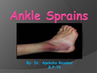
- 2. Introduction: A sprained ankle also known as, twisted ankle, rolled ankle, ankle injury or ankle ligament injury, is a common medical condition where there is (complete or partial) trauma to ligaments due to adduction or abduction violence that causes pain and disability depending on the degree of injury to the
- 3. Incidence: • Ankle sprains make about 15% of all athletic injuries. • These are particularly common in Basketball, Volleyball, Soccer players and Ballet dancers. • Most patients fully recover, but an estimated 20-40% develop chronic symptoms of pain and instability. • Incidence is increased in a) Individuals with varus malallignment of lower limbs. b) Individuals with calf muscle tightness
- 4. Relevant Anatomy • The ankle joint is a uniaxial joint which permits one degree of freedom; Dorsiflexion and Plantar flexion. • The stability of joint depends on the inherent constraints provided by the bony configuration and the active and passive soft tissue restraints. • Talocrural joint: Proximal articulating surface is formed by inferior surface of tibia, tibial malleolus,and fibular malleolus known as MORTISE. Distal articulating surface is
- 5. • Body of talus has 3 articular surfaces or facets: 1. Central portion: TROCHLEA. 2. Medial Facet. 3. Lateral facet. • During dorsiflexion the large anterior portion of the trochlea sits in the mortise, and during this the mortise expands. • During plantar flexion the narrow posterior portion of
- 6. • Active soft tissue restraint depends on the muscle tendon units involved in movement and support of the joint. • Passive support of the ankle is provided by the capsule of the joint which is in turn reinforced by the medial(deltoid ligament) and lateral collateral ligaments, posterior ligaments and the syndesmosis. • The lateral collateral ligament complex is the structure consisting of 3 sets of fibers: anterior and posterior talo fibular ligaments and third the calcaneo fibular ligament from
- 7. Mechanism of injury • The mechanism of injury is usually inversion of the plantar flexed foot, which involves an isolated tear of Anterior Talofibular Ligament, followed by a combined tear of the Anterior Talo fibular and the Calcaneofibular ligament.
- 8. Classification of Lateral Collateral Ligament Sprain GRADE 1 sprain: Mild ankle sprain a) Some stretching of ligaments with no macroscopic tear. b) No joint instability. c) Mild pain. d) There may be mild swelling or tenderness e) Some joint stiffness or difficulty walking or running. f) No functional impairment.
- 9. GRADE 2 sprain: Moderate ankle sprain. a) Partial tear of the ligament. b) Moderate swelling and tenderness. c) Moderate to severe pain, stiffness and difficulty in walking. d) Some loss of joint function. e) Mild joint instability. f) Minor bruising may be evident.
- 10. GRADE 3 sprain: Severe Sprain 1. Complete tear of the ligaments(ATFL and CFL) 2. Severe swelling and ecchymosis and tenderness. 3. Severe pain initially followed later by no pain. 4. Inability to bear weight on the extremity. 5. Mechanical joint
- 11. Diagnosis: • An inversion injury is commonly associated with a tearing sensation or a pop felt by the patient over the lateral ankle. • Swelling can be immediate in grades 2 and 3 sprains, and the initial intense pain subsides after a few hours, only to return more intensely as the hemorrhage continues 6 to 12 hours after injury.
- 12. Assessment: Aims: 1. To assess the degree of instability. 2. Grade of ligament damage. 3. Identify any reduction in range of motion or reduced strength. 4. Identify any other additional or associated injuries such as an avulsion fracture where a piece of bone at the end of a ligament has come away from the main
- 13. History Taking: • How did it happen? • Was there any pain at the time? • Was the pain sudden onset or gradual? • Was there any swelling and was it sudden onset or gradual? - a sudden swelling often indicates a bleeding into the joint rather than a gradual increase in synovial fluid within the joint. • Did you hear any noises? - this could indicate ligaments tearing or bone breaking. • Did you apply any emergency procedures such as RICE? • Is there anything you do which makes it worse / better?
- 14. Physical Examination: Active movements • The patient moves the foot from plantar flexion to dorsiflexion. • Looking for reduction in normal range of movement and any pain in performing these movements. • Then repeat moving from eversion to inversion
- 15. Passive movements: • The therapist moves the ankle and foot from plantar flexion to dorsi flexion and then inversion to eversion looking again at range of movement, comparing one foot with the other and painful movements. • Any pain at the extreme range of inversion may indicate ligament damage as it is the ligament that is being stressed. • The anterior drawer test is a special test which assesses the integrity of the ankle ligaments, particularly the anterior Talofibular ligament
- 16. Anterior drawer test • It’s a test for ligament instability. • Grasp the patient’s foot at the heel and pull forward while maintaining the tibia in a fixed position with the other hand at the anterior distal tibia. Translation greater than 3mm or difference in anterior translation from the asymptomatic ankle suggests the tear of the ATFL. • Excessive anteroposterior translation of the tibia on the
- 17. Resisted movements: • The therapist gently resists the movements as they try to move the ankle from inversion to eversion. • Pain when performing this test may be an indication of tendon damage or inflammation (possibly peroneal tendons) as it is the tendons connecting muscle to bone that are
- 18. Syndesmosis injury: Disruption of the syndesmosis ligament complex (tibiofibular ligaments and interosseous membrane). Rupture of the syndesmosis is often associated with deltoid ligament rupture ,and fracture of fibula is common. Mechanism may be pronation and eversion of the foot combined with internal rotation of tibia on a fixed foot, such as occurs in football players who have an external rotation force applied to the foot (stepped on) while lying
- 19. Clinical features: • Point tenderness and pain are located primarily on the anterior aspect of the syndesmosis. • The patient is unable to bear weight. • This injury is more severe than ankle sprains, with more pain swelling and difficulty in weight bearing. • Two tests are performed to diagnose this injury: 1. Squeeze test 2. External rotation test.
- 20. Squeeze test: • It is used to evaluate the syndesmotic ligaments of the ankle. • Performed by grasping the anterior of the leg proximally and squeezing the tibia and fibula, thus compressing the interosseous ligaments. • If the injury exists, the patient complains of distal ankle pain at the joint.
- 21. External rotation test: • Performed with patient’s ankle in neutral position and the knee flexed 90 degrees. Stabilizing the tibia and fibula with one hand, the therapist rotates the ankle with other hand. • Pain in the syndesmotic area indicates injury to the same.
- 22. Radiographic evaluation: Radiographs should include 3 views of the ankle: a) AP view b) Lateral view c) Mortise views To rule out fractures of the medial and lateral malleoli, the talus, and the fifth metatarsal base.
- 23. References: 1. Clinical Orthopaedic Rehabilitation (second edition) by S.Brent Brotzman, M.D. 2. Outline of Fractures(including joint injuries) (eleventh edition) by John Crawford Adams and David L. Hamblen. 3. Taber’s Cyclopedic medical dictionary.(20th edition) 4. Joint structure and Function a Comprehensive analysis. (fourth edition)by Cynthia C. Norkin 5. Google search.