

Recomendados
Más contenido relacionado
La actualidad más candente
La actualidad más candente (19)
Similar a Synchrotron absas
Similar a Synchrotron absas (20)
Último
Último (20)
Synchrotron absas
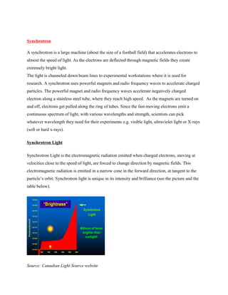
- 1. Synchrotron A synchrotron is a large machine (about the size of a football field) that accelerates electrons to almost the speed of light. As the electrons are deflected through magnetic fields they create extremely bright light. The light is channeled down beam lines to experimental workstations where it is used for research. A synchrotron uses powerful magnets and radio frequency waves to accelerate charged particles. The powerful magnet and radio frequency waves accelerate negatively charged electron along a stainless steel tube, where they reach high speed. As the magnets are turned on and off, electrons get pulled along the ring of tubes. Since the fast-moving electrons emit a continuous spectrum of light, with various wavelengths and strength, scientists can pick whatever wavelength they need for their experiments e.g. visible light, ultraviolet light or X-rays (soft or hard x-rays). Synchrotron Light Synchrotron Light is the electromagnetic radiation emitted when charged electrons, moving at velocities close to the speed of light, are forced to change direction by magnetic fields. This electromagnetic radiation is emitted in a narrow cone in the forward direction, at tangent to the particle’s orbit. Synchrotron light is unique in its intensity and brilliance (see the picture and the table below). Source: Canadian Light Source website
- 2. Another uniqueness of synchrotron light is that it can be generated across the range of the electromagnetic spectrum including visible, infra-red, ultra-violet, 'soft' and 'hard' x-ray and microwaves. Synchrotron scientists are trying to push synchrotron light farther into the X-ray and microwave regions and the brightness has improved over years as shown in the picture above. The table below shows the brightness of synchrotron light compared to other lights. Brightness is measured by roughly calculating the number of photons that strike a 1mm² samples during one second exposure. Comparative brightness of lights from various sources Type of light Photons per second per mm2 Synchrotron Light 10 000 000 000 000 000 000 Sunlight 10 000 000 000 000 Candle 1 000 000 000 Medical X-ray 10 000 000 Other useful properties of synchrotron light are: - high energy beams to penetrate deeper into matter - small wavelengths permit the studying of tiny features, e.g. bonds in molecules; Nano scale objects - synchrotron beams can be coherent and/or polarized, permitting specific experiments - the synchrotron beam can be made to flash at a very high frequency, giving the light a time structure. Synchrotron light and electromagnetic spectrum The electromagnetic spectrum below shows wavelengths of radiation emitted by various sources and the names of the radiations based on their wavelength frequencies and sizes and lots more.Synchrotron light is not just limited to Synchrotrons. As mentioned above, the real name
- 3. of Synchrotron light is electromagnetic radiation. Anything that emits any form of electromagnetic radiation from microwaves to x-rays is emitting a form of Synchrotron light. The only difference is that Synchrotrons makes the radiation up to a billion times more brilliant than the normal radiation emitted by other man-made objects. Synchrotron in detail What are the parts of a Synchrotron? 1. Electron Gun
- 4. Electron Gun (odec.ca, n.d) A screen near the button is given a short, strong positive charge 125 times per second, which pulls the negative charge particles, the electrons, away from the disk. This procedure is similar to that found in a television picture tube. 2. Linear Accelerator (LINAC) Linear Accelerator The Electron gun feeds into the LINAC. Microwave radio frequency fields provide energy to accelerate the electrons to almost the speed of light. The speed of the electrons is approximately 3x10⁸ m/s. 3. Booster Ring (odec.ca,n.d)
- 5. As the electrons circulate in the Booster ring, they receive a boost in energy from approximately 250 MeV (Mega electron volt) to approximately 2.9 to 6 GeV (Giga electron volts) — power equivalent to about 2 billion flashlight batteries — from microwaves generated in a Radio Frequency Cavity. 4. Storage Ring, Beam line, and End Station (odec.ca,n.d) When the electrons have enough energy to produce light, an injection system transfers them from the booster ring to the storage ring. The process of transferring electrons from booster to storage ring occurs approximately once per second up to 600 cycles (about 10 minutes) as required to reach an average circulating current of. Once in the storage ring, the electrons will circulate for four to twelve hours producing photons every time the 6800kg dipole magnets change the direction of the flow of electrons. While the ring looks circular, it is really a series of 12 straight
- 6. sections. After each turn there is a photon port to allow the light to travel down the beam lines to the research stations. The magnets and steerage ring are shown in the picture. Applications of synchrotron • Developing "designer" molecules for pharmaceutical drugs • Optimization of seed oil biotechnology • Improving yields of plant natural health products • Continuing cancer and diabetes research • Watching living cells as they react to drugs. • Medicine and Pharmaceuticals Medicine and Pharmaceuticals Decoding proteins: Reducing need for insulin for diabetic patients Canadian Scientist Dr. Gerald Audette and his team are trying to reduce the need for insulin for diabetic patients using synchrotron light to study properties of proteins involved in glucose metabolism. Two proteins interacting to each other Dr. Audette's team is using powerful international synchrotron while waiting for the Canadian Light Source (CLS) beam lines to be commissioned. They are trying to invent a drug that will stop or more precisely reduce glucose creating proteins from interacting each other inside a diabetic patient without creating any side effects to the patient.
- 7. Micromachining Scientists are using synchrotron light to manufacture tiny machine parts. An everyday example is inkjet printer heads. So next time you print a document using inkjet printer heads remember you are using technology created by synchrotron light. Below is a picture of an inkjet printer head. Inkjet printer head What are the most import advantages/characteristics of synchrotron radiation compared to lab X-ray sources? The most important advantage of synchrotron radiation over a laboratory X-ray source is its brilliance. A synchrotron source like the ESRF has a brilliance that is more than a billion times higher than a laboratory source. Brilliance is a term that describes both the brightness and the angular spread of the beam. The difference between the two sources can be likened to the difference between a laser beam and a light bulb. Higher brilliance lets us see more detail in the material under study e.g. there is a greater precision in the diffraction of light from a crystal where both the angle and the intensity is significant and recorded by a detector. Cutting edge parts, equipments, techniques used The monochromators used are typically periodic, diffracting elements to select given energies of radiation. - Gratings are used for energies (roughly) below 1 keV. - For higher energies crystals are used which are natural three-dimensional "gratings", mainly high quality silicon crystals. For the ESRF, typical energies are between 5 and 60 keV, but there exists beam lines using lower and higher energies.
- 8. - There exists a further possibility of X-ray optical elements that allows one to select a narrow energy band. These elements are multilayers, a periodic arrangement of rather thin double layers, usually a metal and a lower density material. Prisms could be used in the visible range; however synchrotrons are mainly dedicated to producing much higher photon energies that is radiation with much shorter wavelengths. So prisms are not used to our knowledge in synchrotrons like the ESRF. Influence in nano science applications Is it possible with synchrotron X-rays to detect particulate matter in the Nano-range in human fluids (such as blood) and tell at the same time the composition of the particles (heavy metals, carbon etc.)? X-ray fluorescence imaging can be used to detect trace levels of heavy elements such as metals (but not carbon). If the nanoparticles contain elements such as Pt, Au, Cd, Ag or lanthanides that are not generally present in body fluids then trace quantities of these nanoparticles can be detected. The ESRF X-ray Nano probe can be used to visualize nanoparticles or clusters of nanoparticles if they are 100 nm or larger. Controversies The use of synchrotron radiation as a source for single crystal x-ray diffraction studies has recently been the subject of considerable discussion and controversy. In contrast to conventional sources, the polychromatic radiation produced by synchrotron emission can be monochromated over a large range of wavelengths to give an intense x-ray beam. References: 1.How does a synchrotron work? , viewed on 26th march 2012, http://www.odec.ca/projects/2005/shar5a0/public_html/how_does_a_synchrotron_work.htm#electro n_gun, 2.Applications of Synchrotron Light , viewed on 26th march 2012, http://www.odec.ca/projects/2005/shar5a0/public_html/applications_of_synchrotron_ligh.htm 3.Applications of Synchrotron radiation to protein crystallography, viewed on 26th march 2012, http://www.pnas.org/content/73/1/128.full.pdf
- 9. 4.New Approach to quantum corrections in synchrotron radation, viewed on 26th march 2012,http://www.sciencedirect.com/science/article/pii/0003491678901422 5. What is synchrotron light? , viewed on 26th march 2012,http://www.odec.ca/projects/2005/shar5a0/public_html/what_is_synchrotron_light.htm
- 10. 4.New Approach to quantum corrections in synchrotron radation, viewed on 26th march 2012,http://www.sciencedirect.com/science/article/pii/0003491678901422 5. What is synchrotron light? , viewed on 26th march 2012,http://www.odec.ca/projects/2005/shar5a0/public_html/what_is_synchrotron_light.htm