Dengue, dengue hemorrhagic fever, dengue shock syndrome
•
5 recomendaciones•658 vistas

Everything all about dengue.

Recomendados
Más contenido relacionado
La actualidad más candente
Similar a Dengue, dengue hemorrhagic fever, dengue shock syndrome
Similar a Dengue, dengue hemorrhagic fever, dengue shock syndrome (20)
Último
Último (20)
Dengue, dengue hemorrhagic fever, dengue shock syndrome
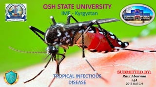
- 1. IMF - Kyrgyztan SUBMITTED BY: Ravi Abarnaa 14A 2016 BATCH
- 3. CONTENTS Introduction Dengue virus Epidemiology Etiology Pathophysiology Classification Clinical presentation Diagnosis Complications Management Prevention Vaccines
- 4. INTRODUCTION • Dengue fever is an acute infectious viral disease, also known as breakbone fever • Dengue is transmitted by mosquitoes of the genus. • Dengue hemorrhagic fever is a fatal manifestation of dengue virus that manifest with bleeding diathesis and hypovolemic shock.
- 5. DENGUE VIRUS • Flavi viruses: RNA • Arbovirus group • 4 serotypes – Den 1- 4 • Cycle involves humans and mosquitos • Infection with one virus gives immunity to that serotype only • Causes dengue and dengue hemorrhagic fever
- 6. EPIDEMIOLOGY • First reported epidemics in 1779 –80 in Asia, Africa and North America. • Considered a mild non fatal disease • Epidemics every 10-40 years due to introduction of new serotype • After World War II, pandemic of dengue which began in Southeast Asia, expanded geographical distribution, epidemics with multiple serotypes and emergence of DHF
- 7. • 1980s: a second re-expansion of DHF in Asia with epidemics in India, Sri Lanka and Maldives, Taiwan, PRC; Africa and Americas • Progressively larger epidemics • Primarily urban
- 8. Mainly in Urban and Semi Urban area
- 9. DENGUE GLOBALLY 390 million dengue infections per year 22,000 deaths, mostly among children. South America, South-East Asia and Western Pacific regions are the most seriously affected.
- 10. Why the no. of cases keep increases worldwide ?? Increased air travel Uneffective mosquito control Unreliable drainage systems Increasing population
- 11. ETIOLOGY • Vector - Aedes aegypti* ▪ bite during daytime ▪ Lays egg in clean & stagnant water ▪ Female feeds on blood - Aedes alboticus AEDES EGYPTI AEDES ALBOPTICUS
- 13. PATHOPHYSIOLOGY
- 14. TRANSMISSION
- 15. DENGUE CLASSIFICATION There are actually four dengue clinical syndromes: • Undifferentiated fever; • Classic dengue fever; • Dengue hemorrhagic fever, or DHF; and • Dengue shock syndrome, or DSS. • Dengue shock syndrome is actually a severe form of DHF.
- 16. Classic Dengue Fever Dengue hemorrhagic Fever ( > chances in ? ) Dengue Shock Syndrome In critical phase , Might **Monitor Warning Signs*** Without or without haemorrhage Clinical Manifestation
- 17. PATHOGENESIS OF PRIMARY INFECTION Incubation period : 4-7 days (range 3-14) Primary Dengue Infection – Self Limited May also progress to severe dengue (DHF/DSS) (normally children, elderly & immunocompromised PATHOGENESIS OF SECONDARY INFECTION “Antibody dependent enhancement mechanism” Infection by virus of another serotype Production of non neutralizing antibodies Facilitate entry of virus to monocytes through Fc Receptor
- 18. More Cytokines Released Acute increase in vascular permeability Hypovolaemia or shock or death Dengue Hemorrhagic Fever (DHF) Dengue Shock Syndrome (DSS) may
- 19. Clinical Manifestation Dengue Virus Infection Asymptomatic Symptomatic Undifferentiated fever (viral syndrome) Dengue fever Mostly Without hemorrhage With unusual hemorrhage Dengue hemorrhagic fever (plasma leakage) No shock Dengue shock syndrome Dengue fever Severe Dengue Secondary Infection
- 20. PHASES • Febrile Phase • Critical Phase • Recovery Phase
- 21. Febrile Phase x 7days High fever 40 °C (104 °F) headache generalized arthalgia myalgia petechiae bleeding from mucus membrane. Arash occurs in 50–80%
- 22. Critical Phase x 2days Leukopenia thrombocytopenia. Increase capillary permeability leading to plasma leakage that lead to metabolic acidosis. In children febrile phase is common carries nausea, vomiting, thrombocytopnea.
- 23. Recovery phase x 2-3 days Stabilize hemodynamic status increase urine output overall clinical improvement. Increase in fluid overload can cause cerebral edema.
- 24. SIGN & SYMPTOMS of dengue( Based on WHO ) • Fever, Chills , ( more than 105 ) • headache • Myalgia • Arthralgia • Retro-orbital pain • Deep bone pain – “break bone fever” • Rashes ( appear 4-5 days after fever ) • Positive Tourniquet • Test
- 25. Symptoms – Dengue Fever Positive tourniquet test Goal of the test :- To asses fragility of capillary walls To identify thrombocytopenia In DHF grade 1, a positive tourniquet test serves as the only indicator of haemorrhagic tendency • 20 or more petechiae per 1 square inch. (MOH MALAYSIA 2014)
- 26. WARNING SIGNS • Severe abdominal pain • Persistent vomiting • Vomit with blood • Drowsiness or irritability • Dyspnoea • Swollen lymph node • Prostration • diarrhea Raised HCT, with rapid fall in platelet Fever to hypothermia Mucosal Bleed Liver Enlargement Normal Male Hct 40.7 to 50.3% • Normal Female Hct: 36.1 to 44.3% • The normal number of platelets in the blood is 150,000 to 400,000 platelets per microliter (mcL).
- 27. The 4 WHO Criteria for DHF Fever Hemorrhagic manifestations(Symptoms) Low platelet count (100,000/mm 3 or less Elevated hematocrit ( >20% then normal) or ( > 50% THEN BASELINE)
- 28. Symptoms - Dengue Hemorrhagic Fever (DHF) • petechiae • epistaxis(nose bleed), • gingival bleeding (gum bleed) • Microscopic hematuria.
- 29. SYMPTOMS
- 30. DENGUE SHOCK SYNDROME • It can trigger Dengue Shock Syndrome – Massive bleeding – Death – Dehydration – Febrile convulsion
- 31. DIAGNOSIS History Clinical Lab • History tells us the endemic area, previous dengue infection and etc • Clinical diagnosis are all the symptoms. We can make only provisional diagnosis • Lab Diagnosis is the confirmatory
- 32. Lab Diagnosis – Is the Confirmatory test Tests include 1. Serological Test – ELISA – To Detect Antibody 2. Non Structural Protein (NS1 antigen) Test These 2 tests are most widely used diagnostic test OTHER TEST 1. Virus isolation 2. RT-PCR
- 33. 1. Non Structural Protein (NS1 antigen) Test • Latest diagnostic tool for diagnosing dengue • Useful in the diagnosing in the early phase (3 to 4 of illness) Some times even from second day of illness • But It is not useful after 5 days of illness . • Criteria for primary infection • Postive NS1 antigen • Criteria for secondary infection • Usually Negative NS1 antigen • rarely Can be postive as well
- 34. 2. Serological Test by ELISA – To Detect Antibody (Ig M and Ig G) • Criteria for primary infection Positive IgM after 5 to 7 days of illness Ig G present after 7 days • Criteria for secondary infection Positive Ig G after 5 to 7 days onwards Usually Absence or slight increase in IgM after 5 to 7 days onwards
- 35. Rapid Test Combo Kit • SD BIOLINE Dengue Duo • (To detect Dengue NS1 Ag and IgG/IgM in a single test )
- 37. OTHER TEST • Virus Isolation performed in the lab equipped with tissue culture and other virus isolation facilities. blood should be collected before day 5 of illness – before the formation of neutralizing antibodies. • It may take up to two weeks to complete the test and it is expensive. • PCR can be used as a diagnostic tool in early dengue infection . • It is not recommended as a routine diagnostic test due to limited availability and cost.
- 38. Lab Test for Provisional Diagnosis/ Screening Criteria and disease monitoring purpose Full Blood Count (FBC) White cell count (WCC) shows – 1 Leucopaenia 2 Thrombocytopaenia 3 Normal or raised HCT
- 40. COMPLICATION 1 Febrile phase - Dehydration 2 Critical phase - Shock from plasma leakage: severe haemorrhage; organ impairment = Dengue Shock Syndrome 3 Recovery phase - Hypervolaemia A small percentage of individual who have dengue fever can develop a more serious form of disease • Dengue haemorrhagic fever and disseminated intravascular coagulation • Hepatitis, cerebral haemorrhage or oedema, encephalitis, • cranial nerve palsies, rhabdomyolysis, myocarditis • Vertical transmission if infection within 5 wks of delivery
- 41. MOST OF YOU GET CONFUSED THAT WHAT IS DENGUE HEMMORAHAGIC FEVER AND DENGUE SHOCK SNDROME ALWAYS REMEMBER THEYARE THE COMPLICATION OF DENGUE FEVER
- 42. CONTROL AND PREVENTION • Vector Control • Individual Preventive Measures • Immunization - Sanofi Dengvaxia - All for types
- 43. TREATMENT • No specific treatment , only Supportive therapy • No antiviral agents are of proven value • Fluid replacement and Monitor the Ht and Platelet Count MANAGEMENT • Bleeding prevention & control • Fluid & water replacement • Symptoms relief & fever control
- 44. *Only* for severe cases ( DHF and DSS ) • Close monitoring of hypotension/shock • IV. Infusion of crystalloids/colloids • Oxygen administration • Platelet transfusion • Clotting factors replacement
- 46. PROGNOSIS • For the majority of peoples the people infected with dengue virus fever the prognosis is excellent. • Although they are likely to feel very ill during first 1-2 week of acute illness. • Overall the fatality rate is about 1% for all denge fever infection.
- 47. REFERENCE 1. k. Park , park’s textbook of preventive and social medicine, February 2011, 21st Edition, Published Banarsidas Bhanot Publishers 2. Nettina S. M., Lippincott manual of nursing practice ,2016, 10th edition, Jaypee brothers. 3. James S R , Kristine N, Jean A., Nursing Careof Children: Principles and Practice, 2014, 4th Edition,Elsevier. 4. D. L. Wong, L. F.Whaley, Essentials of Pediatric Nursing ,January 15, 1997,5 th edition,Mosby 5. Mentor : AP. DR. Durgadas , IMS – MSU 6. Book : Lange Microbiology 14th edition 7. Guidelines : MOH Malaysia 2014 and WHO 2014 8. Journal : International Medical Journal Malaysia ( IMJM) 9. Official Portal : Selangor Health Department 10. Online web site : Medscape 11. Picture Source : Flicker , Google Images