SlideShare una empresa de Scribd logo
1 de 83
Learn Chest X-Ray
With Its Normal
Positioning &
Radio-Anatomy
Dr. Santosh Atreya
MD Resident (Phase- A)
Radiology & Imaging
BSMMU from Nepal
Before the procedure…
• Bring a signed form from your doctor when
you visit the X-ray technician.
• Be aware that you may have to hold your
breath for a few seconds during the X-ray,
especially if the X-ray focuses on the stomach
or chest areas.
What do I need to remove for the
procedure?
• Any jewellery around the affected areas.
unless otherwise advised by your doctor.
LUNGS
Introduction
• Radiographic examination of the lungs is
performed for a wide variety of medical
conditions, including primary lung disease and
pulmonary effects of diseases in other organ
systems. Such effects produce significant
changes in the appearance of the lung
parenchyma and may vary over time
depending on the nature and extent of the
disease.
Recommended projections
Examination is performed by means of the following
projections:
• Basic Postero-anterior – erect
• Alternative Antero-posterior – erect
Antero-posterior – semi-erect
Antero-posterior -supine
• Supplementary
Lateral
Postero-anterior – expiration
Apices
Lateral – upper anterior region
Decubitus with horizontal beam
Positioning
• The choice of erect or decubitus technique is
governed primarily by the condition of the patient.
• With the patient erect, positioning is simplified,
control of respiration is more satisfactory, the
gravity effect on the abdominal organs allows for
the disclosure of the maximum area of the lung
tissue & fluid levels are defined more easily.
• Supplementary projections may be required for
specific indications at the request of a clinician or
radiologist.
Postero-anterior – erect
A 35 X 43-cm or 35 X 35-cm cassette is
selected, depending on the size of the
patient.
Position of patient and cassette
• The patient is positioned facing the cassette,
with the chin extended and centered to the
middle of the top of the cassette.
• The median sagittal plane is adjusted at
right-angles to the middle of the cassette. The
shoulders are rotated forward and pressed
downward in contact with the cassette.
• This is achieved by placing the dorsal aspect
of the hands behind and below the hips, with
the elbows brought forward, or by allowing
the arms to encircle the cassette.
….contd
Direction and centering of the X-ray beam
• The horizontal central beam is directed at
right-angles to the cassette at the level of the
eighth thoracic vertebrae (i.e. spinous
process of T7), which is coincident with the
lung midpoint
• The surface marking of T7 spinous process
can be assessed by using the inferior angle of
the scapula before the shoulders are pushed
forward.
• Exposure is made in full normal arrested
inspiration.These days automatic chest film
changer devices is in use.
Essential image
characteristics
• Full lung fields with the scapulae
projected laterally away from the
lung fields.
• The clavicles symmetrical &
equidistant from the spinous
processes & not obscuring the lung
apices.
• The lungs well inflated.
• The costophrenic angles &
diaphragm outlined clearly.
• The mediastinum & heart central &
defined sharply.
• The fine demarcation of the lung
tissues shown from the hilum to
the periphery.
Penetration
• Adequate penetration of
the patient is also required
for a good film.
• On a good PA film, the
thoracic spine disc spaces
should be barely visible
through the heart but bony
details of the spine are not
usually seen.
• On the other hand
penetration is sufficient
then bronchovascular
structures can usually be
seen through the heart.
•
Left, an example of a normal PA film that is
underpenetrated. Right, an overpenetrated PA film.
Underpenetration: Likelihood of missing an
abnormality
overlying by another structure
Overpenetration: results in loss of
visibility of low density lesion
e.g. early consolidation
•Structures seen in a PA
View
Trachea and Bronchi
• The tracheal lumen appears as
a vertical radiolucent shadow.
Around the aortic knuckle it
deviates slightly to the right.
• Normal maximum coronal
diameter: 25mm for males and
21mm for females.
• In proper erect posture
trachea bifurcates at the level
of lower border of 6th thoracic
vertebra
……contd
• The tracheal lumen is
bordered on the right by
a water density stripe,
called the right
paratracheal stripe, it is
seen in 60% of patients,
normally measuring
about less than 5mm.
• Lt paratracheal stripe is
not visualized.
…….contd
• Rt bronchus is shorter,wider and steeper than
left, which is longer, narrow and less oblique.
• .
The Heart
• The central dense shadow seen
on the PA chest film-
mediastinum, heart, spine and
sternum.
• With good centering, 2/3rd of the
cardiac shadow lies to the left of
midline & 1/3rd to the right.
• The normal cardio-thoracic ratio
< 50% in PA film in adult.in
children however, the CTR is
usually <60%.
• .
 Right border of heart is
formed by-
• Superior vena cava
• The right atrium
• Small part of inferior vena
cava
 Left heart border is formed by-
• aortic nuckle
• The pulmonary bay
• The auricle of left atrium
• The left ventricle.
……Contd
• In babies and young
children the normal
thymus is a triangular sail-
shaped structure with
well-defined borders
projecting from one or
both sides of the
mediastinum
Diaphragm
Diaphragm
• Dome shaped sheet of muscle
• Contains right hemidiaphrgm,central tendon
and left hemidiaphragm
• arches over abdominal contents
• Seperating abdomen from chest
….contd
• on inspiration the domes
are at the level of 6th rib
anteriorly and at or below
the 10th rib posteriorly
• In most patients the right
hemidiaphragm is higher
than the left.
Measurements of hyperinflation of
the lungs
• The normal dome of
each hemidiaphragm should
rise at least 1.5cm above a line
connecting the costophrenic
angle posteriorly and
sternophrenic angle anteriorly
OBLIQUE FISSURE OF LEFT LUNG
HORIZONTAL FISSURE
OBLIQUE FISSURE OF RIGHT LUNG
• The horizontal fissure of right lung may be
seen ,on the PA film extending from right
hilum to the region of the 6th rib in the axillary
line.
On a PA radiograph the main lobes overlap, so
the lung is divided into 3 zones separated by
imaginary horizontal lines 1.upper zone 2.mid
zone 3.lower zone.
• The central area on each
side is referred to as the
hilum of lung field. left hilum
is higher than the right.
• Hilum is formed by
pulmonary vessels,bronchus
and bronchopulmonary
lymph nodes .
• Of all the structures in the
hilum only the pulmonary
arteries and upper lobe veins
contribute significantly to the
hilar shadows.
• Normal bronchial walls are
only seen end-on.
• The apex of each lung field projects above the
medial third of clavicle.
• The infero-lateral corner of each lung field is called
lateral costophrenic angle.
• The infero-medial corner of each lung field is called
cardiophrenic angle.
Bones of chest wall
1. Clavicles:
• The radiograph shows almost entire length of
clavicle. The medial end of the clavicle lie
equidistant from the midline of the spinous
process of the 4th thoracic vertebra in proper
position.
• Rotation Can be assessed by observing the
clavicular heads and determining whether they
are equal distance from the spinous process of
the thoracic vertebral bodies.
2. Thoracic vertebrae
The radiopaque bodies of thoracic vertebrae
should be discernible within the cardiovascular
shadow.
3. Ribs
The radiograph shows the head and neck of most
of the ribs and the bodies of the upper 8 to 10 ribs.
4. Scapulae
The scapula project lateral to the supero-lateral
regions of the lung field in proper positioning.
Soft tissues
It is important to confirm the
presence or absence of breast
shadows.The breast may partially
obscure the lung bases.
Nipple shadows are nodular
opacities that are consistent in
shape, size and position
.
• oval or round
– 5-15mm in diameter
– between the 9th and 10th rib
posteriorly or the 5th and 6th
rib anteriorly
• Care is necessary to avoid mis-
interpretation as a neoplasm
or vice versa
LATERAL VIEW
Lateral view
A supplementary lateral
projection may be useful for
localizing the position of a
lesion, encysted pleural fluid
and demonstrating anterior
mediastinal masses not shown
on the postero-anterior
projection.
Position of patient and cassette
• The patient is turned to bring the side
under investigation in contact with the
cassette.
• The median sagittal plane is adjusted
parallel to the cassette.
• The arms are folded over the head or
raised above the head to rest on a
horizontal bar.
• The mid-axillary line is coincident with
the middle of the film, and the cassette is
adjusted to include the apices and the
lower lobes to the level of the first lumbar
vertebra.
Direction and centering of the X-ray
beam
• Direct the horizontal
central ray at right-angles
to the middle of the
cassette at the mid-
axillary line.
Postero-anterior and lateral radiographs of same patient
showing a tumour in the right lower lobe
Structures seen in lateral view
The clear spaces
• There are two clear spaces;retrosternal and
retrocardiac. these correspond to the sites
where the lungs meet behind the sternum and
the heart
• Loss of translucency of these areas indicates
local pathology.
• Normally the retrosternal space is less than 3
cm deep maximum.
Normal lateral film. Note
the retrosternal and
retrocardiac clear spaces
(open arrows) and the
increased translucency of the
lower vertebrae. The axillary
folds (straight black arrows)
and scapulae (curved black
arrows) overlie the lungs. The
tracheal translucency is well
seen (small black arrows)
Trachea
• Tracheal lumen appears as an vertical radiolucent
band which passes down slightly posterior
direction to the T6/7 level of the spine.
• Tracheal lumen is bordered posteriorly by a
water-density stripe that represents the posterior
tracheal wall and collapsed esophagus together
• The right upper lobe bronchus is seen end-on as a
circular structure overlying the lower trachea.
• Lying inferiorly is the left upper lobe bronchus
seen end-on .
Mediastinal viscera
 The chambers of the heart
cast a bulbous shadow
 The anterior wall of the right
ventricle defines the lower
half of the anterior border of
the cardiac shadow
 The posterior wall of left
ventricle defines the lower
third of the posterior border
of the cardiac shadow
 Posterior wall of left atrium
defines the middle third of
the posterior border of the
cardiac shadow
 The 3 segment of thoracic aorta cast a cane
shaped shadow
 The shadow of ascending aorta projects upward
from the superior limit of the anterior border of
the cardiac shadow
 The shadow of aortic arch arches over the cardiac
shadow and into the radiolucent space anterior to
the bodies of the 3rd and 4th thoracic vertebra
 The shadow of descending thoracic aorta parallels
the curved stack of the bodies of the lower
thoracic vertebrae
Diaphragm outline
• Both diaphragms are visible
throughout their length, except
the left anteriorly where it
merges with the heart. A small
segment of the right hemi
diaphragm is effaced by the IVC.
• The posterior costophrenic
angles are acute and small
amounts of pleural fluid may be
detected by blunting of these
angles.
Lungs
• All fissures of lung are clearly seen on lateral
film.
• The horizontal fissure runs anteriorly and
often slightly downwards
• Both oblique fissures commence posteriorly at
the level of T4 /T5,passing through the hilum.
The left ends 5cm behind the anterior
costophrenic angle and right ends just behind
the angle.
Bones and soft tissues of chest wall
• The radiograph shows the manubrium sterni
and body of sternum in profile.
• Shows the body of thoracic vertebrae, images
of the paired pedicles of each vertebra are
seen.
• The T4-T5 intervertebral disc lies at the level
of manubrio-sternal joint.
• The bodies of thoracic vertebra appear
progressively more radiolucent inferiorly.
• ANTERO POSTERIOR VIEW
Antero-posterior – erect
This projection is used as an alternative to the
postero-anterior erect projection for elucidation of an
opacity seen on a postero-anterior, or when the
patient’s shape or medical condition makes it difficult
or unsafe for the patient to stand or sit for the basic
projection.
Position of patient and cassette
• The patient may be standing or sitting with their back against
the cassette, which is supported vertically with the upper
edge of the cassette above the lung apices.
• The median sagittal plane is adjusted at right-angles to the
middle of the cassette.
• The shoulders are brought downward and forward, with the
backs of the hands below the hips and the elbows well
forward, which has the effect of projecting the scapulae clear
of the lung fields.
• In the unwell patient, it may not be possible to perform this
procedure, In this situation, it is preferable that the patient’s
arms are rotated laterally and supported with the palms of
the hands facing forward. In this position, the scapulae are
superimposed on the lungs but the effect of absorption is
less.
Direction and centering of the X-ray
beam
• The horizontal ray is directed first at right-angles
to the cassette and towards the sternal notch.
• The central ray is then angled until it is coincident
with the middle of the cassette. This has the effect
of confining the radiation field to the film, thus
avoiding unnecessary exposure of the eyes.
• The exposure is taken on normal full inspiration.
Normal antero-posterior radiograph
of thorax
• Antero-posterior - supine
Antero-posterior – supine
This projection is selected
when patients are unable to
either stand or sit for the
projections described
previously. The patient is
usually lying supine on a
trolley or bed.
Position of patient and cassette
• With assistance, a cassette is carefully positioned
under the patient’s chest with the upper edge of
the cassette above the lung apices.
• The median sagittal plane is adjusted at right-
angles to the middle of the cassette, and the
patient’s pelvis is checked to ensure that it is not
rotated.
• The arms are rotated laterally and supported by
the side of the trunk. The head is supported on a
pillow, with the chin slightly raised.
Direction and centering of the X-ray
beam
• The central ray is directed first at right-angles
and towards the sternal notch.
• The central ray is then angled until it is
coincident with the middle of the film, thus
avoiding unnecessary exposure to the eyes.
Normal supine
radiograph of thorax
• ANTERO-POSTERIOR SEMI ERECT
Antero-posterior – semi-erect
This semi-recumbent
position is adopted as an
alternative to the antero-
posterior erect projection
when the patient is too ill to
stand or sit erect without
support.
Normal semi-erect radiograph of thorax. The chin
is just superimposed on the upper thorax
DIFFERENCES OF PA & AP VIEW
S.N POSTERIOR ANTERIOR VIEW ANTEROPOSTERIOR VIEW
1. Clavicles are more horizontal,donot project towards the
apices
Projects towards the apices
2. Scapula are away from the lung field Scapula are towards the lung
field.
3. Reduces radiation dose to radiosensetive organs like
eyes,thyroid and breasts.
Radiation exposure
4. Magnification of heart is reduced Heart size is exaggerated .
5. X-rays pass from the posterior to the anterior of the
patient - hence Posterior-Anterior (PA) projection. The
image is viewed as if looking at the patient face-to-face.
X-rays pass from the anterior
to the posterior of the patient
- hence Anterior-Posterior (AP)
projection. The image is still
viewed as if looking at the
patient face-to-face.
6. Compression of breast tissue against the film cassette
reduces the density of tissue around the cp bases
therefore visualizing them more clearly
no any compressions.
•APICES
Apices
• Opacities obscured in
the apical region by
overlying ribs or
clavicular shadows may
be demonstrated by
modification of the
postero-anterior and
antero-posterior
projections.
Direction and centering of the X-ray beam
• With the patient in the position for the postero-anterior
projection, the central ray is angled 30 degrees caudally towards
the seventh cervical spinous process coincident with the sternal
angle.
• With the patient in the position for the antero-posterior
projection, the central ray is angled 30 degrees cephalad
towards the sternal angle.
• With the patient reclining, and the coronal plane at 30 degrees
to the cassette, to enable the nape of the neck to rest against
the upper border of the cassette, the central ray is directed at
right-angles to the film towards the sternal angle. Alternatively,
if the patient is unable to recline 30 degrees, the technique is
adapted, with the patient reclining 15 degrees and the tube
angled 15 degrees cephalad.
Normal postero-anterior 30 degrees caudad
Antero-posterior 30 degrees cephalad,
showing small tumour at the left apex
•LORDOTIC VIEW
Lordotic view
This technique may be used to demonstrate
right middle-lobe collapse or an inter-lobar
pleural effusion. The patient is positioned to
bring the middle-lobe fissure horizontal.
Position of patient and cassette
• The patient is placed for
the postero-anterior
projection.
• Then clasping the sides
of the vertical Bucky, the
patient bends backwards
at the waist.
• The degree of
dorsiflexion varies for
each subject, but in
general it is about 30–40
degrees.
Direction and centering of the X-ray beam
The horizontal ray is directed
at right-angles to the cassette and
towards the middle of the film.
Lordotic postero-anterior radiograph
showing middle lobe collapse
• Decubitus: the patient is lying down. In the decubitus
position,
• the patient may be lying in any of the following positions:
• • Supine (dorsal decubitus): lying on the back.
• • Prone (ventral decubitus): lying face-down.
• • Lateral decubitus: lying on the side. Right lateral decubitus
–
• lying on the right side. Left lateral decubitus – lying on the
• left side.
• • Semi-recumbent: reclining, part way between supine and
sitting
Heart and lungs – fluid level
Postero-anterior or antero-posterior (lateral decubitus)
This projection is used to confirm the presence of fluid. Moving the
patient into a different position causes movement of free fluid, so that
loculation is also detected. It may also be used to demonstrate the
lateral chest wall of the affected side clear of fluid, and to unmask any
underlying lung pathology.
Position of patient and cassette
• The patient is turned on to the lateral decubitus position and, if
possible, raised on to a supporting foam pad.
• A cassette is supported vertically against the anterior chest wall, and
the median sagittal plane is adjusted at right-angles to the cassette.
• The patient’s arms are raised and folded over the head to clear the
chest wall.
• Direction and centring of the X-ray beam
• • Centre to the level of the eighth thoracic
vertebra, with the central
• ray horizontal and directed at right-angles to the
cassette.
• Alternatively, an antero-posterior projection may
be taken, with
• the cassette supported against the posterior
aspect of the patient
Postero-anterior radiograph in lateral
decubitus position showing pleural effusion
with pneumothorax
Patient positioned for postero-anterior chest
(lateral decubitus) projection
Supine(dorsal decubitus)
This projection will show as much as possible of the lung fields, clear of
a fluid level, when the patient is unable to turn on their side.
Position of patient and cassette
• The patient lies supine and, if possible, is raised off the bed on a
supporting foam pad.
• The arms are extended and supported above the head.
• A cassette is supported vertically against the lateral aspect of the
chest of the affected side and adjusted parallel to the median sagittal
plane.
Direction and centring of the X-ray beam
• Centre to the axilla, with the central ray horizontal and directed at
right-angles to the cassette.
Patient positioned for lateral chest (dorsal
decubitus) projection
Lateral radiograph in dorsal decubitus position
showing pleural effusion with pneumothorax
Right anterior oblique
This projection is used to separate the heart, aorta and vertebral
column on a 35x 43-cm film. The projection will also demonstrate the
diameter and the degree of unfolding of the aorta.
Position of patient and cassette
• The patient is initially positioned facing the cassette, which is
supported vertically in the cassette holder with the upper edge above
the lung apices.
• With the right side of the trunk kept in contact with the cassette, the
patient is rotated to bring the left side away from the cassette, so that
the coronal plane forms an angle of 60 degrees to the cassette.
Direction and centring of the X-ray beam
• Direct the horizontal central ray at right-angles to the middle of the
cassette at the level of the sixth thoracic vertebrae, to show the heart,
aortic arch and descending aorta.
Right anterior oblique radiograph
Right anterior oblique
radiograph with barium outlining
the oesophagus
LIMITATIONS OF THE PLAIN CHEST FILM
First,the radiologist may fail to spot a lesion.Felson
reported that 20-30% of significant information
on a chest film may be over-looked by a trained
radiologist.
Second,a disease process may fail to appear as a
visible abnormality on a plain film.examples
includes miliary shadowing,metastasis,infective
process such as tuberculosis,histoplasmosis,and
pneumocystis,bronchiectasis and small pleural
effusions.such lesions are demonstrated earlier
using high resolution CT.
Learn Chest X-Ray With Its Normal Positioning & Radio-Anatomy

Más contenido relacionado

La actualidad más candente

Mammography physics and technique
Mammography  physics and techniqueMammography  physics and technique
Mammography physics and techniqueArchana Koshy
 
CT Procedure OF Abdomen & Pelvis
CT Procedure OF Abdomen & Pelvis CT Procedure OF Abdomen & Pelvis
CT Procedure OF Abdomen & Pelvis Upakar Paudel
 
Anterograde/Retrograde urethrography (RGU/MCU)
Anterograde/Retrograde urethrography (RGU/MCU)Anterograde/Retrograde urethrography (RGU/MCU)
Anterograde/Retrograde urethrography (RGU/MCU)Shubham Singhal
 
HRCT Chest - By Dr. Tinku Joseph
HRCT Chest - By Dr. Tinku JosephHRCT Chest - By Dr. Tinku Joseph
HRCT Chest - By Dr. Tinku JosephDr.Tinku Joseph
 
Radiograpic views for shoulder joint
Radiograpic views  for shoulder jointRadiograpic views  for shoulder joint
Radiograpic views for shoulder jointGanesan Yogananthem
 
Radiographic views of sacrum and coccyx
Radiographic views of sacrum and coccyxRadiographic views of sacrum and coccyx
Radiographic views of sacrum and coccyxChandan Prasad
 
Chest radiography ppt
Chest radiography pptChest radiography ppt
Chest radiography pptNiranjan Jha
 
CT ARTIFACTS.pptx
CT ARTIFACTS.pptxCT ARTIFACTS.pptx
CT ARTIFACTS.pptxdypradio
 
Chest decubitus
Chest decubitusChest decubitus
Chest decubitusSelf
 
Mammography positioning technique for MLO View
Mammography positioning technique for MLO View Mammography positioning technique for MLO View
Mammography positioning technique for MLO View Selin Prasad
 
Interpretation of X-Ray and other imaging
Interpretation of X-Ray and other imagingInterpretation of X-Ray and other imaging
Interpretation of X-Ray and other imagingdrmainuddin
 

La actualidad más candente (20)

Mammography physics and technique
Mammography  physics and techniqueMammography  physics and technique
Mammography physics and technique
 
Positioning of skull
Positioning of skullPositioning of skull
Positioning of skull
 
CT Procedure OF Abdomen & Pelvis
CT Procedure OF Abdomen & Pelvis CT Procedure OF Abdomen & Pelvis
CT Procedure OF Abdomen & Pelvis
 
CT ABDOMEN ANATOMY
 CT ABDOMEN ANATOMY CT ABDOMEN ANATOMY
CT ABDOMEN ANATOMY
 
Orbit imaging anatomy
Orbit imaging anatomyOrbit imaging anatomy
Orbit imaging anatomy
 
Abdomen radiography
Abdomen radiographyAbdomen radiography
Abdomen radiography
 
Anterograde/Retrograde urethrography (RGU/MCU)
Anterograde/Retrograde urethrography (RGU/MCU)Anterograde/Retrograde urethrography (RGU/MCU)
Anterograde/Retrograde urethrography (RGU/MCU)
 
CT CHEST ANATOMY
CT CHEST ANATOMYCT CHEST ANATOMY
CT CHEST ANATOMY
 
HRCT Chest - By Dr. Tinku Joseph
HRCT Chest - By Dr. Tinku JosephHRCT Chest - By Dr. Tinku Joseph
HRCT Chest - By Dr. Tinku Joseph
 
Radiograpic views for shoulder joint
Radiograpic views  for shoulder jointRadiograpic views  for shoulder joint
Radiograpic views for shoulder joint
 
Radiographic views of sacrum and coccyx
Radiographic views of sacrum and coccyxRadiographic views of sacrum and coccyx
Radiographic views of sacrum and coccyx
 
Abdominal CT scan
Abdominal CT scanAbdominal CT scan
Abdominal CT scan
 
Chest radiography ppt
Chest radiography pptChest radiography ppt
Chest radiography ppt
 
CT ARTIFACTS.pptx
CT ARTIFACTS.pptxCT ARTIFACTS.pptx
CT ARTIFACTS.pptx
 
Chest decubitus
Chest decubitusChest decubitus
Chest decubitus
 
CT artifact
CT artifact CT artifact
CT artifact
 
Mammography positioning technique for MLO View
Mammography positioning technique for MLO View Mammography positioning technique for MLO View
Mammography positioning technique for MLO View
 
Chest x ray
Chest x rayChest x ray
Chest x ray
 
Basics of CT chest
Basics of CT chestBasics of CT chest
Basics of CT chest
 
Interpretation of X-Ray and other imaging
Interpretation of X-Ray and other imagingInterpretation of X-Ray and other imaging
Interpretation of X-Ray and other imaging
 

Similar a Learn Chest X-Ray With Its Normal Positioning & Radio-Anatomy

Presentation2.pptx technique chest 2
Presentation2.pptx technique chest 2Presentation2.pptx technique chest 2
Presentation2.pptx technique chest 2Yashawant Yadav
 
Normal chest x ray and collapse
Normal chest x ray and collapseNormal chest x ray and collapse
Normal chest x ray and collapseAabid Rahiman
 
Interpretation of chest xray ppt
Interpretation of chest xray pptInterpretation of chest xray ppt
Interpretation of chest xray pptRithwik Karumuri
 
Basic Chest X ray Views - AP, PA & Lateral etc . pptx
Basic Chest X ray Views - AP, PA & Lateral etc . pptxBasic Chest X ray Views - AP, PA & Lateral etc . pptx
Basic Chest X ray Views - AP, PA & Lateral etc . pptxDr Abna J
 
Chest x ray - basics
Chest x ray - basicsChest x ray - basics
Chest x ray - basicsRikin Hasnani
 
X RAY DETERMINATION AND EVALUATION.pptx
X RAY DETERMINATION AND EVALUATION.pptxX RAY DETERMINATION AND EVALUATION.pptx
X RAY DETERMINATION AND EVALUATION.pptxShoaibKhatik3
 
xrayandotherimaging-180902063930.pdf
xrayandotherimaging-180902063930.pdfxrayandotherimaging-180902063930.pdf
xrayandotherimaging-180902063930.pdfEmmanuelOluseyi1
 
Chest radiography positioning and Technique.pptx
Chest radiography positioning and Technique.pptxChest radiography positioning and Technique.pptx
Chest radiography positioning and Technique.pptxDrsmcsideptofradiodi
 
Chest radiology
Chest radiologyChest radiology
Chest radiologyDr Vaziri
 
Thoracic positioning
Thoracic positioningThoracic positioning
Thoracic positioningKushagra Garg
 
Normal chest X ray radiography interpretation
Normal chest X ray radiography interpretationNormal chest X ray radiography interpretation
Normal chest X ray radiography interpretationAkhil Rohan
 
R NKUNA X_RAY INTERPRETATION 2020.pptx Physiotherapy
R NKUNA X_RAY INTERPRETATION 2020.pptx PhysiotherapyR NKUNA X_RAY INTERPRETATION 2020.pptx Physiotherapy
R NKUNA X_RAY INTERPRETATION 2020.pptx PhysiotherapySakhileKhoza2
 
The normal chest BY Dr Nikhil Bansal
The normal chest BY Dr Nikhil BansalThe normal chest BY Dr Nikhil Bansal
The normal chest BY Dr Nikhil BansalNikhil Bansal
 
Chest x rays BY Dr Anoop K R
Chest x rays BY Dr Anoop K RChest x rays BY Dr Anoop K R
Chest x rays BY Dr Anoop K Ranoop k r
 
Chest x ray anatomy - how to interpret chest x-ray (2)
Chest x ray anatomy - how to interpret chest x-ray (2)Chest x ray anatomy - how to interpret chest x-ray (2)
Chest x ray anatomy - how to interpret chest x-ray (2)Yusuf Shieba Elhamd
 

Similar a Learn Chest X-Ray With Its Normal Positioning & Radio-Anatomy (20)

Presentation2.pptx technique chest 2
Presentation2.pptx technique chest 2Presentation2.pptx technique chest 2
Presentation2.pptx technique chest 2
 
Normal chest x ray and collapse
Normal chest x ray and collapseNormal chest x ray and collapse
Normal chest x ray and collapse
 
Interpretation of chest xray ppt
Interpretation of chest xray pptInterpretation of chest xray ppt
Interpretation of chest xray ppt
 
chest.pptx
chest.pptxchest.pptx
chest.pptx
 
Basic Chest X ray Views - AP, PA & Lateral etc . pptx
Basic Chest X ray Views - AP, PA & Lateral etc . pptxBasic Chest X ray Views - AP, PA & Lateral etc . pptx
Basic Chest X ray Views - AP, PA & Lateral etc . pptx
 
Chest x ray - basics
Chest x ray - basicsChest x ray - basics
Chest x ray - basics
 
X RAY DETERMINATION AND EVALUATION.pptx
X RAY DETERMINATION AND EVALUATION.pptxX RAY DETERMINATION AND EVALUATION.pptx
X RAY DETERMINATION AND EVALUATION.pptx
 
chest-x-ray.pptx
chest-x-ray.pptxchest-x-ray.pptx
chest-x-ray.pptx
 
Chest X ray ppt.ppt
Chest X ray ppt.pptChest X ray ppt.ppt
Chest X ray ppt.ppt
 
xrayandotherimaging-180902063930.pdf
xrayandotherimaging-180902063930.pdfxrayandotherimaging-180902063930.pdf
xrayandotherimaging-180902063930.pdf
 
Chest radiography positioning and Technique.pptx
Chest radiography positioning and Technique.pptxChest radiography positioning and Technique.pptx
Chest radiography positioning and Technique.pptx
 
Chest radiology
Chest radiologyChest radiology
Chest radiology
 
Normal chest x ray
Normal chest x rayNormal chest x ray
Normal chest x ray
 
Thoracic positioning
Thoracic positioningThoracic positioning
Thoracic positioning
 
Normal chest X ray radiography interpretation
Normal chest X ray radiography interpretationNormal chest X ray radiography interpretation
Normal chest X ray radiography interpretation
 
R NKUNA X_RAY INTERPRETATION 2020.pptx Physiotherapy
R NKUNA X_RAY INTERPRETATION 2020.pptx PhysiotherapyR NKUNA X_RAY INTERPRETATION 2020.pptx Physiotherapy
R NKUNA X_RAY INTERPRETATION 2020.pptx Physiotherapy
 
The normal chest BY Dr Nikhil Bansal
The normal chest BY Dr Nikhil BansalThe normal chest BY Dr Nikhil Bansal
The normal chest BY Dr Nikhil Bansal
 
Chest x rays BY Dr Anoop K R
Chest x rays BY Dr Anoop K RChest x rays BY Dr Anoop K R
Chest x rays BY Dr Anoop K R
 
Chest x ray anatomy - how to interpret chest x-ray (2)
Chest x ray anatomy - how to interpret chest x-ray (2)Chest x ray anatomy - how to interpret chest x-ray (2)
Chest x ray anatomy - how to interpret chest x-ray (2)
 
Pediatric chest xray
Pediatric chest xrayPediatric chest xray
Pediatric chest xray
 

Más de Dr.Santosh Atreya

Focal liver lesions- in the eye of a radiologist
Focal liver lesions- in the eye of a radiologistFocal liver lesions- in the eye of a radiologist
Focal liver lesions- in the eye of a radiologistDr.Santosh Atreya
 
Chondrosarcoma of skull base,Radiology point of view
Chondrosarcoma of skull base,Radiology point of viewChondrosarcoma of skull base,Radiology point of view
Chondrosarcoma of skull base,Radiology point of viewDr.Santosh Atreya
 
Silent sinus syndrome-In the eye of a Radiologist
Silent sinus syndrome-In the eye of a RadiologistSilent sinus syndrome-In the eye of a Radiologist
Silent sinus syndrome-In the eye of a RadiologistDr.Santosh Atreya
 
Rasmussen encephalitis in the eye of a Radiologist
Rasmussen encephalitis in the eye of a RadiologistRasmussen encephalitis in the eye of a Radiologist
Rasmussen encephalitis in the eye of a RadiologistDr.Santosh Atreya
 
Intussusception in Adults-Submucosal Lipoma at Transverse colon-A rare cause
 Intussusception in Adults-Submucosal Lipoma at Transverse colon-A rare cause Intussusception in Adults-Submucosal Lipoma at Transverse colon-A rare cause
Intussusception in Adults-Submucosal Lipoma at Transverse colon-A rare causeDr.Santosh Atreya
 
Lhermitte Duclos Disease Radiology ll Dysplastic cerebellar gangliocytoma
Lhermitte Duclos Disease Radiology ll Dysplastic cerebellar gangliocytomaLhermitte Duclos Disease Radiology ll Dysplastic cerebellar gangliocytoma
Lhermitte Duclos Disease Radiology ll Dysplastic cerebellar gangliocytomaDr.Santosh Atreya
 
Metabolic & endocrine disorders affecting bone (Radiology)
Metabolic & endocrine disorders affecting bone (Radiology)Metabolic & endocrine disorders affecting bone (Radiology)
Metabolic & endocrine disorders affecting bone (Radiology)Dr.Santosh Atreya
 
Intraventricular mass (Radiology) of a child {A CASE}
Intraventricular mass (Radiology) of a child {A CASE}Intraventricular mass (Radiology) of a child {A CASE}
Intraventricular mass (Radiology) of a child {A CASE}Dr.Santosh Atreya
 
Know "Solitary Pulmonary Nodule" in a simple way !! (Radiology)
Know "Solitary Pulmonary Nodule" in a simple way !! (Radiology)Know "Solitary Pulmonary Nodule" in a simple way !! (Radiology)
Know "Solitary Pulmonary Nodule" in a simple way !! (Radiology)Dr.Santosh Atreya
 
Learn Barium Meal & Follow Through
Learn Barium Meal & Follow ThroughLearn Barium Meal & Follow Through
Learn Barium Meal & Follow ThroughDr.Santosh Atreya
 

Más de Dr.Santosh Atreya (12)

Focal liver lesions- in the eye of a radiologist
Focal liver lesions- in the eye of a radiologistFocal liver lesions- in the eye of a radiologist
Focal liver lesions- in the eye of a radiologist
 
Chondrosarcoma of skull base,Radiology point of view
Chondrosarcoma of skull base,Radiology point of viewChondrosarcoma of skull base,Radiology point of view
Chondrosarcoma of skull base,Radiology point of view
 
Silent sinus syndrome-In the eye of a Radiologist
Silent sinus syndrome-In the eye of a RadiologistSilent sinus syndrome-In the eye of a Radiologist
Silent sinus syndrome-In the eye of a Radiologist
 
Rasmussen encephalitis in the eye of a Radiologist
Rasmussen encephalitis in the eye of a RadiologistRasmussen encephalitis in the eye of a Radiologist
Rasmussen encephalitis in the eye of a Radiologist
 
Intussusception in Adults-Submucosal Lipoma at Transverse colon-A rare cause
 Intussusception in Adults-Submucosal Lipoma at Transverse colon-A rare cause Intussusception in Adults-Submucosal Lipoma at Transverse colon-A rare cause
Intussusception in Adults-Submucosal Lipoma at Transverse colon-A rare cause
 
Lhermitte Duclos Disease Radiology ll Dysplastic cerebellar gangliocytoma
Lhermitte Duclos Disease Radiology ll Dysplastic cerebellar gangliocytomaLhermitte Duclos Disease Radiology ll Dysplastic cerebellar gangliocytoma
Lhermitte Duclos Disease Radiology ll Dysplastic cerebellar gangliocytoma
 
Metabolic & endocrine disorders affecting bone (Radiology)
Metabolic & endocrine disorders affecting bone (Radiology)Metabolic & endocrine disorders affecting bone (Radiology)
Metabolic & endocrine disorders affecting bone (Radiology)
 
Intraventricular mass (Radiology) of a child {A CASE}
Intraventricular mass (Radiology) of a child {A CASE}Intraventricular mass (Radiology) of a child {A CASE}
Intraventricular mass (Radiology) of a child {A CASE}
 
Adrenal Adenoma Radiology
Adrenal Adenoma RadiologyAdrenal Adenoma Radiology
Adrenal Adenoma Radiology
 
Know "Solitary Pulmonary Nodule" in a simple way !! (Radiology)
Know "Solitary Pulmonary Nodule" in a simple way !! (Radiology)Know "Solitary Pulmonary Nodule" in a simple way !! (Radiology)
Know "Solitary Pulmonary Nodule" in a simple way !! (Radiology)
 
Learn Barium Meal & Follow Through
Learn Barium Meal & Follow ThroughLearn Barium Meal & Follow Through
Learn Barium Meal & Follow Through
 
Osteosarcoma (knee joint)
Osteosarcoma (knee joint)Osteosarcoma (knee joint)
Osteosarcoma (knee joint)
 

Último

College Call Girls Pune Mira 9907093804 Short 1500 Night 6000 Best call girls...
College Call Girls Pune Mira 9907093804 Short 1500 Night 6000 Best call girls...College Call Girls Pune Mira 9907093804 Short 1500 Night 6000 Best call girls...
College Call Girls Pune Mira 9907093804 Short 1500 Night 6000 Best call girls...Miss joya
 
Bangalore Call Girls Marathahalli 📞 9907093804 High Profile Service 100% Safe
Bangalore Call Girls Marathahalli 📞 9907093804 High Profile Service 100% SafeBangalore Call Girls Marathahalli 📞 9907093804 High Profile Service 100% Safe
Bangalore Call Girls Marathahalli 📞 9907093804 High Profile Service 100% Safenarwatsonia7
 
Russian Call Girl Brookfield - 7001305949 Escorts Service 50% Off with Cash O...
Russian Call Girl Brookfield - 7001305949 Escorts Service 50% Off with Cash O...Russian Call Girl Brookfield - 7001305949 Escorts Service 50% Off with Cash O...
Russian Call Girl Brookfield - 7001305949 Escorts Service 50% Off with Cash O...narwatsonia7
 
College Call Girls Vyasarpadi Whatsapp 7001305949 Independent Escort Service
College Call Girls Vyasarpadi Whatsapp 7001305949 Independent Escort ServiceCollege Call Girls Vyasarpadi Whatsapp 7001305949 Independent Escort Service
College Call Girls Vyasarpadi Whatsapp 7001305949 Independent Escort ServiceNehru place Escorts
 
Mumbai Call Girls Service 9910780858 Real Russian Girls Looking Models
Mumbai Call Girls Service 9910780858 Real Russian Girls Looking ModelsMumbai Call Girls Service 9910780858 Real Russian Girls Looking Models
Mumbai Call Girls Service 9910780858 Real Russian Girls Looking Modelssonalikaur4
 
Book Call Girls in Yelahanka - For 7001305949 Cheap & Best with original Photos
Book Call Girls in Yelahanka - For 7001305949 Cheap & Best with original PhotosBook Call Girls in Yelahanka - For 7001305949 Cheap & Best with original Photos
Book Call Girls in Yelahanka - For 7001305949 Cheap & Best with original Photosnarwatsonia7
 
Call Girl Service Bidadi - For 7001305949 Cheap & Best with original Photos
Call Girl Service Bidadi - For 7001305949 Cheap & Best with original PhotosCall Girl Service Bidadi - For 7001305949 Cheap & Best with original Photos
Call Girl Service Bidadi - For 7001305949 Cheap & Best with original Photosnarwatsonia7
 
Call Girls Hsr Layout Just Call 7001305949 Top Class Call Girl Service Available
Call Girls Hsr Layout Just Call 7001305949 Top Class Call Girl Service AvailableCall Girls Hsr Layout Just Call 7001305949 Top Class Call Girl Service Available
Call Girls Hsr Layout Just Call 7001305949 Top Class Call Girl Service Availablenarwatsonia7
 
VIP Call Girls Pune Vrinda 9907093804 Short 1500 Night 6000 Best call girls S...
VIP Call Girls Pune Vrinda 9907093804 Short 1500 Night 6000 Best call girls S...VIP Call Girls Pune Vrinda 9907093804 Short 1500 Night 6000 Best call girls S...
VIP Call Girls Pune Vrinda 9907093804 Short 1500 Night 6000 Best call girls S...Miss joya
 
Call Girls ITPL Just Call 7001305949 Top Class Call Girl Service Available
Call Girls ITPL Just Call 7001305949 Top Class Call Girl Service AvailableCall Girls ITPL Just Call 7001305949 Top Class Call Girl Service Available
Call Girls ITPL Just Call 7001305949 Top Class Call Girl Service Availablenarwatsonia7
 
Call Girls Jp Nagar Just Call 7001305949 Top Class Call Girl Service Available
Call Girls Jp Nagar Just Call 7001305949 Top Class Call Girl Service AvailableCall Girls Jp Nagar Just Call 7001305949 Top Class Call Girl Service Available
Call Girls Jp Nagar Just Call 7001305949 Top Class Call Girl Service Availablenarwatsonia7
 
Call Girls Electronic City Just Call 7001305949 Top Class Call Girl Service A...
Call Girls Electronic City Just Call 7001305949 Top Class Call Girl Service A...Call Girls Electronic City Just Call 7001305949 Top Class Call Girl Service A...
Call Girls Electronic City Just Call 7001305949 Top Class Call Girl Service A...narwatsonia7
 
Call Girl Lucknow Mallika 7001305949 Independent Escort Service Lucknow
Call Girl Lucknow Mallika 7001305949 Independent Escort Service LucknowCall Girl Lucknow Mallika 7001305949 Independent Escort Service Lucknow
Call Girl Lucknow Mallika 7001305949 Independent Escort Service Lucknownarwatsonia7
 
Call Girls Kanakapura Road Just Call 7001305949 Top Class Call Girl Service A...
Call Girls Kanakapura Road Just Call 7001305949 Top Class Call Girl Service A...Call Girls Kanakapura Road Just Call 7001305949 Top Class Call Girl Service A...
Call Girls Kanakapura Road Just Call 7001305949 Top Class Call Girl Service A...narwatsonia7
 
High Profile Call Girls Jaipur Vani 8445551418 Independent Escort Service Jaipur
High Profile Call Girls Jaipur Vani 8445551418 Independent Escort Service JaipurHigh Profile Call Girls Jaipur Vani 8445551418 Independent Escort Service Jaipur
High Profile Call Girls Jaipur Vani 8445551418 Independent Escort Service Jaipurparulsinha
 
Low Rate Call Girls Pune Esha 9907093804 Short 1500 Night 6000 Best call girl...
Low Rate Call Girls Pune Esha 9907093804 Short 1500 Night 6000 Best call girl...Low Rate Call Girls Pune Esha 9907093804 Short 1500 Night 6000 Best call girl...
Low Rate Call Girls Pune Esha 9907093804 Short 1500 Night 6000 Best call girl...Miss joya
 
Russian Call Girls Chickpet - 7001305949 Booking and charges genuine rate for...
Russian Call Girls Chickpet - 7001305949 Booking and charges genuine rate for...Russian Call Girls Chickpet - 7001305949 Booking and charges genuine rate for...
Russian Call Girls Chickpet - 7001305949 Booking and charges genuine rate for...narwatsonia7
 
Call Girls Frazer Town Just Call 7001305949 Top Class Call Girl Service Avail...
Call Girls Frazer Town Just Call 7001305949 Top Class Call Girl Service Avail...Call Girls Frazer Town Just Call 7001305949 Top Class Call Girl Service Avail...
Call Girls Frazer Town Just Call 7001305949 Top Class Call Girl Service Avail...narwatsonia7
 
Call Girls Whitefield Just Call 7001305949 Top Class Call Girl Service Available
Call Girls Whitefield Just Call 7001305949 Top Class Call Girl Service AvailableCall Girls Whitefield Just Call 7001305949 Top Class Call Girl Service Available
Call Girls Whitefield Just Call 7001305949 Top Class Call Girl Service Availablenarwatsonia7
 

Último (20)

College Call Girls Pune Mira 9907093804 Short 1500 Night 6000 Best call girls...
College Call Girls Pune Mira 9907093804 Short 1500 Night 6000 Best call girls...College Call Girls Pune Mira 9907093804 Short 1500 Night 6000 Best call girls...
College Call Girls Pune Mira 9907093804 Short 1500 Night 6000 Best call girls...
 
Bangalore Call Girls Marathahalli 📞 9907093804 High Profile Service 100% Safe
Bangalore Call Girls Marathahalli 📞 9907093804 High Profile Service 100% SafeBangalore Call Girls Marathahalli 📞 9907093804 High Profile Service 100% Safe
Bangalore Call Girls Marathahalli 📞 9907093804 High Profile Service 100% Safe
 
Russian Call Girl Brookfield - 7001305949 Escorts Service 50% Off with Cash O...
Russian Call Girl Brookfield - 7001305949 Escorts Service 50% Off with Cash O...Russian Call Girl Brookfield - 7001305949 Escorts Service 50% Off with Cash O...
Russian Call Girl Brookfield - 7001305949 Escorts Service 50% Off with Cash O...
 
College Call Girls Vyasarpadi Whatsapp 7001305949 Independent Escort Service
College Call Girls Vyasarpadi Whatsapp 7001305949 Independent Escort ServiceCollege Call Girls Vyasarpadi Whatsapp 7001305949 Independent Escort Service
College Call Girls Vyasarpadi Whatsapp 7001305949 Independent Escort Service
 
Mumbai Call Girls Service 9910780858 Real Russian Girls Looking Models
Mumbai Call Girls Service 9910780858 Real Russian Girls Looking ModelsMumbai Call Girls Service 9910780858 Real Russian Girls Looking Models
Mumbai Call Girls Service 9910780858 Real Russian Girls Looking Models
 
Book Call Girls in Yelahanka - For 7001305949 Cheap & Best with original Photos
Book Call Girls in Yelahanka - For 7001305949 Cheap & Best with original PhotosBook Call Girls in Yelahanka - For 7001305949 Cheap & Best with original Photos
Book Call Girls in Yelahanka - For 7001305949 Cheap & Best with original Photos
 
Call Girl Service Bidadi - For 7001305949 Cheap & Best with original Photos
Call Girl Service Bidadi - For 7001305949 Cheap & Best with original PhotosCall Girl Service Bidadi - For 7001305949 Cheap & Best with original Photos
Call Girl Service Bidadi - For 7001305949 Cheap & Best with original Photos
 
Call Girls Hsr Layout Just Call 7001305949 Top Class Call Girl Service Available
Call Girls Hsr Layout Just Call 7001305949 Top Class Call Girl Service AvailableCall Girls Hsr Layout Just Call 7001305949 Top Class Call Girl Service Available
Call Girls Hsr Layout Just Call 7001305949 Top Class Call Girl Service Available
 
VIP Call Girls Pune Vrinda 9907093804 Short 1500 Night 6000 Best call girls S...
VIP Call Girls Pune Vrinda 9907093804 Short 1500 Night 6000 Best call girls S...VIP Call Girls Pune Vrinda 9907093804 Short 1500 Night 6000 Best call girls S...
VIP Call Girls Pune Vrinda 9907093804 Short 1500 Night 6000 Best call girls S...
 
Call Girls ITPL Just Call 7001305949 Top Class Call Girl Service Available
Call Girls ITPL Just Call 7001305949 Top Class Call Girl Service AvailableCall Girls ITPL Just Call 7001305949 Top Class Call Girl Service Available
Call Girls ITPL Just Call 7001305949 Top Class Call Girl Service Available
 
Call Girls Jp Nagar Just Call 7001305949 Top Class Call Girl Service Available
Call Girls Jp Nagar Just Call 7001305949 Top Class Call Girl Service AvailableCall Girls Jp Nagar Just Call 7001305949 Top Class Call Girl Service Available
Call Girls Jp Nagar Just Call 7001305949 Top Class Call Girl Service Available
 
Call Girls Electronic City Just Call 7001305949 Top Class Call Girl Service A...
Call Girls Electronic City Just Call 7001305949 Top Class Call Girl Service A...Call Girls Electronic City Just Call 7001305949 Top Class Call Girl Service A...
Call Girls Electronic City Just Call 7001305949 Top Class Call Girl Service A...
 
Call Girl Lucknow Mallika 7001305949 Independent Escort Service Lucknow
Call Girl Lucknow Mallika 7001305949 Independent Escort Service LucknowCall Girl Lucknow Mallika 7001305949 Independent Escort Service Lucknow
Call Girl Lucknow Mallika 7001305949 Independent Escort Service Lucknow
 
Call Girls Kanakapura Road Just Call 7001305949 Top Class Call Girl Service A...
Call Girls Kanakapura Road Just Call 7001305949 Top Class Call Girl Service A...Call Girls Kanakapura Road Just Call 7001305949 Top Class Call Girl Service A...
Call Girls Kanakapura Road Just Call 7001305949 Top Class Call Girl Service A...
 
High Profile Call Girls Jaipur Vani 8445551418 Independent Escort Service Jaipur
High Profile Call Girls Jaipur Vani 8445551418 Independent Escort Service JaipurHigh Profile Call Girls Jaipur Vani 8445551418 Independent Escort Service Jaipur
High Profile Call Girls Jaipur Vani 8445551418 Independent Escort Service Jaipur
 
Low Rate Call Girls Pune Esha 9907093804 Short 1500 Night 6000 Best call girl...
Low Rate Call Girls Pune Esha 9907093804 Short 1500 Night 6000 Best call girl...Low Rate Call Girls Pune Esha 9907093804 Short 1500 Night 6000 Best call girl...
Low Rate Call Girls Pune Esha 9907093804 Short 1500 Night 6000 Best call girl...
 
Russian Call Girls Chickpet - 7001305949 Booking and charges genuine rate for...
Russian Call Girls Chickpet - 7001305949 Booking and charges genuine rate for...Russian Call Girls Chickpet - 7001305949 Booking and charges genuine rate for...
Russian Call Girls Chickpet - 7001305949 Booking and charges genuine rate for...
 
Escort Service Call Girls In Sarita Vihar,, 99530°56974 Delhi NCR
Escort Service Call Girls In Sarita Vihar,, 99530°56974 Delhi NCREscort Service Call Girls In Sarita Vihar,, 99530°56974 Delhi NCR
Escort Service Call Girls In Sarita Vihar,, 99530°56974 Delhi NCR
 
Call Girls Frazer Town Just Call 7001305949 Top Class Call Girl Service Avail...
Call Girls Frazer Town Just Call 7001305949 Top Class Call Girl Service Avail...Call Girls Frazer Town Just Call 7001305949 Top Class Call Girl Service Avail...
Call Girls Frazer Town Just Call 7001305949 Top Class Call Girl Service Avail...
 
Call Girls Whitefield Just Call 7001305949 Top Class Call Girl Service Available
Call Girls Whitefield Just Call 7001305949 Top Class Call Girl Service AvailableCall Girls Whitefield Just Call 7001305949 Top Class Call Girl Service Available
Call Girls Whitefield Just Call 7001305949 Top Class Call Girl Service Available
 

Learn Chest X-Ray With Its Normal Positioning & Radio-Anatomy

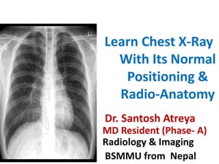
  • 1. Learn Chest X-Ray With Its Normal Positioning & Radio-Anatomy Dr. Santosh Atreya MD Resident (Phase- A) Radiology & Imaging BSMMU from Nepal
  • 2. Before the procedure… • Bring a signed form from your doctor when you visit the X-ray technician. • Be aware that you may have to hold your breath for a few seconds during the X-ray, especially if the X-ray focuses on the stomach or chest areas.
  • 3. What do I need to remove for the procedure? • Any jewellery around the affected areas. unless otherwise advised by your doctor.
  • 5. Introduction • Radiographic examination of the lungs is performed for a wide variety of medical conditions, including primary lung disease and pulmonary effects of diseases in other organ systems. Such effects produce significant changes in the appearance of the lung parenchyma and may vary over time depending on the nature and extent of the disease.
  • 6. Recommended projections Examination is performed by means of the following projections: • Basic Postero-anterior – erect • Alternative Antero-posterior – erect Antero-posterior – semi-erect Antero-posterior -supine • Supplementary Lateral Postero-anterior – expiration Apices Lateral – upper anterior region Decubitus with horizontal beam
  • 7. Positioning • The choice of erect or decubitus technique is governed primarily by the condition of the patient. • With the patient erect, positioning is simplified, control of respiration is more satisfactory, the gravity effect on the abdominal organs allows for the disclosure of the maximum area of the lung tissue & fluid levels are defined more easily. • Supplementary projections may be required for specific indications at the request of a clinician or radiologist.
  • 8. Postero-anterior – erect A 35 X 43-cm or 35 X 35-cm cassette is selected, depending on the size of the patient. Position of patient and cassette • The patient is positioned facing the cassette, with the chin extended and centered to the middle of the top of the cassette. • The median sagittal plane is adjusted at right-angles to the middle of the cassette. The shoulders are rotated forward and pressed downward in contact with the cassette. • This is achieved by placing the dorsal aspect of the hands behind and below the hips, with the elbows brought forward, or by allowing the arms to encircle the cassette.
  • 9. ….contd Direction and centering of the X-ray beam • The horizontal central beam is directed at right-angles to the cassette at the level of the eighth thoracic vertebrae (i.e. spinous process of T7), which is coincident with the lung midpoint • The surface marking of T7 spinous process can be assessed by using the inferior angle of the scapula before the shoulders are pushed forward. • Exposure is made in full normal arrested inspiration.These days automatic chest film changer devices is in use.
  • 10. Essential image characteristics • Full lung fields with the scapulae projected laterally away from the lung fields. • The clavicles symmetrical & equidistant from the spinous processes & not obscuring the lung apices. • The lungs well inflated. • The costophrenic angles & diaphragm outlined clearly. • The mediastinum & heart central & defined sharply. • The fine demarcation of the lung tissues shown from the hilum to the periphery.
  • 11. Penetration • Adequate penetration of the patient is also required for a good film. • On a good PA film, the thoracic spine disc spaces should be barely visible through the heart but bony details of the spine are not usually seen. • On the other hand penetration is sufficient then bronchovascular structures can usually be seen through the heart. •
  • 12. Left, an example of a normal PA film that is underpenetrated. Right, an overpenetrated PA film. Underpenetration: Likelihood of missing an abnormality overlying by another structure Overpenetration: results in loss of visibility of low density lesion e.g. early consolidation
  • 14. Trachea and Bronchi • The tracheal lumen appears as a vertical radiolucent shadow. Around the aortic knuckle it deviates slightly to the right. • Normal maximum coronal diameter: 25mm for males and 21mm for females. • In proper erect posture trachea bifurcates at the level of lower border of 6th thoracic vertebra
  • 15. ……contd • The tracheal lumen is bordered on the right by a water density stripe, called the right paratracheal stripe, it is seen in 60% of patients, normally measuring about less than 5mm. • Lt paratracheal stripe is not visualized.
  • 16. …….contd • Rt bronchus is shorter,wider and steeper than left, which is longer, narrow and less oblique. • .
  • 17. The Heart • The central dense shadow seen on the PA chest film- mediastinum, heart, spine and sternum. • With good centering, 2/3rd of the cardiac shadow lies to the left of midline & 1/3rd to the right. • The normal cardio-thoracic ratio < 50% in PA film in adult.in children however, the CTR is usually <60%. • .
  • 18.  Right border of heart is formed by- • Superior vena cava • The right atrium • Small part of inferior vena cava  Left heart border is formed by- • aortic nuckle • The pulmonary bay • The auricle of left atrium • The left ventricle.
  • 19. ……Contd • In babies and young children the normal thymus is a triangular sail- shaped structure with well-defined borders projecting from one or both sides of the mediastinum
  • 21. Diaphragm • Dome shaped sheet of muscle • Contains right hemidiaphrgm,central tendon and left hemidiaphragm • arches over abdominal contents • Seperating abdomen from chest
  • 22. ….contd • on inspiration the domes are at the level of 6th rib anteriorly and at or below the 10th rib posteriorly • In most patients the right hemidiaphragm is higher than the left.
  • 23. Measurements of hyperinflation of the lungs • The normal dome of each hemidiaphragm should rise at least 1.5cm above a line connecting the costophrenic angle posteriorly and sternophrenic angle anteriorly
  • 24. OBLIQUE FISSURE OF LEFT LUNG HORIZONTAL FISSURE OBLIQUE FISSURE OF RIGHT LUNG
  • 25. • The horizontal fissure of right lung may be seen ,on the PA film extending from right hilum to the region of the 6th rib in the axillary line. On a PA radiograph the main lobes overlap, so the lung is divided into 3 zones separated by imaginary horizontal lines 1.upper zone 2.mid zone 3.lower zone.
  • 26.
  • 27. • The central area on each side is referred to as the hilum of lung field. left hilum is higher than the right. • Hilum is formed by pulmonary vessels,bronchus and bronchopulmonary lymph nodes . • Of all the structures in the hilum only the pulmonary arteries and upper lobe veins contribute significantly to the hilar shadows. • Normal bronchial walls are only seen end-on.
  • 28. • The apex of each lung field projects above the medial third of clavicle. • The infero-lateral corner of each lung field is called lateral costophrenic angle. • The infero-medial corner of each lung field is called cardiophrenic angle.
  • 29. Bones of chest wall 1. Clavicles: • The radiograph shows almost entire length of clavicle. The medial end of the clavicle lie equidistant from the midline of the spinous process of the 4th thoracic vertebra in proper position. • Rotation Can be assessed by observing the clavicular heads and determining whether they are equal distance from the spinous process of the thoracic vertebral bodies.
  • 30. 2. Thoracic vertebrae The radiopaque bodies of thoracic vertebrae should be discernible within the cardiovascular shadow. 3. Ribs The radiograph shows the head and neck of most of the ribs and the bodies of the upper 8 to 10 ribs. 4. Scapulae The scapula project lateral to the supero-lateral regions of the lung field in proper positioning.
  • 31. Soft tissues It is important to confirm the presence or absence of breast shadows.The breast may partially obscure the lung bases. Nipple shadows are nodular opacities that are consistent in shape, size and position .
  • 32. • oval or round – 5-15mm in diameter – between the 9th and 10th rib posteriorly or the 5th and 6th rib anteriorly • Care is necessary to avoid mis- interpretation as a neoplasm or vice versa
  • 34. Lateral view A supplementary lateral projection may be useful for localizing the position of a lesion, encysted pleural fluid and demonstrating anterior mediastinal masses not shown on the postero-anterior projection.
  • 35. Position of patient and cassette • The patient is turned to bring the side under investigation in contact with the cassette. • The median sagittal plane is adjusted parallel to the cassette. • The arms are folded over the head or raised above the head to rest on a horizontal bar. • The mid-axillary line is coincident with the middle of the film, and the cassette is adjusted to include the apices and the lower lobes to the level of the first lumbar vertebra.
  • 36. Direction and centering of the X-ray beam • Direct the horizontal central ray at right-angles to the middle of the cassette at the mid- axillary line.
  • 37. Postero-anterior and lateral radiographs of same patient showing a tumour in the right lower lobe
  • 38. Structures seen in lateral view
  • 39. The clear spaces • There are two clear spaces;retrosternal and retrocardiac. these correspond to the sites where the lungs meet behind the sternum and the heart • Loss of translucency of these areas indicates local pathology. • Normally the retrosternal space is less than 3 cm deep maximum.
  • 40. Normal lateral film. Note the retrosternal and retrocardiac clear spaces (open arrows) and the increased translucency of the lower vertebrae. The axillary folds (straight black arrows) and scapulae (curved black arrows) overlie the lungs. The tracheal translucency is well seen (small black arrows)
  • 41. Trachea • Tracheal lumen appears as an vertical radiolucent band which passes down slightly posterior direction to the T6/7 level of the spine. • Tracheal lumen is bordered posteriorly by a water-density stripe that represents the posterior tracheal wall and collapsed esophagus together • The right upper lobe bronchus is seen end-on as a circular structure overlying the lower trachea. • Lying inferiorly is the left upper lobe bronchus seen end-on .
  • 42. Mediastinal viscera  The chambers of the heart cast a bulbous shadow  The anterior wall of the right ventricle defines the lower half of the anterior border of the cardiac shadow  The posterior wall of left ventricle defines the lower third of the posterior border of the cardiac shadow  Posterior wall of left atrium defines the middle third of the posterior border of the cardiac shadow
  • 43.  The 3 segment of thoracic aorta cast a cane shaped shadow  The shadow of ascending aorta projects upward from the superior limit of the anterior border of the cardiac shadow  The shadow of aortic arch arches over the cardiac shadow and into the radiolucent space anterior to the bodies of the 3rd and 4th thoracic vertebra  The shadow of descending thoracic aorta parallels the curved stack of the bodies of the lower thoracic vertebrae
  • 44. Diaphragm outline • Both diaphragms are visible throughout their length, except the left anteriorly where it merges with the heart. A small segment of the right hemi diaphragm is effaced by the IVC. • The posterior costophrenic angles are acute and small amounts of pleural fluid may be detected by blunting of these angles.
  • 45. Lungs • All fissures of lung are clearly seen on lateral film. • The horizontal fissure runs anteriorly and often slightly downwards • Both oblique fissures commence posteriorly at the level of T4 /T5,passing through the hilum. The left ends 5cm behind the anterior costophrenic angle and right ends just behind the angle.
  • 46.
  • 47. Bones and soft tissues of chest wall • The radiograph shows the manubrium sterni and body of sternum in profile. • Shows the body of thoracic vertebrae, images of the paired pedicles of each vertebra are seen. • The T4-T5 intervertebral disc lies at the level of manubrio-sternal joint. • The bodies of thoracic vertebra appear progressively more radiolucent inferiorly.
  • 49. Antero-posterior – erect This projection is used as an alternative to the postero-anterior erect projection for elucidation of an opacity seen on a postero-anterior, or when the patient’s shape or medical condition makes it difficult or unsafe for the patient to stand or sit for the basic projection.
  • 50. Position of patient and cassette • The patient may be standing or sitting with their back against the cassette, which is supported vertically with the upper edge of the cassette above the lung apices. • The median sagittal plane is adjusted at right-angles to the middle of the cassette. • The shoulders are brought downward and forward, with the backs of the hands below the hips and the elbows well forward, which has the effect of projecting the scapulae clear of the lung fields. • In the unwell patient, it may not be possible to perform this procedure, In this situation, it is preferable that the patient’s arms are rotated laterally and supported with the palms of the hands facing forward. In this position, the scapulae are superimposed on the lungs but the effect of absorption is less.
  • 51. Direction and centering of the X-ray beam • The horizontal ray is directed first at right-angles to the cassette and towards the sternal notch. • The central ray is then angled until it is coincident with the middle of the cassette. This has the effect of confining the radiation field to the film, thus avoiding unnecessary exposure of the eyes. • The exposure is taken on normal full inspiration.
  • 54. Antero-posterior – supine This projection is selected when patients are unable to either stand or sit for the projections described previously. The patient is usually lying supine on a trolley or bed.
  • 55. Position of patient and cassette • With assistance, a cassette is carefully positioned under the patient’s chest with the upper edge of the cassette above the lung apices. • The median sagittal plane is adjusted at right- angles to the middle of the cassette, and the patient’s pelvis is checked to ensure that it is not rotated. • The arms are rotated laterally and supported by the side of the trunk. The head is supported on a pillow, with the chin slightly raised.
  • 56. Direction and centering of the X-ray beam • The central ray is directed first at right-angles and towards the sternal notch. • The central ray is then angled until it is coincident with the middle of the film, thus avoiding unnecessary exposure to the eyes.
  • 59. Antero-posterior – semi-erect This semi-recumbent position is adopted as an alternative to the antero- posterior erect projection when the patient is too ill to stand or sit erect without support.
  • 60. Normal semi-erect radiograph of thorax. The chin is just superimposed on the upper thorax
  • 61. DIFFERENCES OF PA & AP VIEW
  • 62. S.N POSTERIOR ANTERIOR VIEW ANTEROPOSTERIOR VIEW 1. Clavicles are more horizontal,donot project towards the apices Projects towards the apices 2. Scapula are away from the lung field Scapula are towards the lung field. 3. Reduces radiation dose to radiosensetive organs like eyes,thyroid and breasts. Radiation exposure 4. Magnification of heart is reduced Heart size is exaggerated . 5. X-rays pass from the posterior to the anterior of the patient - hence Posterior-Anterior (PA) projection. The image is viewed as if looking at the patient face-to-face. X-rays pass from the anterior to the posterior of the patient - hence Anterior-Posterior (AP) projection. The image is still viewed as if looking at the patient face-to-face. 6. Compression of breast tissue against the film cassette reduces the density of tissue around the cp bases therefore visualizing them more clearly no any compressions.
  • 64. Apices • Opacities obscured in the apical region by overlying ribs or clavicular shadows may be demonstrated by modification of the postero-anterior and antero-posterior projections.
  • 65. Direction and centering of the X-ray beam • With the patient in the position for the postero-anterior projection, the central ray is angled 30 degrees caudally towards the seventh cervical spinous process coincident with the sternal angle. • With the patient in the position for the antero-posterior projection, the central ray is angled 30 degrees cephalad towards the sternal angle. • With the patient reclining, and the coronal plane at 30 degrees to the cassette, to enable the nape of the neck to rest against the upper border of the cassette, the central ray is directed at right-angles to the film towards the sternal angle. Alternatively, if the patient is unable to recline 30 degrees, the technique is adapted, with the patient reclining 15 degrees and the tube angled 15 degrees cephalad.
  • 66. Normal postero-anterior 30 degrees caudad
  • 67. Antero-posterior 30 degrees cephalad, showing small tumour at the left apex
  • 69. Lordotic view This technique may be used to demonstrate right middle-lobe collapse or an inter-lobar pleural effusion. The patient is positioned to bring the middle-lobe fissure horizontal.
  • 70. Position of patient and cassette • The patient is placed for the postero-anterior projection. • Then clasping the sides of the vertical Bucky, the patient bends backwards at the waist. • The degree of dorsiflexion varies for each subject, but in general it is about 30–40 degrees.
  • 71. Direction and centering of the X-ray beam The horizontal ray is directed at right-angles to the cassette and towards the middle of the film.
  • 73. • Decubitus: the patient is lying down. In the decubitus position, • the patient may be lying in any of the following positions: • • Supine (dorsal decubitus): lying on the back. • • Prone (ventral decubitus): lying face-down. • • Lateral decubitus: lying on the side. Right lateral decubitus – • lying on the right side. Left lateral decubitus – lying on the • left side. • • Semi-recumbent: reclining, part way between supine and sitting
  • 74. Heart and lungs – fluid level Postero-anterior or antero-posterior (lateral decubitus) This projection is used to confirm the presence of fluid. Moving the patient into a different position causes movement of free fluid, so that loculation is also detected. It may also be used to demonstrate the lateral chest wall of the affected side clear of fluid, and to unmask any underlying lung pathology. Position of patient and cassette • The patient is turned on to the lateral decubitus position and, if possible, raised on to a supporting foam pad. • A cassette is supported vertically against the anterior chest wall, and the median sagittal plane is adjusted at right-angles to the cassette. • The patient’s arms are raised and folded over the head to clear the chest wall.
  • 75. • Direction and centring of the X-ray beam • • Centre to the level of the eighth thoracic vertebra, with the central • ray horizontal and directed at right-angles to the cassette. • Alternatively, an antero-posterior projection may be taken, with • the cassette supported against the posterior aspect of the patient
  • 76. Postero-anterior radiograph in lateral decubitus position showing pleural effusion with pneumothorax Patient positioned for postero-anterior chest (lateral decubitus) projection
  • 77. Supine(dorsal decubitus) This projection will show as much as possible of the lung fields, clear of a fluid level, when the patient is unable to turn on their side. Position of patient and cassette • The patient lies supine and, if possible, is raised off the bed on a supporting foam pad. • The arms are extended and supported above the head. • A cassette is supported vertically against the lateral aspect of the chest of the affected side and adjusted parallel to the median sagittal plane. Direction and centring of the X-ray beam • Centre to the axilla, with the central ray horizontal and directed at right-angles to the cassette.
  • 78. Patient positioned for lateral chest (dorsal decubitus) projection Lateral radiograph in dorsal decubitus position showing pleural effusion with pneumothorax
  • 79. Right anterior oblique This projection is used to separate the heart, aorta and vertebral column on a 35x 43-cm film. The projection will also demonstrate the diameter and the degree of unfolding of the aorta. Position of patient and cassette • The patient is initially positioned facing the cassette, which is supported vertically in the cassette holder with the upper edge above the lung apices. • With the right side of the trunk kept in contact with the cassette, the patient is rotated to bring the left side away from the cassette, so that the coronal plane forms an angle of 60 degrees to the cassette. Direction and centring of the X-ray beam • Direct the horizontal central ray at right-angles to the middle of the cassette at the level of the sixth thoracic vertebrae, to show the heart, aortic arch and descending aorta.
  • 80.
  • 81. Right anterior oblique radiograph Right anterior oblique radiograph with barium outlining the oesophagus
  • 82. LIMITATIONS OF THE PLAIN CHEST FILM First,the radiologist may fail to spot a lesion.Felson reported that 20-30% of significant information on a chest film may be over-looked by a trained radiologist. Second,a disease process may fail to appear as a visible abnormality on a plain film.examples includes miliary shadowing,metastasis,infective process such as tuberculosis,histoplasmosis,and pneumocystis,bronchiectasis and small pleural effusions.such lesions are demonstrated earlier using high resolution CT.