
Recomendados
Más contenido relacionado
La actualidad más candente
La actualidad más candente (20)
Destacado
Similar a Benign & precancerous tumors of female genital organs
Similar a Benign & precancerous tumors of female genital organs (20)
Más de berbets
Más de berbets (20)
Benign & precancerous tumors of female genital organs
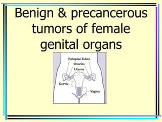
- 1. Benign & precancerous tumors of female genital organs
- 2. Benign & precancerous tumors of vulva
- 3. Urethral Caruncle • A urethral caruncle is a small, fleshy outgrowth of the distal edge of the urethra. • The tissue of the caruncle is soft, smooth, friable, and bright red and initially appears as an eversion of the urethra • They occur most frequently in postmenopausal women and must be differentiated from urethral carcinomas. • Urethral caruncles are believed to arise from an ectropion of the posterior urethral wall associated with retraction and atrophy of the postmenopausal vagina.
- 4. • The growth of the caruncle is secondary to chronic irritation or infection. • Histologically the caruncle is composed of transitional and stratified squamous epithelium with a loose connective tissue • Frequently subdivided by their histologic appearance into papillomatous, granulomatous, and angiomatous varieties. • Many women are asymptomatic, whereas others experience dysuria, frequency, and urgency. Sometimes the caruncle produces point tenderness after contact with undergarments or during intercourse. Ulcerative lesions usually produce spotting on contact more commonly than hematuria.
- 5. Urethral Caruncle • Initial therapy is oral or topical estrogen and avoidance of irritation. • If the caruncle does not regress or is symptomatic, it may be destroyed by cryosurgery, laser therapy, fulguration, or operative excision. • Following operative destruction, a Foley catheter should be left in place for 48 to 72 hours.
- 6. Papilloma • Warty sessile growth arises most usually from labium major.
- 7. Cysts • The most common large cyst of the vulva is a cystic dilation of an obstructed Bartholin's duct. • Approximately 2% of new gynecologic patients present with an asymptomatic Bartholin's duct cyst. • Treatment is not necessary in women younger than 40 unless the cyst becomes infected or enlarges enough to produce symptoms.
- 8. Hydradenoma • The hidradenoma is a rare, small, benign vulvar tumor that originates from apocrine sweat glands of the inner surface of the labia majora and nearby perineum. Occasionally, they may originate from eccrine sweat glands. • For unknown reasons, they are discovered exclusively in white women between the ages of 30 and 70, most commonly in the fourth decade of life. These tumors have not been reported prior to puberty. Hidradenomas may be cystic or solid. Approximately 50% of hidradenomas are less than 1 cm in diameter. • These tumors have well-defined capsules • Treatment - surgical
- 9. Lipoma • Lipomas are benign, slow-growing, circumscribed tumors of fat cells arising from the subcutaneous tissue of the vulva • The largest vulvar lipoma reported in the literature weighed 44 pounds. • Lipomas are the second most frequent benign vulvar mesenchymal tumor. Because of the fat distribution of the vulva, most lipomas are discovered in the labia majora and are superficial in location. • They are slow growing, and their malignant potential is extremely low.
- 10. Fibromas are the most common benign solid tumors of the vulva. They are more frequent than lipomas, the other common benign tumors of mesenchymal origin. • Fibromas occur in all age groups and most commonly are found in the labia majora. • However, they actually arise from deeper connective tissue. Thus they should be considered as dermatofibromas. • Smaller fibromas are asymptomatic; larger tumors may produce chronic pressure symptoms or acute pain when they degenerate. Treatment is operative removal if the fibromas are symptomatic and/or continue to grow. Occasionally they are removed for cosmetic reasons.
- 11. Leukoplakia
- 12. Lichen sclerosus (kraurosis)
- 13. Behcet’s syndrome • Simultaneous ulcerations of vulva and mouth.
- 14. Benign tumors of vagina • Gaertner cyst of the vagina
- 15. Dysontogenetic Cysts • Dysontogenetic cysts of the vagina are thin- walled, soft cysts of embryonic origin. Whether the cysts arise from the mesonephros (Gartner's duct cyst), the perimesonephrium (müllerian cyst), or the urogenital sinus (vestibular cyst) is predominantly of academic rather than clinical importance. The cysts may be differentiated histologically by the epithelial lining • Most of these benign cysts are asymptomatic, sausage-shaped tumors that are discovered only incidentally during pelvic examination. Small asymptomatic Gartner's duct cysts may be followed conservatively
- 16. Treatment • Operative excision is indicated for chronic symptoms. • Rarely, one of these cysts becomes infected, and if operated on during the acute phase, marsupialization of the cyst is preferred. • Excision of the vaginal cyst may be a much more formidable operation than anticipated. • The cystic structure may extend up into the broad ligament and anatomically be in proximity to the distal course of the ureter.
- 17. Inclusion cysts • Usually result from birth trauma or gynecologic surgery. Often they are discovered in the site of a previous episiotomy or at the apex of the vagina following hysterectomy. • Histologically, inclusion cysts are lined by stratified squamous epithelium. These cysts contain a thick, pale-yellow substance that is oily and formed by degenerating epithelial cells.
- 18. Inclusion cysts • Often these cysts are erroneously called sebaceous cysts in the misbelieve that the central material is sebaceous. • Similar to vulvar inclusion cysts, the etiology is either a small tag of vaginal epithelium buried beneath the surface following a gynecologic or obstetric procedure or a misplaced island of embryonic remnant that was destined to form epithelium. • The majority of inclusion cysts are asymptomatic. If the cyst produces dyspareunia or pain, the treatment is excisional biopsy.
- 19. Vaginal Polyp • This is a rare tumor which can be seen in infants or in adults. The origin from the vaginal mucosa has to be demonstrated to differentiate from much more common urethral caruncles, cervical and uterus polyps.
- 20. Vaginal fibroma • Fibroma of the vagina is a very rare tumor. It may be pedunculated and appear at the introitus. Clinically it is a firm benign noninfiltrating growth.
- 21. Cervix
- 22. Benign cervical lesions Optional precancerous Obligatory lesions precancerous lesions • Cervical erosion • Cervical dyplasia • Leukoplakia (without • Leukoplakia (with atypia) atypia) • Polyps • Erythroplakia • Endometriosis • Adenomatosis • Ectropion, scars • Exo-, endocervicites
- 23. • Intraepithelial neoplasia is a spectrum of premalignant changes in the epithelium of the cervix that histologically show varying degrees of cellular atypia. Numerous terms are used to describe the severity of the atypias, but there is no clearly defined boundary between them. • During reproductive life the squamocolumnar junction is usually on the portio of the cervix near the external os. It may be found farther away from the os during and after pregnancy and usually recedes into the endocervical canal after menopause.
- 24. • Many cases of cervical intraepithelial neoplasia (CIN) do not progress. Some particularly low-grade lesions spontaneously regress, but all have the potential for progression to malignancy. • The risk of progression for CIN I (mild dysplasia— LGSIL) to a higher grade lesion is approximately 16%. • High-grade lesions (carcinoma in situ [CIN III]— HGSIL) are at greater risk for malignant progression and usually are found in larger abnormal transformation zones. • Malignant progression risk is greatest for CIN III, least for CIN I, and intermediate for CIN II. • Carcinoma in situ with gland involvement is treated the same as carcinoma in situ without gland involvement.
- 25. • The precise cause of CIN is not known but appears to be associated with sexual activity and HPV infection. • Females with multiple sex partners are at increased risk for CIN, and males with multiple sex partners increase the risk of neoplasia for a female sex partner. • Cigarette smoking increases the risk of CIN. Increased levels of vitamins A and E may decrease the risk. • Prolonged oral contraceptive use (more than 5 years) is associated with an increased frequency of cervical neoplasia.
- 26. Diagram of cervical epithelium showing various terminologies used to characterize progressive degrees of cervical neoplasia
- 27. Potential Risk Factors for Cervical Neoplasia Epidemiologic Characteristics Other Potential Factors • Early intercourse • Oral contraceptives • Multiple sex partners • Cigarette smoking • Early marriage • Vitamin C • Early childbearing • Prior radiation • Prostitution • Intrauterine DES • Male factors — "high-risk" exposure consort • Lupus erythematosus • Socioeconomic status, • Vitamins A and E, race folates • STD infection Viral Relations • Immune status, including • Papillomavirus HIV infection • Herpesvirus • Cytomegalovirus
- 28. Oncogenic Potential of HPV Types Potential HPV Types • Nononcogenic 6, 11, 42, 43, 44 • Oncogenic 16, 18, 31, 33, 35, 39, 45, 51, 52, 56, 58, 59, 68
- 29. • The false negative rate for properly performed cytology smears is approximately 5% to 20%. • "Rapidly progressing" cervical carcinoma appears primarily due to false-negative smears rather than to a true rapid progression from normal to malignant epithelium. • Abnormal cells on Pap smears occur with increasing frequency in those receiving chemotherapy and in patients with lupus erythematosus. • The colposcope is used to evaluate the cervix if an abnormal Pap smear is present. Usually multiple biopsy specimens of an abnormal transformation zone are needed for an adequate evaluation. • Colposcopic and cytologic findings do not establish a diagnosis; biopsy is necessary.
- 30. Narrow brushes for endocervical sampling. Top, Q-Tip; middle, Cervix Brush (Unimar); bottom, Cytobrush (Medscand).
- 31. Cytology Technique Scrape of exocervix Scrape of endocervix.
- 32. Traditional Classification of Papanicolaou Smear • Normal • Metaplasia • Inflammation • Minimal atypia—koilocytosis • Mild dysplasia (CIN I) • Moderate dysplasia (CIN II) • Severe dysplasia—carcinoma in situ (CIN III) • Invasive carcinoma
- 33. Bethesda Classification (Modified) • Adequacy of smear • Infection type • Squamous abnormalities – Reactive (inflammatory change) – Epithelial cell abnormalities • Atypical type, undetermined • Squamous intraepithelial lesions (SILs) – Low grade: HPV or mild dysplasia (CIN I) – High grade: moderate to severe dysplasia— carcinoma in situ (CIN II-III) – Glandular cells • Atypical and source • Adenocarcinoma and source
- 34. Evaluation of Abnormal Pap Smear
- 35. Antibody-mediated viral neutralization. Neutralizing, conformational isotopes are expressed on the surface of human papillomavirus (HPV) virions. The epitopes (antigens) are recognized by lymphocytes, and specific neutralizing antibodies are generated. These neutralizing antibodies bind specifically to surface epitopes and inhibit viral infection.
- 36. Therapy of Intraepithelial Neoplasia Ablative Treatment • Cryotherapy Three varieties of cryotherapy probes.
- 37. Therapy of Intraepithelial Neoplasia Ablative Treatment Laser Therapy • The laser has been widely used in conjunction with the colposcope. • The energy from the laser beam is absorbed by water with resultant vaporization of the target tissue. • The laser beam is controlled by a small "joystick," and the spot size of the laser can be varied but is usually less than 1 mm. • Usually therapy is carried to a depth of 5 to 7 mm and a power density of over 600 W/cm2 • the complications of pain and bleeding are also related to the power density and depth of treatment.
- 38. Cautery • Electrocautery was the mainstay of outpatient therapy of CIN before the advent of cryosurgery, laser therapy, and the LEEP procedure. • The treatment can be accomplished with a hot wire unit generating heat to the cervix or an electrodiathermy unit, which requires current to be passed through the tissues and electrical grounding of the patient. • The treatment is carried out with sufficient depth to destroy cervical glands. • An electrocautery unit is less expensive than the laser and appears able to yield comparable therapy results to cryosurgery but is infrequently used today.
- 39. Excisional Therapy • Conization • if the colposcopic examination is unsatisfactory, • if there is uncertainty regarding the presence of invasive disease, • if there is neoplasm in the endocervix, • if the cells seen on cytologic examination are not adequately explained by the biopsy specimens • if the biopsy suggests the possibility of microinvasion • if invasion is suspected but cannot be confirmed, conization is mandatory because the proper diagnosis of microinvasion cannot be made from a biopsy specimen. • excisional therapy is also carried out when childbearing function is to be maintained or when a patient prefers therapy less extensive than hysterectomy and is willing to adhere to a strict protocol for follow-up.
- 40. TECHNIQUE • COLD KNIFE CON • LASER CONIZATION. • LOOP ELECTROEXCISION PROCEDURE (LEEP) Examples of electrodes used for a LEEP procedure.
- 41. A, Cone biopsy for CIN of exocervix. Limits of lesion were identified colposcopically. B, Cone biopsy for endocervical disease. Limits of lesions were not seen colposcopically.
- 42. • The goal of treatment in CIN is eradication of all abnormal tissue. • Laser therapy, cryotherapy, and electrocautery have been reported to have equivalent results and lead to eradication of the lesions in about 90% of the patients with carcinoma in situ after initial therapy. • Cervical stenosis, infertility, and premature birth may result from excisional therapy of CIN if large areas of the endocervix are destroyed. Limiting the cone or LEEP height to less than 1.5 to 2.0 cm decreases this risk. • Conization for the therapy of CIN is as effective as hysterectomy, especially if the margins are free of disease.
- 43. • Evaluation of the abnormal Pap smear in pregnancy is conducted primarily to rule out the presence of invasive carcinoma. CIN is evaluated and treated in the postpartum period. • Some CIN lesions discovered during pregnancy spontaneously regress postpartum. • The risk of long-term development (up to 10 years) of intraepithelial neoplasia following initial therapy is about 3%. • Most short-term recurrences of intraepithelial neoplasia occur within 1 to 2 years after initial treatment. • Patients treated for CIN should have annual cytology indefinitely.
- 44. Ulcer of the cervix • A true ulcer with loss of epithelial covering is seen in the anterior lip of cervix
- 45. Lacerations • Cervical lacerations frequently occur with both normal and abnormal deliveries. • Lacerations may occur in non-pregnant women with mechanical dilation of the cervix. • Obstetric lacerations vary from minor superficial tears to extensive full-thickness lacerations at 3 and 9 o'clock, respectively, which may extend into the broad ligament. In gynecology the atrophic cervix of the postmenopausal woman predisposes to the complication of cervical laceration when the cervix is mechanically dilated for a diagnostic dilation and curettage. • Acute cervical lacerations bleed and should be sutured. • Cervical lacerations that are not repaired may give the external os of the cervix a fish-mouthed appearance; however, they are usually asymptomatic.
- 46. Lacerations • The use of laminaria tents to slowly soften and dilate the cervix before mechanical instrumentation of the endometrial cavity has reduced the magnitude of iatrogenic cervical lacerations. • Furthermore, the practice of routine inspection of the cervix, stabilized with one or more ring forceps, following every second- or third-trimester delivery has enabled physicians to discover and repair extensive cervical lacerations. • Lacerations should be palpated to determine the extent of cephalad extension of the tear. • Extensive cervical lacerations especially those involving the endocervical stroma may lead to incompetence of the cervix during a subsequent pregnancy.
- 47. Ectropion
- 48. Cervical polyp • Endocervical and cervical polyps are the most common benign neoplastic growths of the cervix. • Cervical polyps usually present as a single polyp, but multiple polyps do occur occasionally. The majority are smooth, soft, reddish-purple to cherry red, and fragile. They readily bleed when touched. Endocervical polyps may be single or multiple and are a few millimeters to 4 cm in diameter. • The classic symptom of an endocervical polyp is intermenstrual bleeding, especially following contact such as coitus or a pelvic examination. Sometimes an associated leukorrhea emanates from the infected cervix. Many endocervical polyps are asymptomatic and recognized for the first time during a routine speculum examination. Often the polyp seen on inspection is difficult to palpate because of its soft consistency. • Histologically the surface epithelium of the polyp is columnar or squamous epithelium, depending on the site of origin and the degree of squamous metaplasia
- 49. Cervical polyp
- 50. Cervical Myomas • Cervical myomas are smooth, firm masses that are similar to myomas of the fundus. • A cervical myoma is usually a solitary growth in contrast to uterine myomas, which in general, are multiple. • Depending on the series, 3% to 8% of myomas are categorized as cervical myomas. • Because of the relative paucity of smooth muscle fibers in the cervical stroma, the majority of myomas that appear to be cervical actually arise from the isthmus of the uterus.
- 51. Fibroma of the cervix
- 52. Cervical Myomas • Most cervical myomas are small and asymptomatic. When symptoms do occur, they are dependent on the direction in which the enlarging myoma expands. The expanding myoma produces symptoms secondary to mechanical pressure on adjacent organs. Cervical myomas may produce dysuria, urgency, urethral or ureteral obstruction, dyspareunia, or obstruction of the cervix. • Occasionally a cervical myoma may become pedunculated and protrude through the external os of the cervix. These prolapsed myomas are often ulcerated and infected. A very large cervical myoma may produce distortion of the cervical canal and upper vagina. Rarely, a cervical myoma causes dystocia during childbirth. • The diagnosis of a cervical myoma is by inspection and palpation.
- 54. UTERUS
- 55. Endometrial Polyp • Endometrial polyps are localized overgrowths of endometrial glands and stroma that project beyond the surface of the endometrium. • They are soft, pliable, and may be single or multiple. Most polyps arise from the fundus of the uterus. • Polypoid hyperplasia is a benign condition in which numerous small polyps are discovered throughout the endometrial cavity. • Endometrial polyps vary from a few millimeters to several centimeters in diameter, and it is possible for a single large polyp to fill the endometrial cavity. • Endometrial polyps may have a broad base (sessile) or be attached by a slender pedicle (pedunculated).
- 56. Endometrial Polyp • The majority of endometrial polyps are asymptomatic. Those that are symptomatic are associated with a wide range of abnormal bleeding patterns. No single abnormal bleeding pattern is diagnostic for polyps; however, menorrhagia, premenstrual and postmenstrual staining, and scanty postmenstrual spotting are the most common. Occasionally a pedunculated endometrial polyp with a long pedicle may protrude from the external cervical os. Sometimes large endometrial polyps may contribute to infertility. • Polyps are succulent and velvety, with a large central vascular core. The color is usually gray or tan but may occasionally be red or brown. Histologically an endometrial polyp has three components: endometrial glands, endometrial stroma, and central vascular channels
- 57. Endometrial Polyp • Malignant change, when found in an endometrial polyp, is usually curable, and the endometrial carcinoma is most often of a low stage and grade. • It is interesting that benign polyps have been found in approximately 20% of uteri removed for endometrial carcinoma. Recently, unusual polyps have been described in association with chronic administration of the nonsteroidal anti-estrogen tamoxifen. • The incidence of endometrial abnormalities associated with chronic tamoxifen therapy is polyps 20% to 35%, endometrial hyperplasia 2% to 4%, and endometrial carcinoma 1% to 2%. • The management of endometrial polyps is removal by curettage or via the hysteroscope. • Because of the frequent association of endometrial polyps and other endometrial pathology, it is important to examine histologically both the polyp and the associated endometrial lining. Polyps, because of their mobility, often tend to elude the curette.
- 58. Tiny hysteroscopic scissors, about as big around as the ink tube on a standard writing pen, are used to cut the stalk. Photo taken during Hysteroscopy of a small endometrial polyp. Notice the stalk.
- 59. Leiomyomas • Leiomyomas, also called myomas, are benign tumors of muscle cell origin. • These tumors are often referred to by their popular names, fibroids or fibromyomas, but both terms are semantic misnomers if one is referring to the cell of origin. • Most leiomyomas contain varying amounts of fibrous tissue, which is believed to be secondary to degeneration of some of the smooth muscle cells. • Leiomyomas are the most frequent pelvic tumors, with the highest prevalence occurring during the fifth decade of a woman's life. • Although leiomyomas arise throughout the body in any structure containing smooth muscle, in the pelvis the majority are found in the corpus of the uterus.
- 60. • Occasionally, leiomyomas may be found in the fallopian tube or the round ligament, and approximately 5% of uterine myomas originate from the cervix. • Myomas may be single but most often are multiple. Myomas are discovered in one of four white women and one of two black women. • They vary greatly in size from microscopic to multinodular uterine tumors that may weigh more than 50 pounds and literally fill the patient's abdomen. • Myomas are more prone to grow and become symptomatic in nulliparous women. The question as to why some women develop myomas while others do not is unanswered. However, genetic determinants definitely contribute to their development. Symptomatic uterine leiomyomas are the primary indication for approximately 30% of all hysterectomies. • Initially most myomas develop from the myometrium, beginning as intramural myomas. As they grow, they remain attached to the myometrium with a pedicle of varying width and thickness.
- 61. • Myomas are classed into subgroups by their relative anatomic relationship and position to the layers of the uterus. • The three most common types of myomas are intramural, subserous, and submucous, with special nomenclature for broad ligament and parasitic myomas. • Continued growth in one direction determines which myomas will be located just below the endometrium (submucosal) and which will be found just beneath the serosa (subserosal) • The most common symptoms related to myomas are pressure from an enlarging pelvic mass, pain including dysmenorrhea, and abnormal uterine bleeding. The severity of symptoms is usually related to the number, location, and size of the myomas. However, the majority of women with uterine myomas are asymptomatic.
- 63. • Laparoscopic view of a uterus with a pedunculated posterior myoma • A fibroid in this location should not affect chances for pregnancy or miscarriage • However, if it were pushing into the cavity of the uterus, it might cause problems
- 64. Diagnosis • The majority of uterine myomas may be diagnosed by pelvic examination, difficult cases will benefit from ultrasound examination or a search for concentric calcifications on an abdominal x-ray film. • There are several recent reports of computed tomography (CT) and magnetic resonance imaging (MRI) studies of uterine myomas. • However, these imaging techniques are more expensive than ultrasound. • Until CT and MRI can distinguish between benign and malignant myomas, they will rarely be ordered in routine clinical management of myomas.
- 65. Treatment • The management of a woman with small, asymptomatic myomas is judicious observation. When the tumor is first discovered, it is appropriate to perform a pelvic examination at 6-month intervals to determine the rate of growth. The majority of women will not need an operation, especially those women in the perimenopausal period, where the condition usually improves with diminishing levels of circulating estrogens. • Women with abnormal bleeding and leiomyomas should be investigated thoroughly for concurrent problems such as endometrial hyperplasia. If their symptoms do not improve with conservative management, operative therapy may be considered. The choice between a myomectomy and hysterectomy is usually determined by the patient's age, parity, and most important, future reproductive plans.
- 66. • Classic indications for a myomectomy include: – a rapidly expanding pelvic mass, – persistent abnormal bleeding, – pain or pressure, – enlargement of an asymptomatic myoma to more than 8 cm in a woman who has not completed childbearing.
- 67. • Two associated but rare diseases should be noted: intravenous leiomyomatosis and leiomyomatosis peritonealis disseminata. Intravenous leiomyomatosis is a rare condition in which benign smooth muscle fibers invade and slowly grow into the venous channels of the pelvis. The tumor grows by direct extension and grossly appears like a "spaghetti" tumor. Only 25% of tumors extend beyond the broad ligament; however, case reports exist of tumor growth into the vena cava and right heart. • Leiomyomatosis peritonealis disseminata (LPD) is a benign disease with multiple small nodules over the surface of the pelvis and abdominal peritoneum. Grossly, LPD mimics disseminated carcinoma. However, histologic examination demonstrates benign-appearing myomas. This disorder is usually associated with a recent pregnancy.
- 68. Ovary
- 69. Adenomatoid Tumors • The most prevalent benign tumor of the oviduct is the angiomyoma or adenomatoid tumor. • They are small, gray-white, circumscribed nodules, 1 to 2 cm in diameter. • These tumors are usually unilateral and present as small nodules just under the tubal serosa. • These small nodules do not produce pelvic symptoms or signs. • These benign tumors also are found below the serosa of the fundus of the uterus and the broad ligament. • Microscopically they are composed of small tubules lined by a low cuboidal or flat epithelium. Histologic studies have established that the thin-walled channels that comprise these tumors are of mesothelial origin. • These tumors do not become malignant; however, they may be mistaken for a low-grade neoplasm when initially viewed during a frozen-section evaluation.
- 70. Follicular Cysts • Follicular cysts are by far the most frequent cystic structures in normal ovaries. • The cysts are frequently multiple and may vary from a few millimeters to as large as 15 cm in diameter. • However, a normal follicle may physiologically become cystic, and therefore it is important to have a minimal diameter for a follicular cyst. • This diameter is generally considered to be between 2.5 and 3 cm. Follicular cysts are not neoplastic and are believed to be dependent on gonadotrophins for growth.
- 71. • Enlarged polycystic ovary following laparoscopic cauterization Follicular cysts are translucent, thin walled, and are filled with a watery, clear to straw-colored fluid. If a small opening in the capsule of the cyst suddenly develops, the cyst fluid under pressure will squirt out. These cysts are situated in the ovarian cortex, and sometimes they appear as translucent domes on the surface of the ovary.
- 72. Corpus Luteum Cysts • Corpus luteum cysts are less common than follicular cysts, but clinically they are more important. • Corpus luteum cysts may be associated with either normal endocrine function or prolonged secretion of progesterone. The associated menstrual pattern may be normal, delayed menstruation, or amenorrhea. • Most corpus luteum cysts are small, the average diameter being 4 cm. • Corpus luteum cysts vary from being asymptomatic masses to those causing catastrophic and massive intraperitoneal bleeding associated with rupture. • Many corpus luteum cysts produce dull, unilateral, lower abdominal and pelvic pain. The enlarged ovary is moderately tender on pelvic examination. Depending on the amount of progesterone secretion associated with cysts, the menstrual bleeding may be normal or delayed several days to weeks with subsequent menorrhagia.
- 73. • Corpus luteum cyst with thickened cyst wall and definite lutein cell lining recognized by its color. Cyst is filled with hemorrhagic gelatinous material.
- 74. Benign Cystic Teratoma (Dermoid Cyst, Mature Teratoma) • Benign ovarian teratomas are usually cystic structures that on histologic examination contain elements from all three germ cell layers. • The word teratoma was first advanced by Virchow and translated literally means "monstrous growth." • Teratomas of the ovary may be benign or malignant. Although dermoid is a misnomer, it is the most common term used to describe the benign cystic tumor, composed of mature cells, whereas the malignant variety is composed of immature cells (immature teratoma). • Dermoid is a descriptive term in that it emphasizes the preponderance of ectodermal tissue with some mesodermal and rare endodermal derivatives. • Malignant teratomas that are immature are usually solid with some cystic areas and histologically contain immature or embryonic-appearing tissue.
- 75. • From 50% to 60% of dermoids are asymptomatic and are discovered during a routine pelvic examination, coincidentally visualized by an abdominal x-ray or ultrasound examination, or found incidentally at laparotomy. • Presenting symptoms of dermoids include pain, and the sensation of pelvic pressure. • Specific complications of dermoid cysts include torsion, rupture, infection, hemorrhage, and malignant degeneration. • Three medical diseases also may be associated with dermoid cysts: thyrotoxicosis, carcinoid syndrome, and autoimmune hemolytic anemia. Torsion of a dermoid is the most frequent complication
- 76. • Benign cystic teratoma. This section from the tumor demonstrates areas of hair (dark arrows) and solid sebaceous material (S).
- 77. Fibroma • Fibromas are the most common benign, solid neoplasms of the ovary. Their malignant potential is low, less than 1%. These tumors comprise approximately 5% of benign ovarian neoplasms and approximately 20% of all solid tumors of the ovary. • The pelvic symptoms that develop with growth of fibromas include pressure and abdominal enlargement, which may be secondary to both the size of the tumor and ascites. • Smaller tumors are asymptomatic because these tumors do not elaborate hormones. Thus there is no change in the pattern of menstrual flow. • Fibromas may be pedunculated and therefore easily palpable during one examination yet difficult to palpate during a subsequent pelvic examination. • Sometimes on pelvic examination the fibromas appear to be softer than a solid ovarian tumor because of the edema and/or occasional cystic degeneration.
- 78. Meigs' syndrome • Meigs' syndrome is the association of an ovarian fibroma, ascites, and hydrothorax. Both the ascites and the hydrothorax resolve after removal of the ovarian tumor. The ascites is caused by transudation of fluid from the ovarian fibroma.
- 79. • Fibroma of ovary. Cut surface shows somewhat edematous, interlacing bundles of connective tissue.
- 80. Transitional Cell Tumors—Brenner Tumors • Brenner tumors are rare, small, smooth, solid, fibroepithelial ovarian tumors that are generally asymptomatic. The semantic classification of neoplasms changes and the current preferred term for benign Brenner tumor is transitional cell tumor. The benign, proliferative (low malignant potential), and malignant forms together comprise approximately 2% of ovarian tumors. • These tumors usually occur in women aged 40 to 60 years. • Grossly, Brenner tumors are smooth, firm, gray-white, solid tumors that grossly resemble fibromas. Similar to fibromas, transitional cell tumors are slow growing • Management of Brenner tumors is operative, with simple excision being the procedure of choice.
- 81. Adenofibroma and Cystadenofibroma • Adenofibromas and cystadenofibromas are closely related. Both of these benign firm tumors consist of fibrous and epithelial components. • The epithelial element is most commonly serous, but histologically may be mucinous and endometrioid or clear cell. • They differ from benign epithelial cystadenomas in that there is a preponderance of connective tissue. • Most pathologists emphasize that at least 25% of the tumor consists of fibrous connective tissue. Obviously, cystadenofibromas have microscopic or occasional macroscopic areas that are cystic. • The varying degree of fibrous stroma and epithelial elements produces a spectrum of tumors, which have resulted in a confusing nomenclature with terms such as papillomas, fibropapillomas, and fibroadenomas.
- 82. Adenofibroma and Cystadenofibroma • Smaller tumors are asymptomatic and are only discovered incidentally during abdominal or pelvic operations. Large tumors may cause pressure symptoms or, rarely, undergo adnexal torsion. • Because adenofibromas are usually discovered in postmenopausal women, the treatment of choice is bilateral salpingo- oophorectomy and total abdominal hysterectomy. Because these tumors are benign and because malignant transformation is rare, simple excision of the tumor and inspection of the contralateral ovary is appropriate in younger women.
- 83. THE END