Linux Foundation Edge _ Overview of FDO Software Components _ Randy at Intel.pdf
Ch5 Ppt Lect
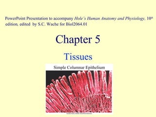
1. Chapter 5 Tissues PowerPoint Presentation to accompany Hole’s Human Anatomy and Physiology, 10 th edition , edited by S.C. Wache for Biol2064.01 Simple Columnar Epithelium
2. You are responsible for the following figures and tables : Tab. 5.1 - Tissues. Tab. 5.4 - Epithelial Tissues (ET) including glandular ET. Tab. 5.3 - Types of glandular exocrine secretions. Tab. 5.7 - Connective Tissues (CT). Tab. 5.8 - Muscle Tissues (MT) and Nervous Tissues (NT). Study all corresponding figures. It is important to correlate structural and functional characteristics of each tissue type. Characteristics of ET Characteristics of CT Characteristics of MT Characteristics of NT
7. secretion lining the ducts of glands simple cuboidal GLANDULAR distensibility lining of urinary bladder and ureters several layers of cells; change shape TRANSITIONAL protection keratinized = epidermis; non-keratinized = lining of vagina, anus, mouth many layers of flattened cells STRATIFIED SQUAMOUS protection, secretion lining of trachea, lining of fallopian tube a single layer of tall cells with scattered nuclei, cilia, & goblet cells PSEUDO- STRATIFIED COLUMNAR protection, absorption, secretion lining of digestive tract single layer of tall cells with basally located nuclei, goblet cells SIMPLE COLUMNAR absorption, secretion linings of kidney tubules, ducts of glands a single layer of cube-shaped cells with large centrally located nuclei SIMPLE CUBOIDAL diffusion, cushioning linings of air sacs, capillaries, lymph vessels, & body cavities single layer of flat cells SIMPLE SQUAMOUS FUNCTION LOCATION STRUCTURE NAME OF ET
34. transport of nutrients, wastes & gases in heart, and blood vessels red and white cells; platelets in plasma BLOOD support, protection, movement, Ca ++ store, hematopoiesis bones concentric circles of calcified matrix BONE shape maintenance plus flexibility external ear, epiglottis above plus elastic fibers ELASTIC CARTILAGE tensile strength, shock absorber intervertebral discs, pubic symphysis less firm than above FIBRO- CARTILAGE support embryo skeleton, costal cartilage, tip of nose, trachea, larynx chondrocytes in lacunae in amorphous matrix HYALINE CARTILAGE durability with stretch lung tissue, wall of aorta matrix of elastic fibers ELASTIC CT tensile strength dermis of skin loose matrix of collagen fibers DENSE IRREGULAR attachment (high tensile strength) tendons, ligaments dense matrix of collagen fibers DENSE REGULAR support basement membrane, lymph organs reticular net of fibers in loose matrix; lymphocytes and reticulocytes RETICULAR insulation, energy store, protection beneath skin, breasts, around kidneys & eyeballs closely packed adipocytes with nuclei pushed to side ADIPOSE cushions, diffusion, inflammation beneath ET (serous membranes around organs & lining cavities) gel-like matrix with fibroblasts, collagen and elastic fibers AREOLAR gives rise to all other CT’s Embryo precursor matrix MESENCHYME FUNCTION LOCATION DESCRIPTION NAME
35.
36.
37.
38.
39.
40.
41.
42.
43.
44.
45.
46.
47.
48.
49.
50.
51.
52.
53.
54.
55.
56.
57.
58.
59.
60.
61. pump blood to lungs and body heart involuntary a network of striated cells with one centrally located nucleus attached by intercalated discs CARDIAC MUSCLE to move substances through passageways (i.e. food, urine, semen), constrict blood vessels, etc walls of visceral hollow organs, irises of eyes, walls of blood vessels involuntary spindle shaped cells with one centrally located nucleus, lacking striations SMOOTH MUSCLE to move bones attached to bones voluntary long, thin fibers with many nuclei and striations SKELETAL MUSCLE FUNCTION LOCATION TYPE OF CONTROL DESCRIPTION OF STRUCTURE MUSCLE TISSUE