SlideShare una empresa de Scribd logo
1 de 64
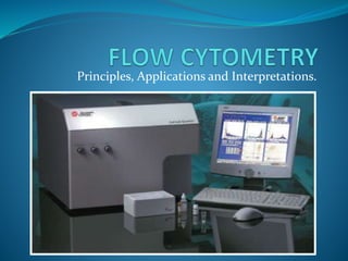
Principles, Applications and Interpretations.
INTRODUCTION 
The concept of flow cytometry has been in existence 
for more than five decades. 
Flow cytometric immunophenotyping (FCI) first 
appeared in clinical laboratories in the 1980s, in the 
wake of the AIDS epidemic. 
Initially utilized to assess CD4 T-cells, the technique 
was soon applied to lymphoid and eventually myeloid 
neoplasms.
 Current flow cytometers have the capability of 
simultaneously measuring multiple parameters of 
individual cells in a cell suspension. 
 Thus, a large number of cell specimens can be 
processed with a quick turnaround time. 
 In addition, flow cytometry is also highly sensitive 
and can detect immunophenotype of cells in a 
specimen with thousands of cells.
The parameters analyzed by flow cytometry include 
 physical properties of cells; the size, cytoplasmic 
granularity, and amount of DNA contents; and 
 cell antigens/markers (surface, cytoplasmic, and 
nuclear) that can be recognized by specific 
antibodies.
By using appropriate antibody panels, flow cytometry 
can reveal 
the cell type (hematopoietic, lymphoid, or 
nonhematopoietic), 
cell lineage (B- and T cells, natural killer cells, 
myeloid/ monocytic cells, neuro/neuroendocrine 
cells, and epithelial cells), 
cell maturation stage (precursors vs. matured cells)
Flow cytometry involves the analysis of the optical 
and fluorescence characteristics of single particle 
(e.g. cells, nuclei, chromosomes) during their 
passage within a narrow, precisely defined liquid 
stream.
For cell analysis, the basic components of a flow 
COMPUTER 
SYSTEM 
ELECTRONIC 
SYSTEM 
OPTICAL 
SYSTEM 
FLOW SYSTEM 
cytometer include:-
SCHEMATIC DIAGRAM OF A FLOW CYTOMETER 
PMT-photomultiplier tubes 
ADC-analogue-to-digital converter
CONCEPT OF SCATTERING 
Physical properties, such as size (represented by 
forward angle light scatter) and internal complexity 
(represented by right-angle scatter) can resolve certain 
cell populations.
 FSC collects light at 180° from the point at which the 
laser beam intersects the cells, usually on a linear scale. 
It is correlated with cell size, and thus can distinguish 
normal lymphocytes (small), monocytes 
(intermediate), and neoplastic cells (generally they are 
large in size). 
 SSC collects right-angle light at 90° and is correlated 
with cytoplasmic granularity and nuclear configuration.
The combination of both FSC and SSC can 
distinguish normal lymphocytes, granulocytes, and 
monocytes. 
 The detection of lymphocytes and monocytes 
provides a reliable internal control to evaluate the size 
of the cells of interest.
IMMUNOPHENOTYPING ANALYSIS 
Requires 
Antibodies. 
Fluorochromes.
ANTIBODY 
Highly specific monoclonal antibodies are used that 
are produced by cloned antibody secreting cells. 
Antibodies are based on cluster of differentiation 
(CD)- a protocol used for identification and distinction 
of cell surface antigens. 
Using CD system we can identify cells by the presence 
or absence of particular surface markers for e.g. CD3+ 
or CD20- etc.
FLUOROCHROMES 
Fluorochromes are substances that can be excited by 
certain light source (such as laser) and emit a fluorescent 
signal at a single wavelength. 
Fluorescent dyes can directly bind to certain cellular 
content, such as DNA and RNA, and allow us to perform 
quantitative analysis on individual cells. 
However, in most cases fluorochromes are conjugated with 
monoclonal antibodies, which specifically target cellular 
antigens/markers.
Characteristics of fluorochromes commonly used in 
flow cytometry . 
FLUOROCHROMES CONJUGATED TO ANTIBODIES EXCITATION WAVELENGTH(NM) EMISSION WAVELENGTH(NM) 
Fluorescein isothiocyanate (FITC) 488 530 
Phycoerythrin (PE) 488 580 
PE-Texas Red 488 615 
PE-Cy5 488 670 
Peridinin chlorophyl protein(PerCP) 488 670 
Allophycocyanin (APC) 633 670 
APC-Cy7 633 767 
Interestingly, although some of them can be excited by the same light source, the 
different fluorochromes may emit fluorescent signals with different 
wavelengths/colors. Thus, multiple fluorochromes can be simultaneously excited by a 
light source and detected by their emission fluorescent signals with different 
wavelengths, respectively.
IMMUNOPHENOTYPING ANALYSIS 
Antibodies conjugated to fluorescent dyes can bind specific 
proteins on cell membranes or inside cells. When labeled cells are 
passed by a light source, the fluorescent molecules are excited to a 
higher energy state. Upon returning to their resting states, the 
fluorochromes emit light energy at higher wavelengths. The use of 
multiple fluorochromes, each with similar excitation wavelengths 
and different emission wavelengths (or “colors”), allows several 
cell properties to be measured simultaneously.
Simultaneous detection of multiple cell 
antigens/markers. 
Multiple cell antigens ( Ag ) are recognized by fluorochromeconjugated specific antibodies ( 
Ab ). Because different fluorochromes have different emission wavelengths/colors, they can 
be simultaneously detected by a flow cytometer. 
FITC fluorescein isothiocyanate; PE phycoerythrin; PerCP peridinin chlorophyll protein; PE-T Red PE-Texas 
Red .
Abnormal/ aberrant antigenic expression can be 
grouped into four basic categories: 
• Abnormally increased or decreased levels of antigenic 
expression (aberrant expression) 
• Gain of antigens not normally expressed in the cell type 
• Expression of antigens not synchronized with normal 
development and maturation stage of the cell type or 
lineage 
• Homogeneous expression of antigen(s) by a cell 
population that normally show more heterogeneous 
expression
SPECIMENS SUITABLE FOR FLOW 
CYTOMETRY 
Theoretically, any specimens from which a single 
cell suspension can be generated are suitable for flow 
cytometry analysis. 
However, a lack of distinct antigens or markers in 
the cells of interest or tissues limits the diagnostic 
value of flow cytometry.
Common specimens suitable for flow 
cytometry analysis include 
Peripheral blood, 
Bone marrow, 
Body fluids, 
Cerebrospinal fluid, 
Urine, 
Lymph node (cells or fresh tissues), 
Any fine-needle aspirates, 
Fresh tissues suspicious for hematopoietic and lymphoid disorders.
SPECIMEN STORAGE 
For blood and bone marrow specimens, anticoagulants 
such as EDTA, heparin, or acid citrate dextrose are needed. 
Fresh tissue specimens are best transported and stored 
in sterile tissue culture medium. 
Although specimens may be stored at room 
temperature, refrigeration is preferred, particularly when 
there is a delay for flow cytometric analysis. 
For flow cytometry analysis, single-cell suspensions of 
the fresh tissues can be achieved by mechanical 
dissociation.
General Notes On Cell Preparation 
1.Single cell suspensions are required for optimal 
staining of samples for flow cytometry. 
2. The narrow bores of the sample injection needle and 
tubing on a flow cytometer will be easily clogged by 
aggregated cells and debris. 
3. Preparation of single cell suspensions from solid 
tissue requires mechanical dissociation and/or 
enzymatic digestion for optimal recovery of cells from 
the tissue.
PROCESSING OF SOLID TISSUES 
1) Tissue is weighed and mechanically and enzymatically 
disaggregated into a single cell suspension. 
2) Collagenase is the most commonly used enzyme, followed by 
dispase and trypsin. 
3) Enzymatic digestion is performed in an incubator or a 
shaking water bath. 
4) Mechanical disaggregation can be accomplished with paired 
scalpels or scissors. 
5) The process often requires centrifugation, harvest of single 
cells and redigestion of tissue fragments.
6) The sample should be visually inspected at all phases of 
tissue digestion. 
7) In the final stage, cell suspensions are passed through a 70 
to 200 micron filter to remove aggregates. 
8) Cell suspensions are then counted and viability is 
determined by a dye exclusion assay, such as trypan blue. 
9) Sample is finally incubated with required antibodies 
attached with fluorochrome in optimal temperature and pH 
and analyzed by flow cytometer.
DNA content Analysis 
The measurement of cellular DNA content by flow 
cytometry uses fluorescent dyes, such as propidium 
iodide, that intercalate into the DNA helical structure. 
The fluorescent signal is directly proportional to the 
amount of DNA in the nucleus and can identify gross 
gains or losses in DNA. 
Abnormal DNA content, also known as “DNA content 
aneuploidy”, can be determined in a tumor cell 
population. 
DNA aneuploidy generally is associated with malignancy
DNA analysis by flow cytometer
Erythrocyte analysis 
Detection and quantification of fetal red cells in 
maternal blood. The use of flow cytometry for the 
detection of fetal cells is much more objective, 
reproducible, and sensitive than the Kleihauer-Betke 
test .
Diagnosis of PNH 
Conventional laboratory tests for the diagnosis of PNH 
include the sugar water test and the Ham’s acid hemolysis test . 
Antibodies to CD55 and CD59 are specific for decay-accelerating 
factor and membrane-inhibitor of reactive lysis, 
respectively, and can be analyzed by flow cytometry to make a 
definitive diagnosis of PNH.
Reticulocyte analysis 
Reticulocyte counts are based on identification of residual 
ribosomes and RNA in immature nonnucleated red blood cells 
by using supravital stain. The flow cytometric enumeration 
of reticulocytes uses fluorescent dyes that bind the residual 
RNA, such as thiazole orange . 
A region has been drawn on the red cells in the scatter plot. The other 
major cluster in the scatter plot are the platelets. 
The histogram was gated on the red cells and the regions on it delineate 
cells with high (H), medium (M) and low (L) fluorescence corresponding 
to increasing reticulocyte maturity. N marks nucleated red cells.
In the blood bank, flow cytometry can be used as a 
complementary or replacement test for red cell 
immunology, including RBC-bound 
immunoglobulins and red cell antigens. Flow 
cytometry has been used to accurately identify and 
phenotype the recipient’s red cells. 
Flow cytometry is being used increasingly in the 
blood bank to assess leukocyte contamination in 
leukocyte-reduced blood products .
Leukocyte Analysis 
Perhaps the best example of simultaneous analysis of 
multiple characteristics by flow cytometry involves the 
immunophenotyping of leukemias and 
lymphomas. 
WHO classification has divided non-Hodgkin 
lymphoma into B-cell and T/NK cell subtypes, which 
are further subclassified into precursor and peripheral 
lymphomas. 
Immunophenotyping by flow cytometry (FCM) is an 
essential aid for accurately diagnosing and 
prognosticating leukemia and lymphoma.
The ability to analyze multiple cellular characteristics, 
along with new antibodies and gating strategies, has 
substantially enhanced the utility of flow cytometry in 
the diagnosis of leukemias and lymphomas. 
Different leukemias and lymphomas often have subtle 
differences in their antigen profiles that make them 
ideal for analysis by flow cytometry. 
B cell: CD5, CD10, CD19, CD20, CD45, Kappa, Lambda; 
T cell: CD2, CD3, CD4, CD5, CD7, CD8, CD45, CD56; 
Myelomonocytic: CD7, CD11b, CD13, CD14, CD15, CD16, CD33, CD34, CD45, 
CD56, CD117, HLA-DR; 
Plasma cell: CD19, CD38, CD45, CD56, CD138
Diagnosis of B-Cell Lymphomas by using 
specific antibodies in flow cytometer
Identification of lineage specific antigens for perfect 
diagnosis
Immunologic monitoring of HIV-infected 
patients is a mainstay of the clinical flow cytometry 
and provides the best possible way for enumeration of 
CD4+ T lymphocytes and HIV viral load.
Flow cytometry can be used for lymphoma 
phenotyping of fine needle aspirates, and is a 
powerful adjunct to cytologic diagnosis. 
Neutropenia may be immune or nonimmune in 
nature. Immune neutropenia may result from 
granulocytespecific autoantibodies, granulocyte-specific 
alloantibodies, or transfusion-related anti- 
HLA antibodies. Flow cytometry can readily identify 
anti-neutrophil antibodies that are either bound to 
granulocytes or free in plasma and confirm the origin 
of neutropenia, possibly eliminating the need for a 
bone marrow procedure.
Functional deficiencies of leukocytes can be 
assessed by flow cytometry. Assays for oxidative burst, 
phagocytosis, opsonization, adhesion, and structure 
are available. 
One of the clinical example is LAD type I is caused by 
a genetic deficiency of β2 -integrins, which are 
heterodimers of CD11 and CD18. This deficiency leads 
to a loss of neutrophil and monocyte migration.
The high sensitivity and capacity for simultaneous 
analysis of multiple characteristics make flow 
cytometry useful for the detection of minimal 
residual disease, especially if abnormal patterns of 
antigen expression are present.
Platelet analysis 
Flow cytometry is an excellent method for direct 
analysis of platelet-bound antibodies, and it has 
also been shown to be of benefit in detection of free 
plasma antibodies in ITP. 
The reticulated platelet count can be quantified by 
flow cytometry in order to assess the rate of 
thrombopoiesis. This measurement can separate 
unexplained thrombocytopenias into those with 
increased destruction and those with defects in 
platelet production.
The pathogenesis and molecular defects of many 
primary thrombocytopathies are well known and 
relate to defects in structural or functional glycoproteins, 
such as the abnormal expression of gpIIb/IIIa in 
Glanzmann thrombasthenia and gpIb in Bernard- 
Soulier disease. 
Flow cytometry is a rapid and useful method of 
obtaining a diagnosis.
Other applications 
Flow cytometry is indicated in the evaluation of serous effusions 
and CSF, including aqueous or vitreous humor of patients 
with a history of hematolymphoid neoplasia. 
Flow cytometry assists in the differential diagnosis between 
plasma cell myeloma and monoclonal gammopathies of 
undetermined significance by determining the percentage of 
aberrant or clonal plasma cells of all bone marrow plasma cells. 
Flow cytometry is useful in diagnostic evaluation of unexplained 
marrow plasmacytosis by assessing phenotypically aberrant or 
clonal plasma cells and its ability to detect other underlying 
monoclonal B-cell process.
Tissue-based lymphoid neoplasias commonly affect 
lymph nodes, spleen, mucosa-associated lymphoid 
tissue, skin, or nonlymphoid solid organs resulting in 
masses or organomegaly. 
Flow cytometry is extremely useful in the diagnosis and 
subclassification of tissue-based lymphoid neoplasias,, 
organomegaly and tissue infiltrates.
Basic parameters and Windows of 
cell population 
Forward light scatter (FSC) and side light scatter (SSC) . FSC collects 
light at 180° from the point at which the laser beam intersects the cells .It is 
correlated with cell size.and thus can distinguish normal lymphocytes 
(small), monocytes (intermediate), and neoplastic cells (generally they are 
large in size). 
SSC collects right-angle light at 90° and is correlated with cytoplasmic 
granularity and nuclear configuration. 
The combination of both FSC and SSC can distinguish normal lymphocytes, 
granulocytes, and monocytes. 
The detection of lymphocytes and monocytes provides a reliable internal 
control to evaluate the size of the cells of interest.
CD45 & SSC 
As the first step, it is most important to determine whether the cells of 
interest are hematopoietic. 
Generally speaking, all hematopoietic/lymphoid cells express CD45 
antigens (CD45+). 
Thus, a histogram of CD45 on a logarithmic scale vs. SSC on a linear 
scale is indispensable as a starting point of flow cytometry analysis. 
Based on antigen expression, cells are divided into CD45+ and CD45– 
groups. 
Among the CD45+ group, the cells can further separated into 
subgroups (windows in the histogram) based on expression levels of 
CD45 and intensity of cytoplasmic granularity.
CD45 negativity is seen in non-haemopoietic 
cells, erythroid 
precursors and abnormal plasma cells. 
Weak positive in blasts. 
More CD45 positivity more the 
brightness
Concept of Gating in brief 
Gating is the most important first step in 
immunophenotyping analysis. 
It is critical particularly in a specimen that contains 
mixed cell populations, such as bone marrow aspirate. 
Gating sets upper and lower limits on the type and 
amount of material that passes through. 
It is used to separate a sub-population from 
heterogeneous population. 
It permits very specific questions to be asked about a 
particular population.
Types of Gating 
1. By cell distribution in the CD45 vs. SSC. This is most useful 
in a specimen containing mixed cell populations . The grouped 
cells in individual windows represent different cell lineages. 
2. By cell size: In FSC vs. SSC histograms, neoplastic cells 
(usually large in size) can be gated by using lymphocytes (small) 
and monocytes (intermediate) as an internal size control . Once 
the cells of interest are gated, further analysis of cell lineage can 
be performed. 
3. By cell lineage-specific antigens (immunophenotype): If 
cells are CD45+ but do not fit into particular windows in the 
CD45 vs. SSC histogram, identification of lineage-specific 
antigen expression is needed
Gating the Lymphocytes 
A region, R1, has been drawn around the lymphocytes (A). 
In B, the lymphocytes are coloured red. 
In C a gate has been set to show only the cells in R1
Gating the Monocytes 
A region, R2, has been drawn around the monocytes (A). 
In B, the monocytes are coloured blue. 
In C a gate has been set to show only the cells in R2
Gating the T-Lymphocytes
Quadrant regions showing the percentage of cells in each 
sub-population
QUALITY CONTROL IN FLOW CYTOMETER 
There are several kinds of quality controls. 
First the flow cytometer itself must be evaluated for proper 
function.This is usually accomplished with standardized 
fluorescent beads. These give very precise, reproducible 
patterns, which quickly assess instrument function. 
A second quality control material is used to set up the 
appropriate instrument settings for the type of staining used. 
These can be beads or antibody-stained cells. 
The third level of quality control is a control substance that 
mimics actual specimens. These controls are available 
commercially and usually consist of stabilized blood, 
sometimes with added tissue-culture cells that mimic a specific 
cancer cell.
Comparison of immunophenotypic techniques . 
FLOW CYTOMETRY IMMUNOHISTOCHEMISTRY 
Shorter turnaround time (minutes to hours) Longer turnaround time (hours to days) 
Less subjective result interpretation Subjective result interpretation 
Quantitative results Semiquantitative results 
Multiple antibodies/fluorochromes per test Usually limited to a single antibody per slide 
Greater antibody selection Fewer antibodies available 
Data/results can be electronically transferred Slides can be shipped by mail or courier service 
Need fresh cells or tissue Can use fixed/archived tissue 
Limited morphologic correlation Architectural and cytologiccorrelation 
Cannot assess nonviable cells Can assess nonviable “ghost” cells
CONCLUSION 
Flow cytometry is a powerful technique for correlating 
multiple characteristics on single cells. This qualitative 
and quantitative technique has made the transition 
from a research tool to standard clinical testing. 
Smaller, less expensive instruments and an increasing 
number of clinically useful antibodies are creating 
more opportunities for routine clinical laboratories to 
use flow cytometry in the diagnosis and management 
of disease 
And last but not the least, keeping pace with scientific 
and clinical advancements is the need of hour.

Más contenido relacionado

La actualidad más candente

Flow cytometry in diagnostics
Flow cytometry in diagnosticsFlow cytometry in diagnostics
Flow cytometry in diagnosticsKaberi Nath
 
Flow Cytometry - basics, principles and applications
Flow Cytometry - basics, principles and applicationsFlow Cytometry - basics, principles and applications
Flow Cytometry - basics, principles and applicationsAnkit Raiyani
 
Flowcytometry by asif
Flowcytometry by asifFlowcytometry by asif
Flowcytometry by asifAsif Zeb
 
Flow Cytometry- Presentation
Flow Cytometry- PresentationFlow Cytometry- Presentation
Flow Cytometry- PresentationBIDISHA MANDAL
 
fish- Fluorescence in situ hybridization
fish- Fluorescence in situ hybridization fish- Fluorescence in situ hybridization
fish- Fluorescence in situ hybridization gaurav raja
 
Next Generation Sequencing (NGS)
Next Generation Sequencing (NGS)Next Generation Sequencing (NGS)
Next Generation Sequencing (NGS)LOGESWARAN KA
 
Presentation on flow cytometry1
Presentation on flow cytometry1Presentation on flow cytometry1
Presentation on flow cytometry1Nagendra sharma
 
MTT cell proliferation assay
MTT cell proliferation assay MTT cell proliferation assay
MTT cell proliferation assay Chander K Negi
 
priciples and applications Immunohistochemistry
priciples and applications Immunohistochemistry priciples and applications Immunohistochemistry
priciples and applications Immunohistochemistry Markos Tadele
 
A Beginner's Guide to Flow Cytometry
A Beginner's Guide to Flow CytometryA Beginner's Guide to Flow Cytometry
A Beginner's Guide to Flow CytometryExpedeon
 

La actualidad más candente (20)

Flow cytometry in diagnostics
Flow cytometry in diagnosticsFlow cytometry in diagnostics
Flow cytometry in diagnostics
 
Flow Cytometry (FCM)
Flow Cytometry (FCM) Flow Cytometry (FCM)
Flow Cytometry (FCM)
 
Flow Cytometry
Flow CytometryFlow Cytometry
Flow Cytometry
 
Flow cytometry
Flow cytometryFlow cytometry
Flow cytometry
 
Immunocytochemistry Protocol
Immunocytochemistry ProtocolImmunocytochemistry Protocol
Immunocytochemistry Protocol
 
Flow Cytometry - basics, principles and applications
Flow Cytometry - basics, principles and applicationsFlow Cytometry - basics, principles and applications
Flow Cytometry - basics, principles and applications
 
Flow cytometry
Flow cytometryFlow cytometry
Flow cytometry
 
Flowcytometry by asif
Flowcytometry by asifFlowcytometry by asif
Flowcytometry by asif
 
Flow Cytometry- Presentation
Flow Cytometry- PresentationFlow Cytometry- Presentation
Flow Cytometry- Presentation
 
fish- Fluorescence in situ hybridization
fish- Fluorescence in situ hybridization fish- Fluorescence in situ hybridization
fish- Fluorescence in situ hybridization
 
Flow Cytometry
Flow CytometryFlow Cytometry
Flow Cytometry
 
Flow cytometry
Flow cytometryFlow cytometry
Flow cytometry
 
Introduction to Flow Cytometry
Introduction to Flow CytometryIntroduction to Flow Cytometry
Introduction to Flow Cytometry
 
Next Generation Sequencing (NGS)
Next Generation Sequencing (NGS)Next Generation Sequencing (NGS)
Next Generation Sequencing (NGS)
 
Flow cytometry training garvan
Flow cytometry training garvanFlow cytometry training garvan
Flow cytometry training garvan
 
Cish
CishCish
Cish
 
Presentation on flow cytometry1
Presentation on flow cytometry1Presentation on flow cytometry1
Presentation on flow cytometry1
 
MTT cell proliferation assay
MTT cell proliferation assay MTT cell proliferation assay
MTT cell proliferation assay
 
priciples and applications Immunohistochemistry
priciples and applications Immunohistochemistry priciples and applications Immunohistochemistry
priciples and applications Immunohistochemistry
 
A Beginner's Guide to Flow Cytometry
A Beginner's Guide to Flow CytometryA Beginner's Guide to Flow Cytometry
A Beginner's Guide to Flow Cytometry
 

Similar a Flow cytometry

Flow cytometry for cell componenet analysis
Flow cytometry for cell componenet analysisFlow cytometry for cell componenet analysis
Flow cytometry for cell componenet analysisRAJA GOPAL
 
Rohan gupta 2015 b1ab651p facs
Rohan gupta 2015 b1ab651p   facsRohan gupta 2015 b1ab651p   facs
Rohan gupta 2015 b1ab651p facsRohan Gupta
 
Immunophinotyping raju
Immunophinotyping rajuImmunophinotyping raju
Immunophinotyping rajurajusehrawat
 
Applications of Flow Cytometry | Cell Analysis
Applications of Flow Cytometry | Cell AnalysisApplications of Flow Cytometry | Cell Analysis
Applications of Flow Cytometry | Cell AnalysisUniversity of The Punjab
 
Ezhil1[1][1].Ppt
Ezhil1[1][1].PptEzhil1[1][1].Ppt
Ezhil1[1][1].Pptelangoelil
 
FLOW CYTOMETRY -SUNANDA ARYA.pptx
FLOW CYTOMETRY -SUNANDA ARYA.pptxFLOW CYTOMETRY -SUNANDA ARYA.pptx
FLOW CYTOMETRY -SUNANDA ARYA.pptxSunandaArya
 
Immunoflowcytometry, the basics and applications.pptx
Immunoflowcytometry, the basics and applications.pptxImmunoflowcytometry, the basics and applications.pptx
Immunoflowcytometry, the basics and applications.pptxAmirRaziq1
 
cell-counters1575.ppt.pptx
cell-counters1575.ppt.pptxcell-counters1575.ppt.pptx
cell-counters1575.ppt.pptxWaqarRaees
 
Cell Counter CBC New.pdf
Cell Counter CBC New.pdfCell Counter CBC New.pdf
Cell Counter CBC New.pdfSumitAhir4
 
Graduate Level Immunology Workshop - Using Antibodies as Research Tools
Graduate Level Immunology Workshop - Using Antibodies as Research ToolsGraduate Level Immunology Workshop - Using Antibodies as Research Tools
Graduate Level Immunology Workshop - Using Antibodies as Research ToolsAndrea Henle
 
Molecular techniques for pathology research - MDX .pdf
Molecular techniques for pathology research - MDX .pdfMolecular techniques for pathology research - MDX .pdf
Molecular techniques for pathology research - MDX .pdfsabyabby
 

Similar a Flow cytometry (20)

Flow cytometry for cell componenet analysis
Flow cytometry for cell componenet analysisFlow cytometry for cell componenet analysis
Flow cytometry for cell componenet analysis
 
Flowcytometry 1
Flowcytometry 1Flowcytometry 1
Flowcytometry 1
 
Rohan gupta 2015 b1ab651p facs
Rohan gupta 2015 b1ab651p   facsRohan gupta 2015 b1ab651p   facs
Rohan gupta 2015 b1ab651p facs
 
Immunophinotyping raju
Immunophinotyping rajuImmunophinotyping raju
Immunophinotyping raju
 
Ezhil1
Ezhil1Ezhil1
Ezhil1
 
Flow cytometry
Flow cytometryFlow cytometry
Flow cytometry
 
Flow cytometry
Flow cytometryFlow cytometry
Flow cytometry
 
Applications of Flow Cytometry | Cell Analysis
Applications of Flow Cytometry | Cell AnalysisApplications of Flow Cytometry | Cell Analysis
Applications of Flow Cytometry | Cell Analysis
 
Ezhil1.Ppt
Ezhil1.PptEzhil1.Ppt
Ezhil1.Ppt
 
Ezhil1
Ezhil1Ezhil1
Ezhil1
 
Ezhil1[1][1].Ppt
Ezhil1[1][1].PptEzhil1[1][1].Ppt
Ezhil1[1][1].Ppt
 
FLOW CYTOMETRY -SUNANDA ARYA.pptx
FLOW CYTOMETRY -SUNANDA ARYA.pptxFLOW CYTOMETRY -SUNANDA ARYA.pptx
FLOW CYTOMETRY -SUNANDA ARYA.pptx
 
Immunoflowcytometry, the basics and applications.pptx
Immunoflowcytometry, the basics and applications.pptxImmunoflowcytometry, the basics and applications.pptx
Immunoflowcytometry, the basics and applications.pptx
 
Flow Cytometry technique
Flow Cytometry technique Flow Cytometry technique
Flow Cytometry technique
 
cell-counters1575.ppt.pptx
cell-counters1575.ppt.pptxcell-counters1575.ppt.pptx
cell-counters1575.ppt.pptx
 
Cell Counter CBC New.pdf
Cell Counter CBC New.pdfCell Counter CBC New.pdf
Cell Counter CBC New.pdf
 
Graduate Level Immunology Workshop - Using Antibodies as Research Tools
Graduate Level Immunology Workshop - Using Antibodies as Research ToolsGraduate Level Immunology Workshop - Using Antibodies as Research Tools
Graduate Level Immunology Workshop - Using Antibodies as Research Tools
 
Lymphoma
LymphomaLymphoma
Lymphoma
 
Patho word
Patho wordPatho word
Patho word
 
Molecular techniques for pathology research - MDX .pdf
Molecular techniques for pathology research - MDX .pdfMolecular techniques for pathology research - MDX .pdf
Molecular techniques for pathology research - MDX .pdf
 

Último

Call Now ☎ 9999965857 !! Call Girls in Hauz Khas Escort Service Delhi N.C.R.
Call Now ☎ 9999965857 !! Call Girls in Hauz Khas Escort Service Delhi N.C.R.Call Now ☎ 9999965857 !! Call Girls in Hauz Khas Escort Service Delhi N.C.R.
Call Now ☎ 9999965857 !! Call Girls in Hauz Khas Escort Service Delhi N.C.R.ktanvi103
 
Call Girls Chandigarh 👙 7001035870 👙 Genuine WhatsApp Number for Real Meet
Call Girls Chandigarh 👙 7001035870 👙 Genuine WhatsApp Number for Real MeetCall Girls Chandigarh 👙 7001035870 👙 Genuine WhatsApp Number for Real Meet
Call Girls Chandigarh 👙 7001035870 👙 Genuine WhatsApp Number for Real Meetpriyashah722354
 
Hubli Call Girls 👙 6297143586 👙 Genuine WhatsApp Number for Real Meet
Hubli Call Girls 👙 6297143586 👙 Genuine WhatsApp Number for Real MeetHubli Call Girls 👙 6297143586 👙 Genuine WhatsApp Number for Real Meet
Hubli Call Girls 👙 6297143586 👙 Genuine WhatsApp Number for Real MeetCall Girls Service
 
❤️Call girls in Jalandhar ☎️9876848877☎️ Call Girl service in Jalandhar☎️ Jal...
❤️Call girls in Jalandhar ☎️9876848877☎️ Call Girl service in Jalandhar☎️ Jal...❤️Call girls in Jalandhar ☎️9876848877☎️ Call Girl service in Jalandhar☎️ Jal...
❤️Call girls in Jalandhar ☎️9876848877☎️ Call Girl service in Jalandhar☎️ Jal...chandigarhentertainm
 
Krishnagiri call girls Tamil aunty 7877702510
Krishnagiri call girls Tamil aunty 7877702510Krishnagiri call girls Tamil aunty 7877702510
Krishnagiri call girls Tamil aunty 7877702510Vipesco
 
ooty Call Girls 👙 6297143586 👙 Genuine WhatsApp Number for Real Meet
ooty Call Girls 👙 6297143586 👙 Genuine WhatsApp Number for Real Meetooty Call Girls 👙 6297143586 👙 Genuine WhatsApp Number for Real Meet
ooty Call Girls 👙 6297143586 👙 Genuine WhatsApp Number for Real MeetCall Girls Service
 
Call Girl Raipur 📲 9999965857 whatsapp live cam sex service available
Call Girl Raipur 📲 9999965857 whatsapp live cam sex service availableCall Girl Raipur 📲 9999965857 whatsapp live cam sex service available
Call Girl Raipur 📲 9999965857 whatsapp live cam sex service availablegragmanisha42
 
Punjab❤️Call girls in Mohali ☎️7435815124☎️ Call Girl service in Mohali☎️ Moh...
Punjab❤️Call girls in Mohali ☎️7435815124☎️ Call Girl service in Mohali☎️ Moh...Punjab❤️Call girls in Mohali ☎️7435815124☎️ Call Girl service in Mohali☎️ Moh...
Punjab❤️Call girls in Mohali ☎️7435815124☎️ Call Girl service in Mohali☎️ Moh...Sheetaleventcompany
 
Call Girls Service Faridabad 📲 9999965857 ヅ10k NiGhT Call Girls In Faridabad
Call Girls Service Faridabad 📲 9999965857 ヅ10k NiGhT Call Girls In FaridabadCall Girls Service Faridabad 📲 9999965857 ヅ10k NiGhT Call Girls In Faridabad
Call Girls Service Faridabad 📲 9999965857 ヅ10k NiGhT Call Girls In Faridabadgragmanisha42
 
Chandigarh Call Girls 👙 7001035870 👙 Genuine WhatsApp Number for Real Meet
Chandigarh Call Girls 👙 7001035870 👙 Genuine WhatsApp Number for Real MeetChandigarh Call Girls 👙 7001035870 👙 Genuine WhatsApp Number for Real Meet
Chandigarh Call Girls 👙 7001035870 👙 Genuine WhatsApp Number for Real Meetpriyashah722354
 
Ozhukarai Call Girls 👙 6297143586 👙 Genuine WhatsApp Number for Real Meet
Ozhukarai Call Girls 👙 6297143586 👙 Genuine WhatsApp Number for Real MeetOzhukarai Call Girls 👙 6297143586 👙 Genuine WhatsApp Number for Real Meet
Ozhukarai Call Girls 👙 6297143586 👙 Genuine WhatsApp Number for Real MeetCall Girls Service
 
VIP Call Girl Sector 32 Noida Just Book Me 9711199171
VIP Call Girl Sector 32 Noida Just Book Me 9711199171VIP Call Girl Sector 32 Noida Just Book Me 9711199171
VIP Call Girl Sector 32 Noida Just Book Me 9711199171Call Girls Service Gurgaon
 
Russian Call Girls Kota * 8250192130 Service starts from just ₹9999 ✅
Russian Call Girls Kota * 8250192130 Service starts from just ₹9999 ✅Russian Call Girls Kota * 8250192130 Service starts from just ₹9999 ✅
Russian Call Girls Kota * 8250192130 Service starts from just ₹9999 ✅gragmanisha42
 
Call Girls Thane Just Call 9907093804 Top Class Call Girl Service Available
Call Girls Thane Just Call 9907093804 Top Class Call Girl Service AvailableCall Girls Thane Just Call 9907093804 Top Class Call Girl Service Available
Call Girls Thane Just Call 9907093804 Top Class Call Girl Service AvailableDipal Arora
 
Bareilly Call Girls 👙 6297143586 👙 Genuine WhatsApp Number for Real Meet
Bareilly Call Girls 👙 6297143586 👙 Genuine WhatsApp Number for Real MeetBareilly Call Girls 👙 6297143586 👙 Genuine WhatsApp Number for Real Meet
Bareilly Call Girls 👙 6297143586 👙 Genuine WhatsApp Number for Real MeetCall Girls Service
 
Jalna Call Girls 👙 6297143586 👙 Genuine WhatsApp Number for Real Meet
Jalna Call Girls 👙 6297143586 👙 Genuine WhatsApp Number for Real MeetJalna Call Girls 👙 6297143586 👙 Genuine WhatsApp Number for Real Meet
Jalna Call Girls 👙 6297143586 👙 Genuine WhatsApp Number for Real MeetCall Girls Service
 
Call Girl Raipur 📲 9999965857 ヅ10k NiGhT Call Girls In Raipur
Call Girl Raipur 📲 9999965857 ヅ10k NiGhT Call Girls In RaipurCall Girl Raipur 📲 9999965857 ヅ10k NiGhT Call Girls In Raipur
Call Girl Raipur 📲 9999965857 ヅ10k NiGhT Call Girls In Raipurgragmanisha42
 
Call Girls Hyderabad Just Call 9907093804 Top Class Call Girl Service Available
Call Girls Hyderabad Just Call 9907093804 Top Class Call Girl Service AvailableCall Girls Hyderabad Just Call 9907093804 Top Class Call Girl Service Available
Call Girls Hyderabad Just Call 9907093804 Top Class Call Girl Service AvailableDipal Arora
 
VIP Call Girl Sector 10 Noida Call Me: 9711199171
VIP Call Girl Sector 10 Noida Call Me: 9711199171VIP Call Girl Sector 10 Noida Call Me: 9711199171
VIP Call Girl Sector 10 Noida Call Me: 9711199171Call Girls Service Gurgaon
 

Último (20)

Call Now ☎ 9999965857 !! Call Girls in Hauz Khas Escort Service Delhi N.C.R.
Call Now ☎ 9999965857 !! Call Girls in Hauz Khas Escort Service Delhi N.C.R.Call Now ☎ 9999965857 !! Call Girls in Hauz Khas Escort Service Delhi N.C.R.
Call Now ☎ 9999965857 !! Call Girls in Hauz Khas Escort Service Delhi N.C.R.
 
Call Girls Chandigarh 👙 7001035870 👙 Genuine WhatsApp Number for Real Meet
Call Girls Chandigarh 👙 7001035870 👙 Genuine WhatsApp Number for Real MeetCall Girls Chandigarh 👙 7001035870 👙 Genuine WhatsApp Number for Real Meet
Call Girls Chandigarh 👙 7001035870 👙 Genuine WhatsApp Number for Real Meet
 
Hubli Call Girls 👙 6297143586 👙 Genuine WhatsApp Number for Real Meet
Hubli Call Girls 👙 6297143586 👙 Genuine WhatsApp Number for Real MeetHubli Call Girls 👙 6297143586 👙 Genuine WhatsApp Number for Real Meet
Hubli Call Girls 👙 6297143586 👙 Genuine WhatsApp Number for Real Meet
 
❤️Call girls in Jalandhar ☎️9876848877☎️ Call Girl service in Jalandhar☎️ Jal...
❤️Call girls in Jalandhar ☎️9876848877☎️ Call Girl service in Jalandhar☎️ Jal...❤️Call girls in Jalandhar ☎️9876848877☎️ Call Girl service in Jalandhar☎️ Jal...
❤️Call girls in Jalandhar ☎️9876848877☎️ Call Girl service in Jalandhar☎️ Jal...
 
Krishnagiri call girls Tamil aunty 7877702510
Krishnagiri call girls Tamil aunty 7877702510Krishnagiri call girls Tamil aunty 7877702510
Krishnagiri call girls Tamil aunty 7877702510
 
ooty Call Girls 👙 6297143586 👙 Genuine WhatsApp Number for Real Meet
ooty Call Girls 👙 6297143586 👙 Genuine WhatsApp Number for Real Meetooty Call Girls 👙 6297143586 👙 Genuine WhatsApp Number for Real Meet
ooty Call Girls 👙 6297143586 👙 Genuine WhatsApp Number for Real Meet
 
Call Girl Raipur 📲 9999965857 whatsapp live cam sex service available
Call Girl Raipur 📲 9999965857 whatsapp live cam sex service availableCall Girl Raipur 📲 9999965857 whatsapp live cam sex service available
Call Girl Raipur 📲 9999965857 whatsapp live cam sex service available
 
Punjab❤️Call girls in Mohali ☎️7435815124☎️ Call Girl service in Mohali☎️ Moh...
Punjab❤️Call girls in Mohali ☎️7435815124☎️ Call Girl service in Mohali☎️ Moh...Punjab❤️Call girls in Mohali ☎️7435815124☎️ Call Girl service in Mohali☎️ Moh...
Punjab❤️Call girls in Mohali ☎️7435815124☎️ Call Girl service in Mohali☎️ Moh...
 
9316020077📞Goa Call Girls Numbers, Call Girls Whatsapp Numbers Goa
9316020077📞Goa  Call Girls  Numbers, Call Girls  Whatsapp Numbers Goa9316020077📞Goa  Call Girls  Numbers, Call Girls  Whatsapp Numbers Goa
9316020077📞Goa Call Girls Numbers, Call Girls Whatsapp Numbers Goa
 
Call Girls Service Faridabad 📲 9999965857 ヅ10k NiGhT Call Girls In Faridabad
Call Girls Service Faridabad 📲 9999965857 ヅ10k NiGhT Call Girls In FaridabadCall Girls Service Faridabad 📲 9999965857 ヅ10k NiGhT Call Girls In Faridabad
Call Girls Service Faridabad 📲 9999965857 ヅ10k NiGhT Call Girls In Faridabad
 
Chandigarh Call Girls 👙 7001035870 👙 Genuine WhatsApp Number for Real Meet
Chandigarh Call Girls 👙 7001035870 👙 Genuine WhatsApp Number for Real MeetChandigarh Call Girls 👙 7001035870 👙 Genuine WhatsApp Number for Real Meet
Chandigarh Call Girls 👙 7001035870 👙 Genuine WhatsApp Number for Real Meet
 
Ozhukarai Call Girls 👙 6297143586 👙 Genuine WhatsApp Number for Real Meet
Ozhukarai Call Girls 👙 6297143586 👙 Genuine WhatsApp Number for Real MeetOzhukarai Call Girls 👙 6297143586 👙 Genuine WhatsApp Number for Real Meet
Ozhukarai Call Girls 👙 6297143586 👙 Genuine WhatsApp Number for Real Meet
 
VIP Call Girl Sector 32 Noida Just Book Me 9711199171
VIP Call Girl Sector 32 Noida Just Book Me 9711199171VIP Call Girl Sector 32 Noida Just Book Me 9711199171
VIP Call Girl Sector 32 Noida Just Book Me 9711199171
 
Russian Call Girls Kota * 8250192130 Service starts from just ₹9999 ✅
Russian Call Girls Kota * 8250192130 Service starts from just ₹9999 ✅Russian Call Girls Kota * 8250192130 Service starts from just ₹9999 ✅
Russian Call Girls Kota * 8250192130 Service starts from just ₹9999 ✅
 
Call Girls Thane Just Call 9907093804 Top Class Call Girl Service Available
Call Girls Thane Just Call 9907093804 Top Class Call Girl Service AvailableCall Girls Thane Just Call 9907093804 Top Class Call Girl Service Available
Call Girls Thane Just Call 9907093804 Top Class Call Girl Service Available
 
Bareilly Call Girls 👙 6297143586 👙 Genuine WhatsApp Number for Real Meet
Bareilly Call Girls 👙 6297143586 👙 Genuine WhatsApp Number for Real MeetBareilly Call Girls 👙 6297143586 👙 Genuine WhatsApp Number for Real Meet
Bareilly Call Girls 👙 6297143586 👙 Genuine WhatsApp Number for Real Meet
 
Jalna Call Girls 👙 6297143586 👙 Genuine WhatsApp Number for Real Meet
Jalna Call Girls 👙 6297143586 👙 Genuine WhatsApp Number for Real MeetJalna Call Girls 👙 6297143586 👙 Genuine WhatsApp Number for Real Meet
Jalna Call Girls 👙 6297143586 👙 Genuine WhatsApp Number for Real Meet
 
Call Girl Raipur 📲 9999965857 ヅ10k NiGhT Call Girls In Raipur
Call Girl Raipur 📲 9999965857 ヅ10k NiGhT Call Girls In RaipurCall Girl Raipur 📲 9999965857 ヅ10k NiGhT Call Girls In Raipur
Call Girl Raipur 📲 9999965857 ヅ10k NiGhT Call Girls In Raipur
 
Call Girls Hyderabad Just Call 9907093804 Top Class Call Girl Service Available
Call Girls Hyderabad Just Call 9907093804 Top Class Call Girl Service AvailableCall Girls Hyderabad Just Call 9907093804 Top Class Call Girl Service Available
Call Girls Hyderabad Just Call 9907093804 Top Class Call Girl Service Available
 
VIP Call Girl Sector 10 Noida Call Me: 9711199171
VIP Call Girl Sector 10 Noida Call Me: 9711199171VIP Call Girl Sector 10 Noida Call Me: 9711199171
VIP Call Girl Sector 10 Noida Call Me: 9711199171
 

Flow cytometry

  • 1. Principles, Applications and Interpretations.
  • 2.
  • 3. INTRODUCTION The concept of flow cytometry has been in existence for more than five decades. Flow cytometric immunophenotyping (FCI) first appeared in clinical laboratories in the 1980s, in the wake of the AIDS epidemic. Initially utilized to assess CD4 T-cells, the technique was soon applied to lymphoid and eventually myeloid neoplasms.
  • 4.  Current flow cytometers have the capability of simultaneously measuring multiple parameters of individual cells in a cell suspension.  Thus, a large number of cell specimens can be processed with a quick turnaround time.  In addition, flow cytometry is also highly sensitive and can detect immunophenotype of cells in a specimen with thousands of cells.
  • 5. The parameters analyzed by flow cytometry include  physical properties of cells; the size, cytoplasmic granularity, and amount of DNA contents; and  cell antigens/markers (surface, cytoplasmic, and nuclear) that can be recognized by specific antibodies.
  • 6. By using appropriate antibody panels, flow cytometry can reveal the cell type (hematopoietic, lymphoid, or nonhematopoietic), cell lineage (B- and T cells, natural killer cells, myeloid/ monocytic cells, neuro/neuroendocrine cells, and epithelial cells), cell maturation stage (precursors vs. matured cells)
  • 7.
  • 8. Flow cytometry involves the analysis of the optical and fluorescence characteristics of single particle (e.g. cells, nuclei, chromosomes) during their passage within a narrow, precisely defined liquid stream.
  • 9. For cell analysis, the basic components of a flow COMPUTER SYSTEM ELECTRONIC SYSTEM OPTICAL SYSTEM FLOW SYSTEM cytometer include:-
  • 10. SCHEMATIC DIAGRAM OF A FLOW CYTOMETER PMT-photomultiplier tubes ADC-analogue-to-digital converter
  • 11. CONCEPT OF SCATTERING Physical properties, such as size (represented by forward angle light scatter) and internal complexity (represented by right-angle scatter) can resolve certain cell populations.
  • 12.  FSC collects light at 180° from the point at which the laser beam intersects the cells, usually on a linear scale. It is correlated with cell size, and thus can distinguish normal lymphocytes (small), monocytes (intermediate), and neoplastic cells (generally they are large in size).  SSC collects right-angle light at 90° and is correlated with cytoplasmic granularity and nuclear configuration.
  • 13. The combination of both FSC and SSC can distinguish normal lymphocytes, granulocytes, and monocytes.  The detection of lymphocytes and monocytes provides a reliable internal control to evaluate the size of the cells of interest.
  • 14.
  • 15. IMMUNOPHENOTYPING ANALYSIS Requires Antibodies. Fluorochromes.
  • 16. ANTIBODY Highly specific monoclonal antibodies are used that are produced by cloned antibody secreting cells. Antibodies are based on cluster of differentiation (CD)- a protocol used for identification and distinction of cell surface antigens. Using CD system we can identify cells by the presence or absence of particular surface markers for e.g. CD3+ or CD20- etc.
  • 17. FLUOROCHROMES Fluorochromes are substances that can be excited by certain light source (such as laser) and emit a fluorescent signal at a single wavelength. Fluorescent dyes can directly bind to certain cellular content, such as DNA and RNA, and allow us to perform quantitative analysis on individual cells. However, in most cases fluorochromes are conjugated with monoclonal antibodies, which specifically target cellular antigens/markers.
  • 18. Characteristics of fluorochromes commonly used in flow cytometry . FLUOROCHROMES CONJUGATED TO ANTIBODIES EXCITATION WAVELENGTH(NM) EMISSION WAVELENGTH(NM) Fluorescein isothiocyanate (FITC) 488 530 Phycoerythrin (PE) 488 580 PE-Texas Red 488 615 PE-Cy5 488 670 Peridinin chlorophyl protein(PerCP) 488 670 Allophycocyanin (APC) 633 670 APC-Cy7 633 767 Interestingly, although some of them can be excited by the same light source, the different fluorochromes may emit fluorescent signals with different wavelengths/colors. Thus, multiple fluorochromes can be simultaneously excited by a light source and detected by their emission fluorescent signals with different wavelengths, respectively.
  • 19. IMMUNOPHENOTYPING ANALYSIS Antibodies conjugated to fluorescent dyes can bind specific proteins on cell membranes or inside cells. When labeled cells are passed by a light source, the fluorescent molecules are excited to a higher energy state. Upon returning to their resting states, the fluorochromes emit light energy at higher wavelengths. The use of multiple fluorochromes, each with similar excitation wavelengths and different emission wavelengths (or “colors”), allows several cell properties to be measured simultaneously.
  • 20. Simultaneous detection of multiple cell antigens/markers. Multiple cell antigens ( Ag ) are recognized by fluorochromeconjugated specific antibodies ( Ab ). Because different fluorochromes have different emission wavelengths/colors, they can be simultaneously detected by a flow cytometer. FITC fluorescein isothiocyanate; PE phycoerythrin; PerCP peridinin chlorophyll protein; PE-T Red PE-Texas Red .
  • 21. Abnormal/ aberrant antigenic expression can be grouped into four basic categories: • Abnormally increased or decreased levels of antigenic expression (aberrant expression) • Gain of antigens not normally expressed in the cell type • Expression of antigens not synchronized with normal development and maturation stage of the cell type or lineage • Homogeneous expression of antigen(s) by a cell population that normally show more heterogeneous expression
  • 22.
  • 23.
  • 24. SPECIMENS SUITABLE FOR FLOW CYTOMETRY Theoretically, any specimens from which a single cell suspension can be generated are suitable for flow cytometry analysis. However, a lack of distinct antigens or markers in the cells of interest or tissues limits the diagnostic value of flow cytometry.
  • 25. Common specimens suitable for flow cytometry analysis include Peripheral blood, Bone marrow, Body fluids, Cerebrospinal fluid, Urine, Lymph node (cells or fresh tissues), Any fine-needle aspirates, Fresh tissues suspicious for hematopoietic and lymphoid disorders.
  • 26. SPECIMEN STORAGE For blood and bone marrow specimens, anticoagulants such as EDTA, heparin, or acid citrate dextrose are needed. Fresh tissue specimens are best transported and stored in sterile tissue culture medium. Although specimens may be stored at room temperature, refrigeration is preferred, particularly when there is a delay for flow cytometric analysis. For flow cytometry analysis, single-cell suspensions of the fresh tissues can be achieved by mechanical dissociation.
  • 27. General Notes On Cell Preparation 1.Single cell suspensions are required for optimal staining of samples for flow cytometry. 2. The narrow bores of the sample injection needle and tubing on a flow cytometer will be easily clogged by aggregated cells and debris. 3. Preparation of single cell suspensions from solid tissue requires mechanical dissociation and/or enzymatic digestion for optimal recovery of cells from the tissue.
  • 28. PROCESSING OF SOLID TISSUES 1) Tissue is weighed and mechanically and enzymatically disaggregated into a single cell suspension. 2) Collagenase is the most commonly used enzyme, followed by dispase and trypsin. 3) Enzymatic digestion is performed in an incubator or a shaking water bath. 4) Mechanical disaggregation can be accomplished with paired scalpels or scissors. 5) The process often requires centrifugation, harvest of single cells and redigestion of tissue fragments.
  • 29. 6) The sample should be visually inspected at all phases of tissue digestion. 7) In the final stage, cell suspensions are passed through a 70 to 200 micron filter to remove aggregates. 8) Cell suspensions are then counted and viability is determined by a dye exclusion assay, such as trypan blue. 9) Sample is finally incubated with required antibodies attached with fluorochrome in optimal temperature and pH and analyzed by flow cytometer.
  • 30.
  • 31. DNA content Analysis The measurement of cellular DNA content by flow cytometry uses fluorescent dyes, such as propidium iodide, that intercalate into the DNA helical structure. The fluorescent signal is directly proportional to the amount of DNA in the nucleus and can identify gross gains or losses in DNA. Abnormal DNA content, also known as “DNA content aneuploidy”, can be determined in a tumor cell population. DNA aneuploidy generally is associated with malignancy
  • 32. DNA analysis by flow cytometer
  • 33. Erythrocyte analysis Detection and quantification of fetal red cells in maternal blood. The use of flow cytometry for the detection of fetal cells is much more objective, reproducible, and sensitive than the Kleihauer-Betke test .
  • 34. Diagnosis of PNH Conventional laboratory tests for the diagnosis of PNH include the sugar water test and the Ham’s acid hemolysis test . Antibodies to CD55 and CD59 are specific for decay-accelerating factor and membrane-inhibitor of reactive lysis, respectively, and can be analyzed by flow cytometry to make a definitive diagnosis of PNH.
  • 35. Reticulocyte analysis Reticulocyte counts are based on identification of residual ribosomes and RNA in immature nonnucleated red blood cells by using supravital stain. The flow cytometric enumeration of reticulocytes uses fluorescent dyes that bind the residual RNA, such as thiazole orange . A region has been drawn on the red cells in the scatter plot. The other major cluster in the scatter plot are the platelets. The histogram was gated on the red cells and the regions on it delineate cells with high (H), medium (M) and low (L) fluorescence corresponding to increasing reticulocyte maturity. N marks nucleated red cells.
  • 36. In the blood bank, flow cytometry can be used as a complementary or replacement test for red cell immunology, including RBC-bound immunoglobulins and red cell antigens. Flow cytometry has been used to accurately identify and phenotype the recipient’s red cells. Flow cytometry is being used increasingly in the blood bank to assess leukocyte contamination in leukocyte-reduced blood products .
  • 37. Leukocyte Analysis Perhaps the best example of simultaneous analysis of multiple characteristics by flow cytometry involves the immunophenotyping of leukemias and lymphomas. WHO classification has divided non-Hodgkin lymphoma into B-cell and T/NK cell subtypes, which are further subclassified into precursor and peripheral lymphomas. Immunophenotyping by flow cytometry (FCM) is an essential aid for accurately diagnosing and prognosticating leukemia and lymphoma.
  • 38. The ability to analyze multiple cellular characteristics, along with new antibodies and gating strategies, has substantially enhanced the utility of flow cytometry in the diagnosis of leukemias and lymphomas. Different leukemias and lymphomas often have subtle differences in their antigen profiles that make them ideal for analysis by flow cytometry. B cell: CD5, CD10, CD19, CD20, CD45, Kappa, Lambda; T cell: CD2, CD3, CD4, CD5, CD7, CD8, CD45, CD56; Myelomonocytic: CD7, CD11b, CD13, CD14, CD15, CD16, CD33, CD34, CD45, CD56, CD117, HLA-DR; Plasma cell: CD19, CD38, CD45, CD56, CD138
  • 39.
  • 40. Diagnosis of B-Cell Lymphomas by using specific antibodies in flow cytometer
  • 41. Identification of lineage specific antigens for perfect diagnosis
  • 42. Immunologic monitoring of HIV-infected patients is a mainstay of the clinical flow cytometry and provides the best possible way for enumeration of CD4+ T lymphocytes and HIV viral load.
  • 43. Flow cytometry can be used for lymphoma phenotyping of fine needle aspirates, and is a powerful adjunct to cytologic diagnosis. Neutropenia may be immune or nonimmune in nature. Immune neutropenia may result from granulocytespecific autoantibodies, granulocyte-specific alloantibodies, or transfusion-related anti- HLA antibodies. Flow cytometry can readily identify anti-neutrophil antibodies that are either bound to granulocytes or free in plasma and confirm the origin of neutropenia, possibly eliminating the need for a bone marrow procedure.
  • 44. Functional deficiencies of leukocytes can be assessed by flow cytometry. Assays for oxidative burst, phagocytosis, opsonization, adhesion, and structure are available. One of the clinical example is LAD type I is caused by a genetic deficiency of β2 -integrins, which are heterodimers of CD11 and CD18. This deficiency leads to a loss of neutrophil and monocyte migration.
  • 45. The high sensitivity and capacity for simultaneous analysis of multiple characteristics make flow cytometry useful for the detection of minimal residual disease, especially if abnormal patterns of antigen expression are present.
  • 46.
  • 47. Platelet analysis Flow cytometry is an excellent method for direct analysis of platelet-bound antibodies, and it has also been shown to be of benefit in detection of free plasma antibodies in ITP. The reticulated platelet count can be quantified by flow cytometry in order to assess the rate of thrombopoiesis. This measurement can separate unexplained thrombocytopenias into those with increased destruction and those with defects in platelet production.
  • 48. The pathogenesis and molecular defects of many primary thrombocytopathies are well known and relate to defects in structural or functional glycoproteins, such as the abnormal expression of gpIIb/IIIa in Glanzmann thrombasthenia and gpIb in Bernard- Soulier disease. Flow cytometry is a rapid and useful method of obtaining a diagnosis.
  • 49. Other applications Flow cytometry is indicated in the evaluation of serous effusions and CSF, including aqueous or vitreous humor of patients with a history of hematolymphoid neoplasia. Flow cytometry assists in the differential diagnosis between plasma cell myeloma and monoclonal gammopathies of undetermined significance by determining the percentage of aberrant or clonal plasma cells of all bone marrow plasma cells. Flow cytometry is useful in diagnostic evaluation of unexplained marrow plasmacytosis by assessing phenotypically aberrant or clonal plasma cells and its ability to detect other underlying monoclonal B-cell process.
  • 50. Tissue-based lymphoid neoplasias commonly affect lymph nodes, spleen, mucosa-associated lymphoid tissue, skin, or nonlymphoid solid organs resulting in masses or organomegaly. Flow cytometry is extremely useful in the diagnosis and subclassification of tissue-based lymphoid neoplasias,, organomegaly and tissue infiltrates.
  • 51.
  • 52. Basic parameters and Windows of cell population Forward light scatter (FSC) and side light scatter (SSC) . FSC collects light at 180° from the point at which the laser beam intersects the cells .It is correlated with cell size.and thus can distinguish normal lymphocytes (small), monocytes (intermediate), and neoplastic cells (generally they are large in size). SSC collects right-angle light at 90° and is correlated with cytoplasmic granularity and nuclear configuration. The combination of both FSC and SSC can distinguish normal lymphocytes, granulocytes, and monocytes. The detection of lymphocytes and monocytes provides a reliable internal control to evaluate the size of the cells of interest.
  • 53.
  • 54. CD45 & SSC As the first step, it is most important to determine whether the cells of interest are hematopoietic. Generally speaking, all hematopoietic/lymphoid cells express CD45 antigens (CD45+). Thus, a histogram of CD45 on a logarithmic scale vs. SSC on a linear scale is indispensable as a starting point of flow cytometry analysis. Based on antigen expression, cells are divided into CD45+ and CD45– groups. Among the CD45+ group, the cells can further separated into subgroups (windows in the histogram) based on expression levels of CD45 and intensity of cytoplasmic granularity.
  • 55. CD45 negativity is seen in non-haemopoietic cells, erythroid precursors and abnormal plasma cells. Weak positive in blasts. More CD45 positivity more the brightness
  • 56. Concept of Gating in brief Gating is the most important first step in immunophenotyping analysis. It is critical particularly in a specimen that contains mixed cell populations, such as bone marrow aspirate. Gating sets upper and lower limits on the type and amount of material that passes through. It is used to separate a sub-population from heterogeneous population. It permits very specific questions to be asked about a particular population.
  • 57. Types of Gating 1. By cell distribution in the CD45 vs. SSC. This is most useful in a specimen containing mixed cell populations . The grouped cells in individual windows represent different cell lineages. 2. By cell size: In FSC vs. SSC histograms, neoplastic cells (usually large in size) can be gated by using lymphocytes (small) and monocytes (intermediate) as an internal size control . Once the cells of interest are gated, further analysis of cell lineage can be performed. 3. By cell lineage-specific antigens (immunophenotype): If cells are CD45+ but do not fit into particular windows in the CD45 vs. SSC histogram, identification of lineage-specific antigen expression is needed
  • 58. Gating the Lymphocytes A region, R1, has been drawn around the lymphocytes (A). In B, the lymphocytes are coloured red. In C a gate has been set to show only the cells in R1
  • 59. Gating the Monocytes A region, R2, has been drawn around the monocytes (A). In B, the monocytes are coloured blue. In C a gate has been set to show only the cells in R2
  • 61. Quadrant regions showing the percentage of cells in each sub-population
  • 62. QUALITY CONTROL IN FLOW CYTOMETER There are several kinds of quality controls. First the flow cytometer itself must be evaluated for proper function.This is usually accomplished with standardized fluorescent beads. These give very precise, reproducible patterns, which quickly assess instrument function. A second quality control material is used to set up the appropriate instrument settings for the type of staining used. These can be beads or antibody-stained cells. The third level of quality control is a control substance that mimics actual specimens. These controls are available commercially and usually consist of stabilized blood, sometimes with added tissue-culture cells that mimic a specific cancer cell.
  • 63. Comparison of immunophenotypic techniques . FLOW CYTOMETRY IMMUNOHISTOCHEMISTRY Shorter turnaround time (minutes to hours) Longer turnaround time (hours to days) Less subjective result interpretation Subjective result interpretation Quantitative results Semiquantitative results Multiple antibodies/fluorochromes per test Usually limited to a single antibody per slide Greater antibody selection Fewer antibodies available Data/results can be electronically transferred Slides can be shipped by mail or courier service Need fresh cells or tissue Can use fixed/archived tissue Limited morphologic correlation Architectural and cytologiccorrelation Cannot assess nonviable cells Can assess nonviable “ghost” cells
  • 64. CONCLUSION Flow cytometry is a powerful technique for correlating multiple characteristics on single cells. This qualitative and quantitative technique has made the transition from a research tool to standard clinical testing. Smaller, less expensive instruments and an increasing number of clinically useful antibodies are creating more opportunities for routine clinical laboratories to use flow cytometry in the diagnosis and management of disease And last but not the least, keeping pace with scientific and clinical advancements is the need of hour.