

Recomendados
Más contenido relacionado
La actualidad más candente
La actualidad más candente (20)
Destacado
Destacado (14)
Similar a Viral hepatitis
Similar a Viral hepatitis (20)
Último
Último (20)
Viral hepatitis
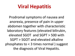
- 1. Viral Hepatitis Prodromal symptoms of nausea and anorexia, presence of pain in upper abdomen together with characteristic laboratory features (elevated bilirubin, elevated SGOT and SGPT > 500 with SGPT > SGOT and elevated alkaline phosphates to < 3 times normal ) suggest the diagnosis of Viral Hepatits.
- 2. Virus Genome Replication Defective DNA Polymerase in Virion HBsAg in Envelope Virus Family HAV ssRNA No No No Picornavirus HBV dsDNA1 No Yes Yes Hepadnavirus HCV ssRNA No No No Flavivirus HDV ssRNA2 Yes No Yes Deltavirus HEV ssRNA No No No Calicivirus
- 3. Features HAV HBV HCV HDV HEV INCUBATION (DAYS) AGE 15-45 Days Children , young adults 30-180 Days Young adults 15-160 Days Any age but > in adults 30-180 Days Any age 14-60 Days Young adults TRANSMISION •Fecal –oral •P.C •Perinatal •Sexual +++ unusual --- + --- +++ +++ ++ --- +++ + + --- +++ + ++ +++ --- --- --- CLINICAL •Severity •Fulminant •Progression to chronicity •Carrier •Cancer •Prognosis Mild 0.1% None None None Excellent Occasionally severe 0.1- 1.0% Occasional (1- 10%) 0.1-%-30% + nt Worse with ag e Moderate 0.1% Common (50- 70%) 1.5 to 3.2% +nt Moderate Occasionally severe 5-20% Common variable variable Acute : good Chronic:poor Mild 1-2% None None None Good PROPHYLAXIS IgG vaccine HBIG None - Unknown
- 4. Hepatitis “A” Virus CLINICAL CAPSULE: HAV is a non enveloped RNA Virus. It is classified in genus Hepatovirus under family Picornavirus. Originally called as enterovirus 72. It is the only human hepatitis virus which can be cultivated in vitro. M/C of hepatits in children. transmission by feco-oral route. Virus is shed in feces during the late Incubation period & prodromal phase of illness.
- 5. LABORATORY DIAGNOSIS OF HAV • “Detection of IgM-specific anti HAV in the blood of an acutely infected patient confirm the diagnosis of hepatitis A.” • IgM appears in acute phase, peaking about 2 weeks after elevation of live enzymes and becomes undetectable withins 3-6 months. • ELISA is the method of choice for measuring antibodies. CLINICAL ASPECTS • Fever, anorexia, and jaundice are common. • ROT: is greatest 2 weeks before to 1 week after the onset of jaundice. • HAV is excreted in clay- colored feces and dark urine for about 2 weeks before the onset of jaundice. • Blood and serum are infective during the brief period of viremia.
- 6. • DIAGNOSIS IgM-specific antibody denotes acute infection. • TREATMENT And PREVENTION No specific tx Passive immunization: Immune serum globulin provides temporary about 80% to 90% protection. Active immunization: Formalin-killed HAV, which is grown in human cell culture, is almost 100% protective
- 9. • HBV belongs to hepadna viridae. • HBV is the only has DNA viruses. • Partial double strand DNA. • HBV contains both DNA- dependent DNA polymerase and RNA-dependent reverse transcriptase. • Found in cytoplasm of infected hepatocytes Structure of HBV: • Serum form pt. of HBV shows 3 types of particle: 1. Dane particle • Double walled spherical structure,42 in diameter. • Complete HBV. • It contains: I. HBcAg II. HBsAg III.HBeAg 2.Spherical particle(MC) • 22nm • HBsAg 3. Filamentous or tubular particle: • 22nm • Identical to spherical is HBs Ag.
- 10. Worldwide distribution of hepatitis B infection. Areas with high prevalence (>8% of population) are in black, and areas with moderate prevalence (2% to 7%) are in gray.
- 12. Antigen HBs Ag Actions : •1st virological marker •Elevation of SGOT/SGPT •Become undetectable 1-2 months after the onset of jaundice. •In chronic HBV infetcion HBsAg remain detectable beyond 6 months. Antibodes : Anti HBsAg Impact: •It become detectable in blood when HBsAg disappears. •Protective AB. •Only marker present after immunization. HBc Ag •It is not detectable because it is enclosed within HBsAg coat. Anti HBcAg •IgM •IgG •IgM: acute or recent infection. •IgG: remote or chronic infection. HBe Ag •Appears shortly after HBsAg. •Indicator of active intraheptic replication and infectivity. Anti HBeAg •Disappearance of HBeAg followed by anti HBe Ag. •It pesence indicates low infectivity & virus infection. HBxAg HCC No A/B Serologic and Virologic Markers of HBV
- 16. Management • High carbohydrate diet. • Domperidome • Silymarin • i.v ornithine (it is amino acid reduced the swelling) • f/u 6 months Anti HBsAg =0 • Liver start regressing --- fulminant hepatic failure(0.1-1%). • Bleeding • Amonia >> in blood >hepatic encephalopathy>coma Tx : Orthoptic liver transplantation. Anti HBsAg + <10 5 copies/ml then wait and watch. >10 5 copies/ml Tx:alfa interferone+lamividine
- 17. Hepatitis “C” Virus • Classified as a new genus Hepacivirus in the family flaviviridae. • The virus can’t be cultured. • Positive-sense RNA genome that encodes three structural and five nonstructural proteins . • Highly heterogeneous virus, hypervariable regions in E2. • Eleven genotypes with multiple subtypes have different geographic distribution. • Genotypes important for therapy response. • HCV replicates in the cytoplasm via negative-sense RNA intermediates • It is the commonest cause of post transfusion hepatitis and chronic hepatitis.
- 18. Laboratory diagnosis of HCV: • The most sensitive and gold standard test in establishing diagnosis is assay for HCV RNA. • HCV-RNA can be detected within a few days of exposure of HCV well before the appearance of A/B ( IgG class) Test for HCV-RNA assays are: • PCR • TMA • B DNA hybridization. • A/B detection by ELISA. The type of A/B are detected: Anti NS-5 (TGA) Anti C22/C33( SGA) Anti C100(FGA) • Hepatitis C is the MCC of Chronic Hepatitis. • 50-80% patients. • Long term prognosis is benign. • Fatigue is the MC symptom. • Immune complex mediated extra hepatic complication are less common than chronic hepatitis B with the exception of essential mixed cryoglobulinemia which is more common in chronic hepatitis C. • Autoimmune play a role in the pathogenesis of chronic hepatitis c. • Lab findings: Characteristics episodic pattern of Aminotransferase activity>aminotransferase level tend to fluctuate. ALT> AST ( after cirrhosis AST>ALT) Presence of Anti LKM1 anti body against p450 2 D6.( HCV microsomal A/B). Anti LKM2 --- (Drug induced A/B) Anti LKM3---(Chronic Hepatits D)
- 20. Inflammation in chronic hepatitis C virus (HCV) infection. Chronic inflammation of the portal area with a lymphoid aggregate in the center can be seen. At the edges of the portal area, the interface between the parenchyma and portal connective tissue, inflammation spreads outward, destroying hepatocytes and expanding the portal tract by piecemeal necrosis.
- 21. TREATMENT: • Pegulated IFN-a + ribavirin • Chronic hepatits is the most common indication for liver transplantation. • MCC of chronic carrier state is HCV. About 50-70% of patients progress to chronic hepatitis. • Chronicity----HCV>HDV>HBV Remember---in HBV infection there are two types of carriers: 1.Super carriers -high titre of HBsAg, HBeAg DNA polymerase and HBV in the circulation. -highly infective. 2.Simple carriers -low titre of HBsAg with negative HBeAg DNA polymerase and HBV. -have low infectivity. HOWEVER • HCV may cause co infection with HBV and co-infection of HCV with HBV increases the rate of development of cirrhosis and the risk of HCC compared with infection. With either virus alone. • HCV can cause liver carcinoma.
- 22. Hepatitis E • Also known as enterically transmitted non-A non-B(NANB) virus or epidemic NANB. • Classified in the genus HERPES virus under the family CALIVIRIDAE. • Single stranded +sense RNA virus. • The viral particles in stool are spherical, 27 to 34 nm in diameter with icosahedral symmetry, and unenveloped, and they exhibit spikes on their surface. • A unique feature is the clinical severity and high case fatality rate of 20-40% in pregnant women, especially in the last trimester of pregnancy. • Characteristically associated with SIMILARITIES IN HAV & HEV: Both have acute onset with mild illness. Both are transmitted by feco-oral route. DIFFERENCES IN HAV & HEV: HAV is more common in children. While HEV is more common in young adult. HEV can cause fulminant hepatitis in pregnant women ( not HAV). Secondary attack rate of HEV is very low (2-3%) as against 10-20% in HAV.
- 23. Hepatitis D or Delta virus • It has been classified in genus Delta virus. • Delta core of HDV is encapsulated by an outer envelope of HBsAg, so require co-operative function of HBV. • Intracellular replication of HDV RNA can occur without HBV but liver injury requires the presence of HBV. • It is defective RNA virus depend on the helper function of HBV for its replication and expression. • It contains defective ssRNA. • It has no independent existence and can survive and replicate only as long as HBV infection persists in the host .
- 24. HDV infection Superinfection • Infected with HBV • Has grave course with more chances of fulminant hepatitis & chronic infection. • IgM anti HDV (+)ve & IgM anti HBc(+)ve Coinfection • Infection occurs simultaneously with HBV. • Comparatively mild course. • IgM anti HDV(+)ve & IgG anti HBc(+)ve.
- 25. • There is no association between HDV and hepatocellular carcinoma • HDV antigen ( Delta antigen) is primarily expressed in liver cell nuclei, where it can be demonstrated by immunofluroscence.
- 26. Distribution of hepatitis E virus infection, among countries in which outbreaks have been identified (shown in black).
- 27. HEPATITIS G • Blood born virus. • It resembles HCV. • Also k/s GB virus C. HGV RNA has been found in patients with: • Acute hepatitis • Chronic hepatitis • Fulminant hepatitis • Hemodialysis • i.v drug addicts. • Blood donor. • Its genome resembles HCV genome except, it lacks a protein corresponding to the core protein of HCV that forms the nucleocapsid. • HGV infection occur independently, it does not require co-infection of HCV.
- 28. Treatment of Hepatitis • Treatment of patients with hepatitis is supportive and directed at allowing hepatocellular damage to resolve and repair itself. • Only HBV and HCV have specific treatments, and those are only partially effective. • Recombinant interferon- and pegylated interferon- are currently the therapy of proven benefit in the treatment of patients chronically infected with HBV or HCV. • With nucleoside and nucleotide analogs, such as lamivudine, HBV DNA levels are reduced, but the virus is rarely eliminated and viral replication resumes in the majority of patients when treatment is stopped. The emergence of drug-resistant virus mutants in long-term therapy is a major problem. • Combination therapy ofinterferon- and ribavirin against chronic hepatitis C gives a sustained response rate of up to 50%, though that therapy is less successful in patients with genotype 1. • Orthotopic liver transplantation is a treatment for chronic hepatitis B and C end- stage liver damage. However, the risk of reinfection on the graft is at least 80% with HBV and 50% with HCV, presumably from extrahepatic reservoirs in the body.
- 29. Drug Nucleoside analogue MOA Viral Spectrum Entecavir Yes Reverse transcriptase inhibitor HBV Foscarnet No Viral polymerase inhibitor HBV Ribavirin Yes Perhaps blocks capping of viral mRNA Respiratory syncytial virus, influenza A and B, Lassa fever, hepatitis C, other Lamivudine (3TC) Yes Reverse transcriptase inhibitor HIV-1, HIV-2, HBV Zalcitabine (ddC) Yes Reverse transcriptase inhibitor HIV-1, HIV-2, HBV Examples of Antiviral Compounds Used for Treatment of Viral Infections
- 30. Clinical and Laboratory Features of Chronic Hepatitis Type of hepatitis Diagnostic test(s) Autoantibodies Therapy Chronic hepatitis B HBsAg, IgG anti-HBc, HBeAg, HBV DNA Uncommon IFN- , PEG;IFN- lamivudine ;adefovir entecavir ;telbivudine ;tenofovir Chronic hepatitis C Anti-HCV, HCV RNA Anti-LKM1a PEG IFN- plus; ribavirin;Telaprevir; Boceprevird Chronic hepatitis D Anti-HDV, HDV RNA, HBsAg, IgG anti-HBc Anti-LKM3 IFN- , PEG ; IFN- c Autoimmune hepatitis ANAb (homogeneous) , anti-LKM1 (±) Hyperglobulinemia ANA, anti-LKM1 anti-SLAe Prednisone, azathioprine Drug-associated — Uncommon Withdraw drug Cryptogenic All negative None Prednisone (?), azathioprine (?)
- 31. Will be grateful for advice.
Notas del editor
- Acute viral hepatitis, moderately severe. There is a lobular disarray with degeneration, apoptosis, and necrosis of liver cells. Disruption of liver cell plates, hypertrophy of Kupffer cells, a predominantly lymphocytic inflammatory infiltrate, and regeneration of surviving liver cells also are seen.
- Schematic diagram of hepatitis B virion.A. The 42-nm particle is the "Dane particle" or the hepatitis B virus. B. The 22-nm particles are the filamentous and circular forms of hepatitis B surface antigen (HbsAg) or protein coat.
- A normal liver (left) shows no signs of scarring. In cirrhosis (right), scar tissue replaces normal liver tissue. Cirrhosis of liver in chronic hepatitis B infection (HBV). This is a needle biopsy of Masson trichrome stain that shows cirrhotic nodules and portion of nodules separated by fibrous scars.