1. INTRODUCTION
Presently, development and research pertaining to
root form dental implants have advanced to the
mature stage. However, the multi-step process of
implant treatment is time-consuming and limited in
terms of patient acceptance. Although the one-stage
implant system provides a partial solution to these
problems, the issue of immediate loading is the next
logical step to execute1)
― and hence the next logical
decision to contend with when considering implant
stability. Immediate loading refers to a restoration
placed in occlusion with the opposing dentition
within 48 hours of implant placement2)
. When the
appropriate implant conditions are present, most
implants can be immediately loaded. Indeed, many
research studies have indicated that the success rate
of immediate loading is good3-4)
. However, primary
implant stability is one of the principal factors that
governs a dentist’s decision on whether a given
implant is suitable for accepting immediate loading.
Presently, few devices and methods are available for
the accurate detection of implant stability immedi-
ately after placement.
The use of resonance frequency (RF) to evaluate
the extent of bone healing with orthopedic treatment
has been studied by many scholars. However, due to
the effects of soft tissue, such techniques remain
unavailable clinically5)
. In dental research, several
recent studies have examined this issue. The results
showed that, in principle, RF can be used to monitor
the process of osseointegration after dental implant
emplacement6-8)
.
Osstell (Integration Diagnostics, G teborgsv ngen,
Sweden), an RF device based on utilizing harmonic
response for monitoring dental implant status,
became commercially available in the year 2000. In
clinical applications, a transducer is attached to the
dental implant fixture and triggered to vibrate by
means of sinusoidal waves. The resonance frequency
value thus obtained is converted to give an Implant
Stability Quotient (ISQ) for analyzing implant
stability9-12)
. Recently, this device has been used in
implant research to perform the following tasks:
compare the success rates between conventional
and early loading of implants13-14)
, evaluate the
survival rates of transmucosal implants immediately
restored with single crowns15)
, monitor the differences
between immediate and standard delayed loading of
implants16-18)
, profile physiological and geometric
factors affecting immediate loaded implants19-20)
, and
measure stability achieved with one-stage surgical
procedures21-22)
. However, clinical investigation has
indicated that temporary attachment, removal, and
sterilization of the transducer are time-consuming
and far from being cost-effective17)
. In addition, the
ISQ value is often influenced by the orientation of
the Osstell’s L-shaped transducer to the alveolar
ridge19,23)
.
Another type of RF analysis, based on impulse
force triggering, has proven useful in terms of
detecting dental implant stability in a series of in
vivo and in vitro experiments24-26)
. In this study,
therefore, the capability of a novel RF detection
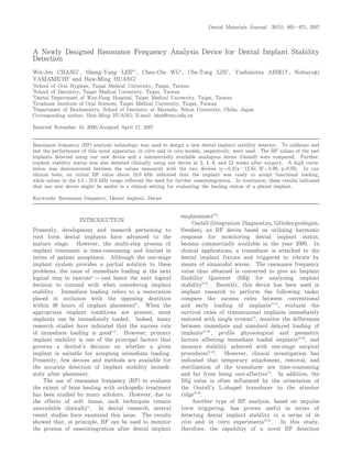
CHANG et al. 665Dental Materials Journal 26(5): 665-671, 2007
A Newly Designed Resonance Frequency Analysis Device for Dental Implant Stability
Detection
Wei-Jen CHANG1
, Sheng-Yang LEE2,3
, Chen-Che WU2
, Che-Tong LIN4
, Yoshimitsu ABIKO5
, Nobuyuki
YAMAMICHI5
and Haw-Ming HUANG4
1
School of Oral Hygiene, Taipei Medical University, Taipei, Taiwan
2
School of Dentistry, Taipei Medical University, Taipei, Taiwan
3
Dental Department of Wan-Fang Hospital, Taipei Medical University, Taipei, Taiwan
4
Graduate Institute of Oral Sciences, Taipei Medical University, Taipei, Taiwan
5
Department of Biochemistry, School of Dentistry at Matsudo, Nihon University, Chiba, Japan
Corresponding author, Haw-Ming HUANG; E-mail: hhm@tmu.edu.tw
Received November 10, 2006/Accepted April 17, 2007
Resonance frequency (RF) analysis technology was used to design a new dental implant stability detector. To calibrate and
test the performance of this novel apparatus, in vitro and in vivo models, respectively, were used. The RF values of the test
implants detected using our new device and a commercially available analogous device (Osstell) were compared. Further,
implant stability status was also detected clinically using our device at 2, 4, 8, and 12 weeks after surgery. A high corre-
lation was demonstrated between the values measured with the two devices (y=0.31x-12.45; R2
=0.98, p<0.05). In our
clinical tests, an initial RF value above 10.0 kHz indicated that the implant was ready to accept functional loading,
while values in the 4.0-10.0 kHz range reflected the need for further osseointegration. In conclusion, these results indicated
that our new device might be useful in a clinical setting for evaluating the healing status of a placed implant.
Keywords: Resonance frequency, Dental implant, Device
2. device based on impulse force triggering for
monitoring dental implant stability was tested. To
reduce measurement operation time, a minimum
contact device that did not require additional installa-
tion and/or disassembly was designed. To validate
the device in terms of implant stability determina-
tion, a series of in vitro and in vivo tests was
performed. Finally, the standard value for this novel
technique, which was to be an indicator of a test
implant’s readiness for immediate loading, was
determined by means of clinical data analysis.
MATERIALS AND METHODS
Device design
As shown in Fig. 1, our new device incorporated a
minimum contact transducer and an attached
handpiece. The handle could be rotated to improve
access in the limited space of the oral cavity
(Fig. 1(a)). The device consisted of two sections of an
electromagnetic coil to provide driving feedback for
the demagnetized iron impact head (Fig. 1(b)). When
the impulse current passed through the first coil
section, the generated electromagnetic field attracted
the impact head and drove it to strike against the
healing abutment. An impact force of 0.18 N was
thereby delivered, as determined by a pressure-
sensitive film (Prescale Pressure Series, Fuji Photo
Film, Tokyo, Japan). The second section then
generated an electromagnetic field in the opposite
direction, retracting the impact head to its original
position. When the impact head strike the test
implant, the resultant vibration was detected via a
piezoelectric microphone and the vibration signal sent
to a spectrum analyzer (resolution: 50 Hz;
Implomates System, Biotech One, Taipei, Taiwan).
The specific resonance frequency of the test implant
was determined from the relatively highest point
with a peak value for vibration amplitude. This
device was used throughout the entire research.
In vitro testing
To validate the experimental device, a series of in
vitro tests was carried out. A commercial pure
titanium dental implant ( 3.75 × 10 mm; 3i
Innovation, FL) with a 6-mm healing abutment was
fixed using a metal clamp stand. RF values for the
test implant were recorded while the clamp torque
was varied (2 to 10 N・ cm in 2 N・ cm increments). In
addition, the clamping level was altered to expose the
implant and alter the vibrational length (0−8 mm in
2 mm steps upward from the base of the implant).
Accuracy of the experimental device was tested by
comparing the results with those obtained using a
conventional excitation method (Osstell). Before RF
detection with Osstell, a piezoelectric transducer was
connected to the dental implant fixture. Transducer
vibration was triggered by 1-V sinusoidal waves in
the 5−15 kHz range. The first RF value detected by
the device was used for implant stability analysis.
Animal study
Five healthy adult beagles (weighing 8−12 kg) were
used as test subjects in the animal study. Three
months prior to implantation, the mandibular first
premolars were bilaterally removed under general
(intravenous injection of 0.5 mg/kg ketamine,
followed by intramuscular injection of 25 mg/kg
pentobarbital) and local anesthesia (2% lidocaine)
(Showa Co., Tokyo, Japan). Prior to dental implant
surgery, the animals were again anesthetized as
described above. Crestal incisions were followed by
flap reflection to expose the alveolar crest. The
A new device for implant stability detection666
Fig. 1 Diagrams of the resonance frequency (RF) detec-
tor used in this study: (a) Designed as a
minimum contact probe, vibration of the test
implant was triggered by impact rod; (b) Sagittal
section of the device showing electromagnetic
driving actuator and non-contacting microphone.
3. surgical sites were prepared using the standard
procedures specified by the dental implant
manufacturer.
Test implants ( 3.75×10 mm; 3i Innovation, FL)
were placed into the drilled holes at the left first
premolar of each animal until their collar margin
reached the boundary of the cortical bone. Right-
side surgical sites without implant placement were
treated as controls. In other words, a total of five
implants were used for the entire experiment. After
implant placement, the flaps were repositioned and
sutured. Profuse cooling with cold normal saline
solution was used throughout the surgical procedure.
After surgery, the animals were fed a soft diet
(Quaker Oats, Peterborough, Ontario, Canada) for
the first two weeks, and long-acting penicillin was
also administered (Penlong XL, Rogar STB, London,
Ontario, Canada) at appropriate intervals during this
period.
Immediately after placement of the implants, RF
was measured using both Osstell and our experimen-
tal device. Test setup was identical to that used for
the in vitro tests above. Measurements were
obtained on the implant samples along the
buccolingual direction at 0, 2, 4, 8, and 12 weeks
after implant surgery. All samples were subjected to
five continuous tests, with results reported as the
mean and standard deviation of the RF values. One-
way analysis of variance was used to test statistical
differences between the test parameters. The
experimental design was approved by the Laboratory
Animal Research Committee, College of Oral
Medicine, Taipei Medical University.
Clinical data collection
Data for continuous RF measurement of 11 implants
were collected from seven patients (one female and
six male patients). All of whom (mean age of 31.4
years, range of 25−48 years at time of surgery) were
fully informed of the study protocol before signing
written agreements. The recruitment criteria were:
no history of oral disease or dental implant surgery.
Edentulous areas were located at the mandibular
premolar or first molar (Fig. 2(a)). The 3i implants
(3i Innovation, FL) were placed in the mandible
according to the manufacturer’s guidelines for a one-
stage procedure (Fig. 2(b)). All the implants were
covered with a 4-mm healing abutment to avoid oral
fluid contamination. Resonance frequencies were
measured using our newly designed apparatus
immediately after the implants were placed (week 0)
and at weeks 2, 4, 8, and 12 after the implantation
surgery (Fig. 2(c)). Detection was performed in the
buccolingual direction. After a healing period of 12
weeks, the patients received their prostheses using
the classic procedure.
RESULTS
Table 1 shows the RF and ISQ values, as measured
in vitro using our novel device and Osstell respec-
tively, for a dental implant with various clamping
torques. The mean experimental RF value increased
from 14.76±0.02 to 15.11±0.02 kHz in the torque
range 2−10 N・ cm. Similarly, under the same test
CHANG et al. 667
Fig. 2 Surgical and test procedures: (a) location of eden-
tulous area; (b) general procedure for implant
placement; and (c) resonance frequencies of test
implants detected using the new device.
4. conditions using the Osstell device, the ISQ value also
increased from 85.67±0.58 to 89.00±0.00. However,
no differences in ISQ value were detected when
clamping torque was increased from 4 to 6 N・ cm
(ISQ=88), and from 8 to 10 N・ cm (ISQ=89). Table
2 lists the measurement data for the various
clamping levels. When the exposed height of the test
implant was increased from 0 to 8 mm, the mean
experimental RF value decreased from 16.10±0.00 to
5.29±0.02 kHz. Similarly, the measured ISQ value
decreased from 88.00± 0.00 to 56.33 ± 1.53 for the
same test conditions. Replotting the measurement
data from Tables 1 and 2 in Fig. 3, a linear correla-
tion was obtained between the RF values of our
newly designed device and ISQ values derived from
Osstell (r=0.991; P<0.01).
All animals used in the in vivo study remained
in excellent health throughout the course of the
experiment. The in vivo data, in the form of
continuous measurements obtained using our novel
device, are plotted in Fig. 4(a). There was a mean
significant decrease of 1.45 kHz between initial
placement and two weeks post surgery (P<0.05), but
a mean significant increase of 2.45 kHz between
weeks 2 and 12 (P<0.01). More importantly, when
the same implants were tested using the Osstell
device, no significant differences were detected
between the ISQ values across the experimental
period (Fig. 4(b)).
Figure 5 shows the results of the clinical investi-
gation, whereby test implants were divided into three
groups. In Group I, the initial RF values were above
9 kHz, remaining high and plateauing at 11 kHz or
more by week 12. In Group II, the initial RF values
of the implants ranged between 3.58 and 5.3 kHz.
These implants remained in excellent health through-
out the course of the experiment and completed the
osseointegration process. By week 12, the RF values
for all the Group II implants were above 9.8 kHz. In
Group III, one implant with an initial RF value of
3.56 kHz did not demonstrate an increasing trend in
RF value after the first two weeks of healing.
Osseointegration of the implant failed, and by week
12 it loosened with a finial RF value of 3.85 kHz.
Our short-term results demonstrated a 91% success
rate during the first 12 weeks.
To assess the relationship between the RF values
of immediate and delayed loaded implants in this
study, the resonance frequency increase ratio (RFIR)
was defined as the ratio between the initial RF value
and the analogous value at week 4. The mean RFIR
of Group II implants (1.98±0.31) was significantly
higher than that of Group I analog (1.09 ± 0.11;
P<0.005). The plot in Fig. 6 revealed a linear
relationship between the RFIR of each test implant
and its initial RF (y= − 0.126x+2.50, R
2
=0.811,
p<0.05).
A new device for implant stability detection668
Torque (N・cm)
2 4 6 8 10
Device
Osstell (ISQ) 85.67 ± 0.58 88.00 ± 0.00 88.00 ± 0.00 89.00 ± 0.00 89.00 ± 0.00
Newly designed device (kHz) 14.76 ± 0.02 14.90 ± 0.00 15.01 ± 0.02 15.10 ± 0.00 15.11 ± 0.02
Data are presented as mean ± SD.
Table 1 Comparison of implant measurement data generated by Osstell and new RFA device for different clamping
torques
Exposed height (mm)
0 2 4 6 8
Device
Osstell (ISQ) 88.00 ± 0.00 77.67 ± 0.00 70.00 ± 0.00 64.67 ± 0.58 56.33 ± 1.53
Newly designed device (kHz) 16.10 ± 0.00 12.31 ± 0.02 9.16 ± 0.02 7.01 ± 0.02 5.29 ± 0.02
Data are presented as mean ± SD.
Table 2 Comparison of implant measurement data from Osstell and new RFA device for different clamping levels
Fig. 3 Relationship of data measured in vitro for the
two devices.
5. DISCUSSION
Osstell was the first commercially available RF device
to test dental implant stability, and the efficacy of
this method for monitoring implant status has been
proven10-12)
. However, it is somewhat inconvenient and
time-consuming
17)
, and it may even affect the
implant-bone interface at the early healing stage.
This is because the Osstell transducer must be
screwed into the test implant with a torque of 10
N・ cm, almost half the force used to place an implant.
In this light, the mechanical effect of disassembling
the healing abutment on the interface in the early
stages of osseointegration should be taken into
consideration. Against this background, our novel
transducer was designed as a minimum contact device
with no torque force required during transducer
application.
In the development of a medical device, accuracy
of the measuring instrument should be an utmost
priority for the engineers involved. To validate the
effectiveness of our newly designed apparatus, in
vitro tests were performed with a view to reducing
the physiological effects of measurement. According
to previous studies, resonance frequency was affected
by bone-implant contact percentage and height of the
exposed implant above the marginal bone6,24,25)
. In this
study, therefore, both factors were simulated and
tested in vitro as in our previous work24)
. As shown
in Fig. 3, a close relationship was demonstrated for
the data obtained using Osstell and our device.
Thus, it seemed reasonable to suggest that our novel
device may be used to monitor changes in implant
RF with various boundary conditions, while offering
significant functional advantages and without
adversely affecting the healing process.
In the present study, the mean RF of the
implants placed in animals decreased during the first
CHANG et al. 669
Fig. 4 Healing curves generated using RF values obtained
from test implants in experimental animals with
(a) experimental device and (b) Osstell. The data
are presented as mean ± SD.
Fig. 5 RF healing curves from test implants in patients
during the 12 weeks post implantation. Solid,
short-dashed, and long-dashed lines denote Groups
I, II, and III (initial RF values >9, 3.5−6, and <4
kHz, respectively).
Fig. 6 Relationship between the initial RF value and RF
increase ratio of the successful implants at four
weeks post surgery. Open and closed circles
denote Group I and Group II implants respectively.
6. two weeks and then increased progressively with
time (Fig. 4). These results were consistent with
other animal RFA14,26)
and reverse torque studies16,27)
.
This initial reduction in stability might correspond
to the remodeling and neo-apposition phases of the
necrotized bone16)
. However, the stability-return
phenomenon was not borne out in our clinical data
(Fig. 5). In fact, demonstrating this phenomenon
using resonance frequency analysis (RFA) remains
controversial. Although a tendency for ISQ to
decrease in the initial stage followed by a stable
increase has been demonstrated in some clinical
studies
14,18-19,28)
, many RFA-based investigations have
not confirmed this phenomenon16,23)
. After testing 276
immediate loaded implants using RFA, Balshi et al.
suggested that bone types II and III were more
consistent in term of stability return, while bone
types I and IV were not19)
.
Primary stability, a function of bone quality,
implant design, and surgical procedure, is one of the
important factors that influences the survival rate of
implants17,20,27)
. It has been reported that primary
stability can serve as a useful predictor of
osseointegration
16)
. Further, it has been demon-
strated that good primary implant stability, as
measured by RFA, did not increase significantly dur-
ing the osseointegration period15,16,29)
. In this study, it
was found that implants with higher stability indeed
had relatively stable RF during the whole healing
period with a mean RFIR of 1.09±0.112 (Figs. 5
and 6). As constant or slightly increased RF values
for immediate loaded implants during the first 4−6
weeks have been reported16)
, it seemed reasonable to
suggest that for implants with initial RF values
exceeding 10 kHz (as measured by our device), these
implants are ready for immediate loading.
A number of researches have proposed that
where the initial ISQ value is above 60, the implant
is ready for immediate loading
17-19)
. However, for
implants with ISQ values below 40, this should serve
as a warning of early failure
18)
. Interestingly, this
proposed functional threshold of ISQ 60 is equivalent
to an RF of 6.10 kHz
18)
, much lower than our
experimentally determined limit of 10 kHz. A
negative relationship has been reported between RF
value and effective vibrational length above bone
level
8,16,24-25)
. Since the Osstell transducer is an L-
shaped device that must be attached to the top of the
test implant, the effective vibrational length of test
implant would thus be greater than that tested using
our device. Accordingly, to compare and exchange
measurement data generated using the Osstell system
(ISQ) and the analogous information from our own
device (in kHz), a transfer function was derived.
As shown in Fig. 3, the relationship between the
two parameters was linear with a ratio of 0.3. As
the relationship formula in Fig. 3 was obtained from
in vitro tests, the constant in the formula should be
modified based on our clinical data. After substitut-
ing 10 kHz and 60 ISQ for y and x in Fig. 3 respec-
tively, a new transfer function was derived: y=0.3x−
8, where y and x are kHz and ISQ values respec-
tively. Based on this function, the early failure
threshold of ISQ 40 for Osstell was 4 kHz using our
novel device. From Fig. 5, the initial and final RF
values of the failed implant (3.56 kHz and 3.85 kHz,
respectively) fit the hypothesis well.
The initial RF values of our test implants
varied (Fig. 5), probably reflecting the marked
interindividual variations in distributions of cortical
and trabecular bone, as well as differences between
various locations within bone
30)
. After analyzing the
RFA data of 120 one-stage implants, Zix et al.
suggested that a single RFA measurement for an
implant at a given time point could not qualify being
a full predictor of its future performance22)
. Repeated
measurements of an implant over a period would
provide a more credible prognosis of its future
status. In this connection, Glauser et al. suggested
that low RFA levels after one and two months
implied risk for future failure
18)
.
In the current study, implants with lower initial
RF values revealed significantly higher RFIR values
(Fig. 6). At this juncture, it must be clarified that
RFIR was defined in this study for early diagnosis
but not for determining the optimal time point for
functional loading. However, it seemed reasonable to
suggest that the time for functional loading of these
implants is when their RF value plateaus or exceeds
9 kHz. Nedir et al. also reported that implants with
lower initial ISQ values demonstrated a larger ISQ
increase during the healing period17)
. In contrast,
higher initial stability was associated with a more
stable ISQ value. In addition, only the implant with
highest stability revealed stability decrease during
the first four weeks before remaining stable. These
results substantially agreed with our own findings.
Based on the results obtained in the present
study, it was therefore reasonable to suggest that
our novel RF detection device was suitable for
evaluating the integrity of bone union during the
osseointegration process. At the same time, our
novel device provided functional superiority without
adversely affecting the restorative process. Amongst
which, the greatest advantage of this new device was
its ergonomic superiority. Thus, we proposed that
our newly designed RFA apparatus had the potential
of delivering substantial benefits to advanced experi-
ments in the future, as well as holding out signifi-
cant promise for future clinical use.
REFERENCES
1) Raghoebar GM, Schoen P, Meijer HJA, Stellingsma
A new device for implant stability detection670
7. K, Vissink A. Early loading of endosseous implants
in the augmented maxilla: a 1-year prospective study.
Clin Oral Impl Res 2003; 14: 697-702.
2) Cochran DL, Morton D, Weber HP. Consensus state-
ments and recommended clinical procedures regarding
loading protocols for endosseous dental implants. Int
J Oral Maxillofac Impl 2004; 19: 109-113
3) Horiuchi K, Uchida H, Yamamoto K, Sugimura M.
Immediate loading of Br nemark system implants
following placement in edentulous patients: a clinical
report. Int J Oral Maxillofac Impl 2000; 15: 824-830.
4) Andersen E, Haanaes HR, Knutsen BM. Immediate
loading of single-tooth ITI implants in the anterior
maxilla: a prospective 5-year pilot study. Clin Oral
Impl Res 2002; 13: 281-287.
5) Nokes LD. The use of low-frequency vibration meas-
urement in orthopaedics. Proc Instn Mech Engrs,
Part H: J Eng Med 1999; 213: 271-290.
6) Meredith N, Alleyne D, Cawley P. Quantitative
determination of the stability of the implant-tissue
interface using resonance frequency analysis. Clin
Oral Impl Res 1996; 7: 261-267.
7) Meredith N. Assessment of implant stability as a
prognostic determinant. Int J Prosthodont 1998; 11:
491-501.
8) Meredith N, Shagaldi F, Alleyne D, Sennerby L,
Cawley P. The application of resonance frequency
measurements to study the stability of titanium
implants during healing in the rabbit tibia. Clin Oral
Impl Res 1997; 8: 234-243.
9) Meredith N. A review of nondestructive test methods
and their application to measure the stability and
osseointegration of bone anchored endosseous
implants. Crit Rev Biomed Eng 1998; 26: 275-291.
10) Cawley P, Pavlakovic B, Alleyne DN, George R, Back
T, Meredith N. The design of a vibration transducer
to monitor the integrity of dental implants. Proc
Instn Mech Engrs, Part H: J Eng Med 1998; 212: 265-
272.
11) Rasmusson L, Meredith N, Cho IH, Sennerby L. The
influence of simultaneous versus delayed placement on
the stability of titanium implants in onlay bone
grafts: A histologic and biomechanic study in the
rabbit. Int J Oral Maxillofac Surg 1999; 28: 224-231.
12) Barewal RM, Oates TW, Meredith N, Cochran DL.
Resonance frequency measurement of implant stabil-
ity in vivo on implants with a sandblasted and acid-
etched surface. Int J Oral Maxillofac Implants 2003;
18: 641-651.
13) Payne AGT, Tawse-Smith A, Duncan WD, Kumara R.
Conventional and early loading of unsplinted ITI
implants supporting mandibular overdentures: two-
year results of a prospective randomized clinical trial.
Clin Oral Impl Res 2002; 13: 603-609.
14) De Smet E, Jaecques S, Vandamme K, Vander Sloten
J, Naert I. Positive effect of early loading on
implant stability in the bi-cortical guinea-pig model.
Clin Oral Impl Res 2005; 16: 402-407.
15) Cornelini R, Cangini F, Covani U, Barone A, Buser
D. Immediate restoration of single-tooth implants in
mandibular molar sites: A 12-month preliminary
report. Int J Oral Maxillofac Implants 2004; 19: 855-
860.
16) Bischof M, Nedir R, Szmukler-Moncler S, Bernard
JP, Samson J. Implant stability measurement of
delayed and immediately loaded implants during
healing. A clinical resonance-frequency analysis
study with sandblasted-and-etched ITI implants. Clin
Oral Impl Res 2004; 15: 529-539.
17) Nedir R, Bischof M, Szmukler-Moncler S, Bernard
JP, Samson J. Predicting osseointegration by means
of implant primary stability. A resonance-frequency
analysis study with delayed and immediately loaded
ITI SLA implants. Clin Oral Impl Res 2004; 15: 520-
528.
18) Glauser R, Sennerby L, Meredith N, Ree A, Lundgren
AK, Gottlow J, Hammerle CHF. Resonance
frequency analysis of implants subjected to immediate
or early functional occlusal loading. Successful vs.
failing implants. Clin Oral Impl Res 2004, 15: 428-
434.
19) Balshi SF, Allen FD, Wolfinger GJ, Balshi TJ. A
resonance frequency analysis assessment of maxillary
and mandibular immediately loaded implants. Int J
Oral Maxillofac Implants 2005; 20: 584-594.
20) Akkocaoglu M, Uysal S, Tekdemir I, Akca K, Cehreli
MC. Implant design and intraosseous stability of
immediately placed implants: A human cadaver study.
Clin Oral Impl Res 2005; 16: 202-209.
21) Monov G, Fuerst G, Tepper G, Watzak G, Zechner W,
Watzek G. The effect of platelet-rich plasma upon
implant stability measured by resonance frequency
analysis in the lower anterior mandibles: A pilot
study. Clin Oral Impl Res 2005; 16: 461-465.
22) Zix J, Kessler-Liechti G, Mericske-Stern R. Stability
measurements of 1-stage implants in the maxilla by
means of resonance frequency analysis: A pilot study.
Int J Oral Maxillofac Implants 2005; 20: 747-752.
23) Kramer FJ, Dempf R, Bremer B. Efficacy of dental
implants placed into fibula-free flaps for orofacial
reconstruction. Clin Oral Impl Res 2005; 16: 80-88.
24) Huang HM, Pan LC, Lee SY, Chiu CL, Fan KH, Ho
KN. Assessing the implant/bone interface by using
natural frequency analysis. Oral Surg Oral Med Oral
Pathol Oral Radiol Endod 2000; 90: 285-291.
25) Huang HM, Lee SY, Yeh CY, Lin CT. Resonance
frequency assessment of dental implant stability with
various bone qualities: a numerical approach. Clin
Oral Impl Res 2002; 13: 65-74.
26) Huang HM, Chiu CL, Yeh CY, Lin CT, Lin LH, Lin
LH, Lee SY. Early detection of implant healing proc-
ess using resonance frequency analysis. Clin Oral
Impl Res 2003; 14: 437-443.
27) Baker D, London RM, O’Neal R. Rate of pull-out
strength gain of dual-etched titanium implants: a
comparative study in rabbits. Int J Oral
Maxillofacial Implants 1999; 14: 722-728.
28) Monov G, Fuerst G, Tepper G, Watzak G, Zechner W,
Watzek G. The effect of platelet-rich plasma upon
implant stability measured by resonance frequency
analysis in the lower anterior mandibles. A pilot
study. Clin Oral Impl Res 2005; 16: 461-465.
29) Friberg B, Sennerby L, Linden B, Grondahl K,
Lekholm U. Stability measurements of one-stage
Branemark implants during healing in mandibles. A
clinical resonance frequency analysis study. Int J
Oral Maxillofac Surg 1999; 28: 266-272.
30) Fanuscu MI, Chang TL. Three-dimensional
morphometric analysis of human cadaver bone:
microstructural data from maxilla and mandible.
Clin Oral Impl Res 2004; 15: 213-218.
CHANG et al. 671