10. The associated proteins mantain the structure of the
chromosome and help control the activities of the genes
Eukaryotic chromosomes consist of chromatin, a
complex of DNA and protein that condenses during cell
division
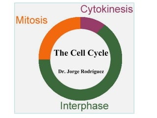
20. Figure 12.6
The cell cycle
INTERPHASE
S
G1, first part of interphase,
followed by S
(M)
P
sis
i
Mi
to
k
to
Cy
MIT
O
sis
ne
(DNA synthesis and
chromosome duplication)
G2
T
HAS IC
E
MITOTIC (M) PHASE, alternate
with interphase
• M phase, daughter cromosomes in daugther
nuclei.
• Cytokinesis, divide the cytoplasm into 2
daugther cells.
• Relative duration of the phases may vary.
22. Figure 12.7a
G2 of Interphase
Centrosomes
(with centriole
pairs)
Prometaphase
Prophase
Fragments
Chromatin Early mitotic Aster
of nuclear
(duplicated) spindle
Centromere envelope
Plasma
membrane
Nucleolus
Nuclear
envelope
• Sobre nuclear rodea el núcleo.
• Nucleoli (s) present.
• 2 centrosomas formados por
duplicación de cada cromosoma.
• Chromosomes not condensed.
Chromosome, consisting
of two sister chromatids
Nonkinetochore
microtubules
Kinetochore
Kinetochore
microtubule
• Chromatin become condensed.
• Envelope fragments.
• Nucleoli dissapear.
• Microtubules invade
nuclear area.
• Each chromosome appears as two
sister chromatids.
• Mitotic spindle begins to form.
Composed by centrosomes and
microtubules.
• Kinetochore appears at
centromeres.
• Kinetochore microtubules
forming.
• Asters, radial arrays of microtubules. • Nonkinetochore
microtubules forms the
• Centrosomes move away.
opposite pole of the
23. Figure 12.7b
Metaphase
Anaphase
Metaphase
plate
Spindle
Telophase and Cytokinesis
Cleavage
furrow
Centrosome at
one spindle pole
Daughter
chromosomes
Nucleolus
forming
Nuclear
envelope
forming
• Two nuclei forms in the cell.
• Centrosomes at opposite poles
of the cell.
• Chromosomes at metaphase
plate.
• Shortest phase.
• Nuclear envelopes arises.
• Cohesins are cleaved.
• Nucleoli reappear.
• Sister chromatids separates. • Chromosomes less condensed.
• Daugther chromosomes
moves towards opposite
poles.
• Depolymerization of microtubules.
• Cell elongates.
• Cytokinesis under way by late
telophase. Formation of cleavage
furrow.
• Mitosis is completed.
25. Figure 12.7a
The Mitotic Spindle: A Closer Look
G2 of Interphase
Centrosomes
(with centriole
pairs)
Chromatin
(duplicated)
Plasma
membrane
Nucleolus
Nuclear
envelope
•
•
•
•
•
Prometaphase
Prophase
Early mitotic
spindle
Aster
Centromere
Chromosome, consisting
of two sister chromatids
Fragments
of nuclear
envelope
Kinetochore
Nonkinetochore
microtubules
Kinetochore
microtubule
Many events of mitosis depend on the mitotic spindle
Begins to form in the cytoplasm during prophase
The mitotic spindle is a structure made of fibers and associated proteins
The spindle polymerize by incorporating subunits of tubulin
Controls chromosome movement during mitosis
26.
27. Figure 12.7a
The Mitotic Spindle: A Closer Look
G2 of Interphase
Centrosomes
(with centriole
pairs)
Chromatin
(duplicated)
Plasma
membrane
Nucleolus
Nuclear
envelope
•
•
•
Prometaphase
Prophase
Early mitotic
spindle
Aster
Centromere
Chromosome, consisting
of two sister chromatids
Fragments
of nuclear
envelope
Kinetochore
Nonkinetochore
microtubules
Kinetochore
microtubule
In animal cells, assembly of spindle microtubules begins in the centrosome, a
subcellular region the microtubule organizing center
– A pair of centrioles is located at the center of the centrosomes, not
essential for cell division
The centrosome replicates during interphase, forming two centrosomes
As spindle microtubules grows, centrosomes migrate to opposite ends of the
cell during prophase and prometaphase
28. Figure 12.7a
The Mitotic Spindle: A Closer Look
G2 of Interphase
Centrosomes
(with centriole
pairs)
Chromatin
(duplicated)
Plasma
membrane
Nucleolus
Nuclear
envelope
•
•
Prometaphase
Prophase
Early mitotic
spindle
Aster
Centromere
Chromosome, consisting
of two sister chromatids
Fragments
of nuclear
envelope
Kinetochore
Nonkinetochore
microtubules
Kinetochore
microtubule
An aster (a radial array of short microtubules) extends from each
centrosome
The spindle includes the centrosomes, the spindle microtubules, and
the asters
29. Figure 12.7a
The Mitotic Spindle: A Closer Look
G2 of Interphase
Centrosomes
(with centriole
pairs)
Chromatin
(duplicated)
Plasma
membrane
Nucleolus
Nuclear
envelope
Prometaphase
Prophase
Early mitotic
spindle
Aster
Centromere
Chromosome, consisting
of two sister chromatids
Fragments
of nuclear
envelope
Kinetochore
Nonkinetochore
microtubules
Kinetochore
microtubule
• During prometaphase, some spindle microtubules attach
to the kinetochores of chromosomes and begin to move the
chromosomes
Kinetochores microtubules
• Kinetochores are protein complexes (2) associated with
centromeres in each sister chromatids
30. Figure 12.7b
Metaphase
Anaphase
Metaphase
plate
Spindle
Centrosome at
one spindle pole
Telophase and Cytokinesis
Cleavage
furrow
Daughter
chromosomes
Nucleolus
forming
Nuclear
envelope
forming
• At metaphase, the chromosomes are all lined up at the
metaphase plate, an imaginary structure at the midway
point between the spindle’s two poles
• The spindle is completed when microtubules of the asters
attach to the plasma membrane
33. Figure 12.7b
Metaphase
Anaphase
Metaphase
plate
Spindle
•
•
•
Centrosome at
one spindle pole
Telophase and Cytokinesis
Cleavage
furrow
Daughter
chromosomes
Nucleolus
forming
Nuclear
envelope
forming
In anaphase, cohesins are cleaved by separases
Sister chromatids separate and move along the kinetochore
microtubules toward opposite ends of the cell
The microtubules shorten by depolymerizing at their kinetochore ends
34. Figure 12.7b
Metaphase
Anaphase
Metaphase
plate
Spindle
•
•
•
•
Centrosome at
one spindle pole
Telophase and Cytokinesis
Cleavage
furrow
Daughter
chromosomes
Nucleolus
forming
Nuclear
envelope
forming
In anaphase, nonkinetochore microtubules from opposite poles overlap and
push against each other, elongating the cell
At the end of anaphase, duplicate chromosomes have arrived at opposite ends
of elongated parent cell
In telophase, genetically identical daughter nuclei form at opposite ends of the
cell
Cytokinesis begins during anaphase or telophase and the spindle eventually
disassembles by depolymerization
36. Figure 12.10a
(a) Cleavage of an animal cell (SEM)
Cleavage furrow
(old metaphase plate)
100 µm
Contractile ring of microfilaments Daughter cells (cytosol, nucleus, organelles,
(actin and myosin)
subcellular structures)
37. (b) Cell plate formation in a plant cell (TEM)
• No cleavage furrow formed.
• Telophase, vesicles from
Golgi move to the middle of the
cell, coalesce and produce a
cell plate.
Vesicles
forming
cell plate
Wall of parent cell
Cell plate
1 µm
New cell wall
Daughter cells
• Vesicles contains cell wall
material and fuses with the
plasma membrane.
• Two daugther cells result.
Figure 12.10b
39. Figure 12.12-1
Origin of
replication
E. coli cell
1 Chromosome
replication
begins.
Cell wall
Plasma membrane
Bacterial chromosome
Two copies
of origin
Chromosome replicates (beginning at the origin
of replication), and the two daughter chromosomes
actively move apart (not understood).
40. Figure 12.12-2
Origin of
replication
E. coli cell
1 Chromosome
replication
begins.
2 Replication
continues.
Cell wall
Plasma membrane
Bacterial chromosome
Two copies
of origin
Origin
Origin
• One copy of each origin at each end of the cell.
• Cell elongates.
41. Figure 12.12-3
Origin of
replication
E. coli cell
1 Chromosome
replication
begins.
2 Replication
continues.
Cell wall
Plasma membrane
Bacterial chromosome
Two copies
of origin
Origin
Origin
3 Replication
finishes.
Plasma membrane grows inward, a new cell wall is
formed.
42. Figure 12.12-4
Origin of
replication
E. coli cell
1 Chromosome
replication
begins.
2 Replication
continues.
3 Replication
finishes.
4 Two daughter
cells result.
Cell wall
Plasma membrane
Bacterial chromosome
Two copies
of origin
Origin
Origin
46. Figure 12.14
EXPERIMENT
Experiment 1
S
G1
S
S
Experiment 2
M
G1
RESULTS
When a cell in the S
phase was fused
with a cell in G1,
the G1 nucleus
immediately entered
the S phase—DNA
was synthesized.
M
M
When a cell in the
M phase was fused with
a cell in G1, the G1
nucleus immediately
began mitosis—a spindle
formed and chromatin
condensed, even though
the chromosome had not
been duplicated.
Conclusion: The results of fusing a G1 with a S or M cell cycle
suggest that molecules present in the cytoplasm during S or
M phase control the progression to those phases.
49. Figure 12.16
G0
G1 checkpoint
G1
(a) Cell receives a go-ahead
signal.
G1
(b) Cell does not receive a
go-ahead signal.
•
For many cells, the G1 checkpoint seems to be the most important
•
If a cell receives a go-ahead signal at the G1 checkpoint, it will usually
complete the S, G2, and M phases and divide
•
If the cell does not receive the go-ahead signal, it will exit the cycle,
switching into a nondividing state called the G0 phase
51. Figure 12.17a
MPF (maturation-promoting factor) is a cyclin-Cdk
complex that triggers a cell’s passage past the G2
checkpoint into the M phase
M
G1
S
G2
M
G1
S
G2
M
G1
MPF activity
Cyclin
concentration
Time
(a) Fluctuation of MPF activity and cyclin concentration
during the cell cycle
The peaks of MPF activity corresponds to the peaks of cyclin
concentration.
Cyclin level rises during the S and G2 , then falls during M
phase.
52. Figure 12.17b
Molecular mechanisms that help regulate the cell cycle
S
G
1
1. Synthesis of cyclin
begins in S and
continue through G2.
Cdk
Degraded
cyclin
Cyclin is
degraded
M
G2
Cdk
G2
checkpoint
MPF
Cyclin
53. Figure 12.17b
Molecular mechanisms that help regulate the cell cycle
G
1
S
Cdk
M
G2
Degraded
cyclin
G2
checkpoint
Cyclin is
degraded
MPF
Cdk
Cyclin
2. Cyclin combines with Cdk, producing MPF.
MPF accumulate, the cell passes the G2
checkpoint and begins mitosis.
54. Molecular mechanisms that help regulate the cell cycle
1
S
G
Figure 12.17b
Cdk
Degraded
cyclin
Cyclin is
degraded
M
G2
G2
checkpoint
MPF
Cdk
Cyclin
3. MPF phosphorylates various proteins.
MPF’s activity peaks during metaphase.
55. Molecular mechanisms that help regulate the cell cycle
S
G
1
Figure 12.17b
Cdk
Degraded
cyclin
Cyclin is
degraded
M
G2
G2
checkpoint
MPF
4. Anaphase, cyclin component of MPF is
degraded, terminating M phase.
The cell enters G1.
Cdk
Cyclin
56. Figure 12.17b
Molecular mechanisms that help regulate the cell cycle
5. G1, degradation of
cyclin continues.
Cdk component of MPF is
recycled.
G
1
S
Cdk
Degraded
cyclin
Cyclin is
degraded
M
G2
G2
checkpoint
MPF
Cdk
Cyclin
57. Figure 12.17b
Molecular mechanisms that help regulate the cell cycle
1. Synthesis of cyclin begins in S
and continue through G2.
5. G1, degradation of cyclin
continues.
Cdk component of MPF is
recycled.
G
1
S
Cdk
Degraded
cyclin
Cyclin is
degraded
4. Anaphase, cyclin
component of MPF is
degraded, terminating M
phase.
The cell enters G1.
M
G2
G2
checkpoint
MPF
3. MPF phosphorylates
various proteins.
MPF’s activity peaks during
metaphase.
Cdk
Cyclin
2.Cyclin combines with Cdk,
producing MPF.
MPF accumulate, the cell passes
the G2 checkpoint and begins
mitosis.
59. Figure 12.18
Scalpels
1 A sample of human
connective tissue is
cut up into small
pieces.
2 Enzymes digest
the extracellular
matrix, resulting in
a suspension of
free fibroblasts.
Petri
dish
3 Cells are transferred to
culture vessels.
Without PDGF
4 PDGF is added
to half the
vessels.
With PDGF
10 µm
61. Figure 12.19
Anchorage dependence, cells anchor to dish surface
and divide.
Density-dependent inhibition, when cell have formed
a single layer, crowded cells stop dividing
Density-dependent inhibition
20 µm
(a) Normal mammalian cells
62. Figure 12.19
20 µm
(b) Cancer cells
Cancer cells, exhibit neither density dependent
inhibition nor anchorage dependence.
65. Figure 12.20
Tumor
Lymph
vessel
Blood
vessel
Glandular
tissue
Cancer
cell
1 A tumor grows
from a single
cancer cell.
Metastatic
tumor
2 Cancer
cells invade
neighboring
tissue.
3 Cancer cells spread
through lymph and
blood vessels to
other parts of the
body.
4 Cancer cells
may survive
and establish
a new tumor
in another part
of the body.