
Recomendados
Recomendados
Más contenido relacionado
La actualidad más candente
La actualidad más candente (7)
Similar a Ceram stents white_paper
Similar a Ceram stents white_paper (20)
Último
Último (20)
Ceram stents white_paper
- 1. STENTS – NEW MATERIALS AND TECHNOLOGIES FOR THE FUTURE Authors: Dr. Phil Jackson & Dr. Chris Pickles 0
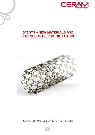
- 2. 1 Introduction Stents are expandable meshed tubes used either to reinforce body vessels possessing weak walls or to increase the internal diameter of a body vessel to allow an improved flow of fluids such as blood or urine. The use of arterial stents in particular has grown significantly over the last 20 years due to an ageing population and to a change in diet which has led to an increase in cardiovascular illness. Estimates vary, but it is predicted that coronary stents will have a market value of $7.2bn by 2012 and will continue to grow at a rate of 6% per annum thereafter. In 2009 over a million US citizens received angioplasty/stent interventions. In this paper we will review some of the technology being used in the development of new stents and how, in particular, computational modelling and material characterisation are helping to improve clinical outcomes. Finally we will look at the future perspectives for next generation stent technology. 2 Stents and coronary disease In the treatment of coronary artery disease, stents offer a less invasive alternative to the Coronary Artery Bypass Graft. It is estimated that for every bypass operation, there are four instances of stents being employed as an alternative approach. Stents are fabricated by laser cutting shaped sections from a metal tube. In use, an opening is made in the patient (groin, arm or neck) and a catheter used to guide a deflated balloon inside a stent to the correct position in the artery. X-rays and dye flow are used to identify the area of the artery suffering from plaque build-up and for associated stent positioning. Once positioned, the balloon is inflated, causing the stent to expand and therefore the plaque to be pushed back against the inner walls. Upon deflation and withdrawal of the balloon the stent remains in place. In some instances, balloon inflation inside the artery is performed without a stent (to assist initial widening) and then a stent is subsequently used. 3 Coronary stent types Prior to the use of stents, angioplasty alone was attempted. However, it was seen that there was a high chance (25-50%) of restenosis (re-thickening in artery walls) occurring. The first stents were Bare Metal Stents (BMS) and still led to a tendency for restenosis, albeit at a lower (15-25% chance) level. Thrombosis (blood clot formation) is also a typical, if relatively infrequent, consequence of stent emplacement. In order to reduce or eliminate these responses, Drug Eluting Stents (DES) were developed and have been shown to be excellent in reducing the re-narrowing of arteries by up to 70%. However, there is claimed to be a longer-term risk of blood clotting when a DES is used, requiring patients to take clot- preventing drugs for 6 months following the operation. Four DES platforms have been approved by the FDA and these represent the current state-of-the-art for clinical use. Whilst the majority of stents are balloon-expanding the development of self-expanding types has been done using so-called memory alloys1 such as Nitinol. This type of stent is often shaped to a diameter greater than the artery it is inserted into. By then crimping and feeding it inside a catheter it can be directed to the relevant part of the artery. Once released from the constraints of the catheter it expands and pushes into the inner arterial wall where it is able to conform to the shape of "non-straight" sections of artery. 1
- 3. 4 Application of supporting technologies in stent developments CERAM has been deeply involved in delivering supporting technology for the development of stents over the last 15 years. In particular this has included the areas of computational modelling and material characterisation. 4.1 Computational modelling Computational modelling can provide useful information to the stent design process. Both Finite Element Analysis (FEA) and Computational Fluid Dynamics (CFD) have been used in the pursuit of improved stent designs. Figure 1: 3-D representation of a stent geometry Figure 2: Meshing associated with a section of the geometry illustrated in figure 1 2
- 4. FEA uses a 3-D representation of a target shape (figure 1) which is "meshed" (figure 2) to divide it into small areas (2D) or volumes (3D). By applying physical data to the model, the impact of external factors (e.g. heat, pressure) on shape or internal stresses can be predicted. Figure 3 shows predicted stress development as a stent is enlarged via pressure applied from within the stent cavity. In the design of stents FEA has been used to explore numerous "what if" scenarios around novel stent geometries, thicknesses and alloy chemistries. Such models can predict how stent diameters increase as a function of balloon pressure. They can also predict properties such as radial strength and stent stiffness. In addition, they can play an important role in designing stents for bifurcation: One option for bifurcation is to expand a stent at the appropriate point in the main vessel to leave a suitably-sized hole in the stent positioned exactly where a second vessel branches off. A second stent can then be fed through the hole and expanded. FEA can predict the size of hole created as a function of balloon pressure. When employing CFD, the mesh is used to represent imaginary points within a given liquid volume. In so doing, the mesh predicts the properties of the fluid passing each point. Predictions can be made about where the fluid moves over time and changes in its physical properties. CFD modelling has been invaluable (in combination with an FEA model of an inserted stent) in predicting the improvements in blood flow attainable via stent interventions. Figure 3: FEA analysis using colour mapping to illustrate stress development during stent expansion 4.2 Material characterisation Stents comprise a range of materials which are invariably modified in some way or other in order to improve functionality and/or biocompatibility. The ability to understand the material chemistry and its physical profile, be it intentionally introduced or merely that of the as-received state, is absolutely fundamental to ensuring efficacy in stent deployment in the patient. Using state-of-the-art material characterisation techniques CERAM has delivered critical insights into material composition in all areas of stent structure, be it the metal substrate or the drug-eluting coating, both pre- and post-use. The metal substrate is invariably an alloy which has been machined in some way. These components are often subject to a cleaning process where detached but re-deposited residues and machining lubricant residues are removed. Validation of the effectiveness of such cleaning processes is carried out by measuring the quantity of such residues still remaining on the surface after cleaning, rinsing and drying and also the quantity of residual cleaning agents. This is carried out using a surface-specific spectroscopic method which is sensitive to all the elements (apart from H) to an accuracy of 0.1 atomic per cent. The method uses a bespoke combinatorial algorithm to generate a single figure cleanliness parameter known as the cleanliness index (CI). 3
- 5. In addition to machining, bare stents are frequently ‘passivated’ both to reduce their corrosion potential and to improve their biocompatibility (this latter for bare metal stents). Passivation is a process of controlled oxidation using either thermal or chemical treatment or a combination of both. It is important to know the depth of the passivation treatment (figure 4) to establish that it has been successfully achieved. This can be High Depth Resolution SIMS Profile measured using surface mass spectrometry operating in depth profiling 1.E+06 60 Ni mode. The substrate surface is 56 Fe continuously sputtered with an ion beam 52 Cr and the material so removed continuously 72 FeO mass analysed to establish the depth of Signal Intensity (Arbitrary Units) 68 CrO 18 O oxidation achieved. The technique is 1.E+05 12 C depth resolved to nanometre precision. Where the stent is to be coated, as with drug-eluting stents, the surface asperity of the bare stent needs to be controlled to 1.E+04 ensure that an even coating can be achieved (whilst retaining sufficient roughness to allow for the effective keying of the coating). This has consequences for the thickness of coating required, as 1.E+03 will be seen below. Surface roughness is 0 5 10 15 measured using a non-contact white light Depth / nm interferometric method which generates a 3D image of the surface with nanometre Figure 4: Secondary Ion Mass resolution in the vertical dimension and Spectrometry (SIMS) analysis, indicating allows an area surface roughness the relative level of key elements as a parameter to be generated for the area function of depth in a passivated metal sampled (figure 5). Surface of coated stent Surface of bare metal stent Figure 5: 3-D profiling (light interferometry) comparing surface roughness of a stent strut before and after coating – 2D image with ‘thermal’ z-axis colour scale 4
- 6. For coated stents, the coating layer thickness (be it primer coat, top coat or both together) can also be measured using white light interferometry. In this application a surface image is generated by focussing alternately on the substrate surface and the coating surface then subtracting the two data sets pixel by pixel to generate a coating thickness 3D image. This can be measured directly to give an average coating thickness or presented as a line profile to show the variation in thickness along, or across/around, the stent dimensions with nanometre resolution. The reader will have gathered that this process generates a 3D roughness image for the outer surface of the coated stent which is, in itself, an important characteristic in relation to the deployment of the stent in the patient. Fig. 6: Thickness Map - Statistical Fig. 7: Line profile along stent axis Method - Measure two ends and - mean thickness 4.4 microns middle with 120o rotation As noted earlier, coated stents carry slow release anti-rejection drug entities which are distributed throughout the coating layer. Such stents are routinely tested in vitro to establish the kinetics of the slow release performance of the coating under repeat elution exposure. The distribution of the drug moieties within the coating - particularly with depth - is of prime importance in establishing the potential efficacy of the slow release mechanism in practice. This can be measured using depth profiling mass spectrometry. The method allows for the continuous depth measurement of drug, coating and substrate simultaneously as the coating is sputtered with an incident ion beam. In this way the depth to which drug molecules are depleted after elution exposure is directly known. In tests at CERAM it has been shown that for certain drug/coating combinations it is possible for drug elution to be restricted to the outermost few microns of the coating despite the fact that several microns of coating need to be applied in order to ensure a minimum coating thickness at all points due to the surface roughness of the substrate. 5 Trends in stent R&D Given that the first bare metal stent entered the market in 1994, the technology is far from mature. At the Transcatheter Cardiovascular Therapeutics conference held in Scranton, USA in November/December 2009 the next generation of stent technologies was discussed. Whilst drug eluting stents will be improved through the use of what are described as more ‘cell friendly’ (i.e. more hydrophobic) coatings and adapted (by using CFD in stent design) to varying wall sheer conditions, the industry is already well down the road of developing bioresorbable coatings and even bioresorbable stents. Such systems are already on limited trial in Europe with FDA trials aimed at 2012 and approval by 2015. Bioresorbable polymeric 5
- 7. stents (Abbott) were recently in the news and represent a highly attractive concept; for example, the ability to avoid potential (metal) stent fracture during restenosis (and associated migration of stent fragments) is a desirable goal. In using all-polymer stents that eventually fully dissolve, a fine balance is required between having a structure that remains strong enough to open the artery for a given time period but which will then dissolve at a desirable rate. There is also the issue of the by-products that are created during biodegradation - they must be formed at a rate that the body can deal with. Polymers are also versatile, in that drug chemistry can be bonded to the polymer backbone, as opposed to being dispersed within the polymer chains. An alternative bioresorbable material is magnesium and its alloys. These have been trialled but shown to offer no particular advantages versus existing BMS, DES. There are still other areas under study including thinner struts which are important in terms of giving improved stent flexibility, a reduced cross-sectional profile within the vessel and potentially reduced restenosis (due to reduced vascular trauma). Cr-Co alloys could offer strength advantages for thinner stents. Sandwich structures, such as a tantalum layer placed between stainless steel layers, could also be a route to greater strength. Manufacturing metal stents via additive layer manufacturing could offer flexibility in terms of more complex geometries. Reducing restenosis remains important and concern that ion release from the metals employed in stents assists restenosis has led to work on alternative coatings to reduce ion release. Silicon carbides, carbon coatings, titanium nitride/oxide and sputtered indium oxide have all been trialled as routes to reducing the incidence of restenosis and/or thrombosis. The need to improve coatings for drug elution (see above) has given rise to research on inorganic coatings. Polymer coatings often require a bond layer to induce compatibility. Parylene coatings can be used to link metal to polybutylmethacrylate co-polymer coatings. Inorganic coating solutions under investigation include nano-porous alumina layers and sol- gel hydroxyapatite layers. A recent paper3 discussing the role of nano-ceramics for drug delivery points, in general, to a number of benefits of these over polymers. Although focussing on particulates, the conclusions are relevant to nano-particulates as coatings. Ceramic nano-particulates are known to have longer biodegradation times, allowing longer-term release of associated drugs. Unlike polymers, ceramic nano-particles do not swell as a result of changes to temperature or pH. Such swelling can be associated with sudden undesirable bursts of released drugs. The high surface area to volume ratio of nano-particles (or nano-porous structures created from particles) offers scope for retaining higher levels of drug. There is currently a great deal of interest in calcium phosphate nano-materials for drug release. There is evidence to show that this chemistry offers sustained release of drugs over significant time periods and the ability to offer discrete “on-off” release triggered by a stimulus such as ultrasonics. CERAM is currently developing novel, nano hydroxyapatites containing one or more substituent chemistries. Although aimed initially at the bone replacement market, this IPR and technology could potentially have applications in the area of drug-delivering stent coatings. Another approach under investigation is to make biodegradable ceramic stents by employing polymer as a binder. Bioceramics of very high porosity of nanometre scale have been successfully developed at CERAM. The challenge is to make the stent degrade at the 6
- 8. right time either naturally or by triggering mechanisms internally and/or externally and to make the degradation in a way so as not to produce any degraded materials that would block the blood vessel. Summary Stents have played a vital role in improving patient quality-of-life without resorting to major surgery. Even with a history of barely 25 years, significant improvements have been made to combat the twin threats of restenosis and thrombosis. It is clear that by embracing polymers and polymer-ceramics as well as metals, together with nanotechnology, stent developments will continue for many years. References 1. T.W. Duerig et al. Min Invas Ther & Allied Technol 2000, 9(3/4) p 235-246 2. O’Brien and Worrall. Acta Biomaterialia Vol 15, p 945-958 3. Lei Yang, B.W.Sheldon, T.J. Webster, Am Cer Soc Bull, Vol 89, No.2, p24-31 7
- 9. Queens Road, Penkhull, Stoke-on-Trent, Staffordshire ST4 7LQ, United Kingdom tel: +44 (0)1782 764428 or +44 (0)845 026 0902 fax: +44 (0)1782 412331 email: enquiries@ceram.com web: www.ceram.com/medical CERAM IS THE TRADING NAME OF CERAM RESEARCH LIMITED. REGISTERED IN ENGLAND. NO.:1960455. REGISTERED OFFICE AS ABOVE. 8