SlideShare una empresa de Scribd logo
1 de 1
Descargar para leer sin conexión
Advanced CT Visualization Soft-
                      ware for Rapid and Reliable De-
                      tection of Cranial Injuries
                      Challenge
                      Computed tomography (CT) has become the benchmark, standard of care diagnostic method for the assessment of patients
                      with traumatic brain injury (TBI). Consequently, reliable and rapid clinical routines are pivotal to obtain robust medical findings
                      within short time periods. Today, in clinical practice, radiologists primarily read and interpret consecutive sectional images
                      “sliced off” along the cranial-caudal axis. Furthermore, the rate and quality of skull fracture and/or haemostasis detection in
                      cranial CT both highly depend on the radiologist’s professional experience. Although the spatial resolution and number of cap-
                      tured images constantly grow with technological progress, transverse section read-out cannot exclude that potentially hazard-
O F F E R


                      ous injuries remain undiagnosed. Thus, it would be beneficial to implement improved and commonly accepted clinical routines
                      which facilitate easier and more accurate CT diagnoses of cranial fractures and/or intracranial haemorrhage.

                      Technology
                      Scientists at the Medical University of Vienna have developed a novel, curved
                      maximum intensity projection (MIP) based, image post-processing algorithm.
                      This algorithm enables transformation of large sets of 3D image data to a modestly
                                                                  manageable amount of 2D images display-
                                                                  ing skull surface and meningeal space. For
                                                                  this purpose a virtual and planar mesh,
                                                                  made up from a predefined array of equi-
                                                                  distant nodal points, is orthogonally posi-
                                                                  tioned relative to the centred cranio-caudal
                                                                  axis and gradually slipped over the skull
                                                                  along said axis. At each “slipping” step the
                                                                  algorithm checks whether a given node
                                                                  position corresponds to a 3D data point
                                                                  equal to or greater than a selected and
                                                                  predefined radiodensity (Hounsfield) value.
                      If such a data point exists, its value is assigned to the respective node position. Said node will then be fixed and not further sunk
T E C H N O L O G Y


                      over the skull/meningeal space. The determination of the surface/space is completed, once the virtual multi-noded mesh has
                      been fully unrolled. Then, the skull/meningeal space can both be unfolded and reprojected into a planar image which deliv-
                      ers an overview-like 2D inspection of the skull surface and meningeal space which can be readily analyzed by radiologists and
                      clinical personnel.

                      Commercial Opportunity
                                                                          TBI affects about 10 million people worldwide annually, and is one of the
                                                                          leading causes of disability and death in the Western world. Despite these
                                                                          numbers, TBI is often considered a silent epidemic with a very low level of
                                                                          awareness. Curved MIPs generated from cranial CT image data have the
                                                                          potential to improve the care of patients with no additional costs or
                                                                          radiation exposure. In addition, their use may have a positive impact on
                                                                          therapeutic management as well as insurance and litigation issues. The
                                                                          curved MIP algorithm enables a significantly higher detection rate than
                                                                          the analysis of axial raw images. Most importantly, the algorithm nearly
                                                                          offsets the experience gap between long-term experts and young
                                                                          professionals. Whereas curved MIP reconstructions show higher sensi-
                                                                          tivity than transverse sections, the latter offer higher specificity. It was
                                                                          found that, despite the very short reading time for MIP reconstructions,
                                                                          fractures detected on the reconstructions should be verified on transverse
                      sections. However, this observation also suggests that the diagnosis may be discontinued, if the reconstructions do not show
                      any fractures. Consequently, reading times could be reduced by up to a factor of 5.

                      Development Status
                      The advantages and efficiency of the software were evaluated and dem-
                      onstrated in two on-site studies involving 200 and 314 consecutive TBI
                      patients, respectively. In the figure below, curved MIPs obtained in the
                      same 85-year-old male patient demonstrate the anatomic background of
                      the study. (a) Internal table of skull with several arterial and venous
                      channels. (b) External table of skull. The image shows only one subtle
                      fracture line (arrows) in the right frontal bone and one in the right zygo-
                      matic arch. The subtle frontal fracture line was reported only when curved
                      MIPs were used and was missed by all readers when transverse sections
                      were analyzed. Further reading: H., Ringl et al., The Skull Unfolded: A
                      Cranial CT Visualization Algorithm for Fast and Easy Detection of Skull
                      Fractures, Radiology, 255 (2010): 553-562.

                      Development Status
                      WO 2011/017730 A2 filed on August 11, 2010 (priority date: August 11, 2009), national stage entered in the US and Europe.

                      Contact
                      Mathias Weiss
                      BDC – The Business Development Company GmbH
                      Pelikanweg 2                                                               Phone: +41 61 270 88 04
                      CH-4054 Basel                                                              Mobile: +41 79 487 47 31
                      Switzerland                                                                Fax: +41 61 270 88 10
                      http://www.bdc-basel.com                                                   mathias.weiss@bdc-basel.com

Más contenido relacionado

La actualidad más candente

La actualidad más candente (10)

Sensors 21-02222-v21
Sensors 21-02222-v21Sensors 21-02222-v21
Sensors 21-02222-v21
 
MNPS. Mevis Neurosurgery Planning System. Overview, Aug. 2018
MNPS.  Mevis Neurosurgery Planning System. Overview, Aug. 2018MNPS.  Mevis Neurosurgery Planning System. Overview, Aug. 2018
MNPS. Mevis Neurosurgery Planning System. Overview, Aug. 2018
 
Optic nerve and retinal
Optic nerve and retinalOptic nerve and retinal
Optic nerve and retinal
 
Magnetom flash 41 *SIEMENS*
Magnetom flash 41 *SIEMENS*Magnetom flash 41 *SIEMENS*
Magnetom flash 41 *SIEMENS*
 
Ct brain
Ct brainCt brain
Ct brain
 
18 08 14
18 08 1418 08 14
18 08 14
 
Brochure Samples
Brochure SamplesBrochure Samples
Brochure Samples
 
의료영역에서의3D 프린팅적용을위한의료영상모델링
의료영역에서의3D 프린팅적용을위한의료영상모델링의료영역에서의3D 프린팅적용을위한의료영상모델링
의료영역에서의3D 프린팅적용을위한의료영상모델링
 
Dynamic optical coherence tomography in dermatology
 Dynamic optical coherence tomography in dermatology Dynamic optical coherence tomography in dermatology
Dynamic optical coherence tomography in dermatology
 
Ax kundenmagazin 14
Ax kundenmagazin 14Ax kundenmagazin 14
Ax kundenmagazin 14
 

Similar a Advanced CT Visualization Software

CBCT IN ORTHODONTICS
CBCT IN ORTHODONTICSCBCT IN ORTHODONTICS
CBCT IN ORTHODONTICSSonu Pandit
 
3D diagnostics in dental medicine - CBCT
3D diagnostics in dental medicine - CBCT3D diagnostics in dental medicine - CBCT
3D diagnostics in dental medicine - CBCTtlauc
 
Comparison of ct and cbct
Comparison of ct and cbct Comparison of ct and cbct
Comparison of ct and cbct Amritha James
 
Susceptibility Weighted Imaging (SWI)
Susceptibility Weighted Imaging (SWI)Susceptibility Weighted Imaging (SWI)
Susceptibility Weighted Imaging (SWI)Nija Panchal
 
CBCT in dental practice
CBCT in dental practiceCBCT in dental practice
CBCT in dental practiceZana Hussein
 
Virtual scanning total joint
Virtual scanning total jointVirtual scanning total joint
Virtual scanning total jointNader Elbokle
 
IMAGE GUIDED SURGERY IN NOSE AND PNS.pptx
IMAGE GUIDED SURGERY IN NOSE AND PNS.pptxIMAGE GUIDED SURGERY IN NOSE AND PNS.pptx
IMAGE GUIDED SURGERY IN NOSE AND PNS.pptxspartonkarthi
 
AI in Ophthalmology | Startup Landscape
AI in Ophthalmology | Startup LandscapeAI in Ophthalmology | Startup Landscape
AI in Ophthalmology | Startup LandscapePetteriTeikariPhD
 
HRT and GDx VCC
HRT and GDx VCCHRT and GDx VCC
HRT and GDx VCCSSSIHMS-PG
 
EPID AND CBCT ON RADIATION THERAPY
EPID AND CBCT ON RADIATION THERAPYEPID AND CBCT ON RADIATION THERAPY
EPID AND CBCT ON RADIATION THERAPYanju k.v.
 
Treatment verification systems in radiation therapy
Treatment verification systems in radiation therapyTreatment verification systems in radiation therapy
Treatment verification systems in radiation therapyanju k.v.
 
diagnostics-12-00961.pdf
diagnostics-12-00961.pdfdiagnostics-12-00961.pdf
diagnostics-12-00961.pdfmarlinadewi2
 

Similar a Advanced CT Visualization Software (20)

CBCT IN ORTHODONTICS
CBCT IN ORTHODONTICSCBCT IN ORTHODONTICS
CBCT IN ORTHODONTICS
 
3D diagnostics in dental medicine - CBCT
3D diagnostics in dental medicine - CBCT3D diagnostics in dental medicine - CBCT
3D diagnostics in dental medicine - CBCT
 
Digital cephalometry
Digital cephalometryDigital cephalometry
Digital cephalometry
 
Neuroradiologia
NeuroradiologiaNeuroradiologia
Neuroradiologia
 
Er36881887
Er36881887Er36881887
Er36881887
 
Ct acute care
Ct acute careCt acute care
Ct acute care
 
8. 22760.pdf
8. 22760.pdf8. 22760.pdf
8. 22760.pdf
 
use of Cbct in dental implant
use of Cbct  in dental implantuse of Cbct  in dental implant
use of Cbct in dental implant
 
Comparison of ct and cbct
Comparison of ct and cbct Comparison of ct and cbct
Comparison of ct and cbct
 
Stereotaxy Brain
Stereotaxy BrainStereotaxy Brain
Stereotaxy Brain
 
Susceptibility Weighted Imaging (SWI)
Susceptibility Weighted Imaging (SWI)Susceptibility Weighted Imaging (SWI)
Susceptibility Weighted Imaging (SWI)
 
CBCT in dental practice
CBCT in dental practiceCBCT in dental practice
CBCT in dental practice
 
Virtual scanning total joint
Virtual scanning total jointVirtual scanning total joint
Virtual scanning total joint
 
IMAGE GUIDED SURGERY IN NOSE AND PNS.pptx
IMAGE GUIDED SURGERY IN NOSE AND PNS.pptxIMAGE GUIDED SURGERY IN NOSE AND PNS.pptx
IMAGE GUIDED SURGERY IN NOSE AND PNS.pptx
 
AI in Ophthalmology | Startup Landscape
AI in Ophthalmology | Startup LandscapeAI in Ophthalmology | Startup Landscape
AI in Ophthalmology | Startup Landscape
 
HRT and GDx VCC
HRT and GDx VCCHRT and GDx VCC
HRT and GDx VCC
 
EPID AND CBCT ON RADIATION THERAPY
EPID AND CBCT ON RADIATION THERAPYEPID AND CBCT ON RADIATION THERAPY
EPID AND CBCT ON RADIATION THERAPY
 
Treatment verification systems in radiation therapy
Treatment verification systems in radiation therapyTreatment verification systems in radiation therapy
Treatment verification systems in radiation therapy
 
Role of CBCT in Forensic Odontology
Role of CBCT in Forensic OdontologyRole of CBCT in Forensic Odontology
Role of CBCT in Forensic Odontology
 
diagnostics-12-00961.pdf
diagnostics-12-00961.pdfdiagnostics-12-00961.pdf
diagnostics-12-00961.pdf
 

Advanced CT Visualization Software

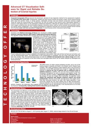
  • 1. Advanced CT Visualization Soft- ware for Rapid and Reliable De- tection of Cranial Injuries Challenge Computed tomography (CT) has become the benchmark, standard of care diagnostic method for the assessment of patients with traumatic brain injury (TBI). Consequently, reliable and rapid clinical routines are pivotal to obtain robust medical findings within short time periods. Today, in clinical practice, radiologists primarily read and interpret consecutive sectional images “sliced off” along the cranial-caudal axis. Furthermore, the rate and quality of skull fracture and/or haemostasis detection in cranial CT both highly depend on the radiologist’s professional experience. Although the spatial resolution and number of cap- tured images constantly grow with technological progress, transverse section read-out cannot exclude that potentially hazard- O F F E R ous injuries remain undiagnosed. Thus, it would be beneficial to implement improved and commonly accepted clinical routines which facilitate easier and more accurate CT diagnoses of cranial fractures and/or intracranial haemorrhage. Technology Scientists at the Medical University of Vienna have developed a novel, curved maximum intensity projection (MIP) based, image post-processing algorithm. This algorithm enables transformation of large sets of 3D image data to a modestly manageable amount of 2D images display- ing skull surface and meningeal space. For this purpose a virtual and planar mesh, made up from a predefined array of equi- distant nodal points, is orthogonally posi- tioned relative to the centred cranio-caudal axis and gradually slipped over the skull along said axis. At each “slipping” step the algorithm checks whether a given node position corresponds to a 3D data point equal to or greater than a selected and predefined radiodensity (Hounsfield) value. If such a data point exists, its value is assigned to the respective node position. Said node will then be fixed and not further sunk T E C H N O L O G Y over the skull/meningeal space. The determination of the surface/space is completed, once the virtual multi-noded mesh has been fully unrolled. Then, the skull/meningeal space can both be unfolded and reprojected into a planar image which deliv- ers an overview-like 2D inspection of the skull surface and meningeal space which can be readily analyzed by radiologists and clinical personnel. Commercial Opportunity TBI affects about 10 million people worldwide annually, and is one of the leading causes of disability and death in the Western world. Despite these numbers, TBI is often considered a silent epidemic with a very low level of awareness. Curved MIPs generated from cranial CT image data have the potential to improve the care of patients with no additional costs or radiation exposure. In addition, their use may have a positive impact on therapeutic management as well as insurance and litigation issues. The curved MIP algorithm enables a significantly higher detection rate than the analysis of axial raw images. Most importantly, the algorithm nearly offsets the experience gap between long-term experts and young professionals. Whereas curved MIP reconstructions show higher sensi- tivity than transverse sections, the latter offer higher specificity. It was found that, despite the very short reading time for MIP reconstructions, fractures detected on the reconstructions should be verified on transverse sections. However, this observation also suggests that the diagnosis may be discontinued, if the reconstructions do not show any fractures. Consequently, reading times could be reduced by up to a factor of 5. Development Status The advantages and efficiency of the software were evaluated and dem- onstrated in two on-site studies involving 200 and 314 consecutive TBI patients, respectively. In the figure below, curved MIPs obtained in the same 85-year-old male patient demonstrate the anatomic background of the study. (a) Internal table of skull with several arterial and venous channels. (b) External table of skull. The image shows only one subtle fracture line (arrows) in the right frontal bone and one in the right zygo- matic arch. The subtle frontal fracture line was reported only when curved MIPs were used and was missed by all readers when transverse sections were analyzed. Further reading: H., Ringl et al., The Skull Unfolded: A Cranial CT Visualization Algorithm for Fast and Easy Detection of Skull Fractures, Radiology, 255 (2010): 553-562. Development Status WO 2011/017730 A2 filed on August 11, 2010 (priority date: August 11, 2009), national stage entered in the US and Europe. Contact Mathias Weiss BDC – The Business Development Company GmbH Pelikanweg 2 Phone: +41 61 270 88 04 CH-4054 Basel Mobile: +41 79 487 47 31 Switzerland Fax: +41 61 270 88 10 http://www.bdc-basel.com mathias.weiss@bdc-basel.com