
Recomendados
Más contenido relacionado
La actualidad más candente
Destacado
Destacado (20)
Similar a Arun article1
Similar a Arun article1 (20)
Arun article1
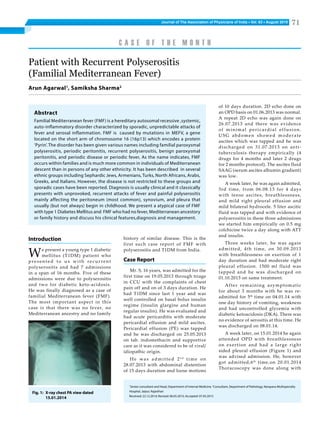
- 1. Journal of The Association of Physicians of India ■ Vol. 63 ■ August 2015 71 history of similar disease. This is the first such case report of FMF with polyserositis and T1DM from India. Case Report Mr. S, 16 years, was admitted for the first time on 19.05.2013 through triage in CCU with the complaints of chest pain off and on of 3 days duration. He had T1DM since last 1 year and was well controlled on basal bolus insulin regime (insulin glargine and human regular insulin). He was evaluated and had acute pericarditis with moderate pericardial effusion and mild ascites. Pericardial effusion (PE) was tapped and he was discharged on 25.05.2013 on tab. indomethacin and supportive care as it was considered to be of viral/ idiopathic origin. He was admitted 2nd time on 28.07.2013 with abdominal distention of 15 days duration and loose motions Patient with Recurrent Polyserositis (Familial Mediterranean Fever) Arun Agarwal1 , Samiksha Sharma2 of 10 days duration. 2D echo done on an OPD basis on 01.06.2013 was normal. A repeat 2D echo was again done on 26.07.2013 and there was evidence of minimal pericardial effusion. USG abdomen showed moderate ascites which was tapped and he was discharged on 31.07.2013 on anti- tuberculosis therapy empirically (4 drugs for 4 months and later 2 drugs for 2 months protocol). The ascites fluid SAAG (serum ascites albumin gradient) was low. A week later, he was again admitted, 3rd time, from 06.08.13 for 4 days with tense ascites, breathlessness, and mild right pleural effusion and mild bilateral hydrocele. 5 liter ascitic fluid was tapped and with evidence of polyserositis in these three admissions we started him empirically on 0.5 mg colchicine twice a day along with ATT and insulin. Three weeks later, he was again admitted, 4th time, on 30.09.2013 with breathlessness on exertion of 1 day duration and had moderate right pleural effusion. 1500 ml fluid was tapped and he was discharged on 01.10.2013 on same treatment. After remaining asymptomatic for about 3 months with he was re- admitted for 5th time on 04.01.14 with one day history of vomiting, weakness and had uncontrolled glycemia with diabetic ketoacidosis (DKA). There was no evidence of serositis at this time. He was discharged on 08.01.14. A week later, on 15.01.2014 he again attended OPD with breathlessness on exertion and had a large right sided pleural effusion (Figure 1) and was advised admission. He, however got admitted,6th time,on 20.01.2014 Thoracoscopy was done along with 1 Senior consultant and Head, Department of Internal Medicine, 2 Consultant, Department of Pathology, Narayana Multispecialty Hospital, Jaipur, Rajasthan Received: 22.12.2014; Revised: 06.05.2015; Accepted: 07.05.2015 C A S E O F T H E M O N T H Introduction We present a young type 1 diabetic mellitus (T1DM) patient who presented to us with recurrent polyserositis and had 7 admissions in a span of 16 months. Five of these admissions were due to polyserositis and two for diabetic keto-acidosis. He was finally diagnosed as a case of familial Mediterranean fever (FMF). The most important aspect in this case is that there was no fever, no Mediterranean ancestry and no family Abstract Familial Mediterranean fever (FMF) is a hereditary autosomal recessive ,systemic, auto-inflammatory disorder characterized by sporadic, unpredictable attacks of fever and serosal inflammation. FMF is caused by mutations in MEFV, a gene located on the short arm of chromosome 16 (16p13) which encodes a protein ‘Pyrin’. The disorder has been given various names including familial paroxysmal polyserositis, periodic peritonitis, recurrent polyserositis, benign paroxysmal peritonitis, and periodic disease or periodic fever, As the name indicates, FMF occurs within families and is much more common in individuals of Mediterranean descent than in persons of any other ethnicity. It has been described in several ethnic groups including Sephardic Jews, Armenians, Turks, North Africans, Arabs, Greeks, and Italians. However, the disease is not restricted to these groups and sporadic cases have been reported. Diagnosis is usually clinical and it classically presents with unprovoked, recurrent attacks of fever and painful polyserositis mainly affecting the peritoneum (most common), synovium, and pleura that usually (but not always) begin in childhood. We present a atypical case of FMF with type 1 Diabetes Mellitus and FMF who had no fever, Mediterranean ancestory or family history and discuss his clinical features,diagnosis and management. Fig. 1: X-ray chest PA view dated 15.01.2014
- 2. Journal of The Association of Physicians of India ■ Vol. 63 ■ August 201572 Fig. 2: Biopsy of pleural nodule (45 x views), H and E stain. Arrow show chronic non-specific inflammation and fibroblasts Fig. 3: X-ray chest PA view dated 05.03.2014 pleural biopsy from a suspicious looking nodule on parietal pleura along with a chest tube insertion. On histopathology (Figure 2) it was chronic non-specific inflammation with negative AFB and PAS stain. By now he had completed 6 months of ATT and he was discharged on colchicine and insulin on 24.01.2014. He again presented on 5th march 2014 with mild breathlessness of 2 days duration and was diagnosed to have moderate right side pleural effusion (Figure 3). By this time we had a strong suspicion of him having FMF in view of 5 episodes of recurrent serositis (polyserositis on three occasions), being asymptomatic between these admissions (attacks), not having any other underlying condition that would explain the symptoms (infective, inflammatory, immunological or malignancy) and probably partial response to colchicine. He had no family history of similar illness, had one younger brother who was reportedly healthy, and there was no history of consanguinity among his parents. He is a resident of Jaipur (a non-Mediterranean region). At this time we did not admit him and instead we increased colchicine dose to 1 mg thrice a day. Two weeks later on 20th march 2014, on follow-up he was asymptomatic and had no evidence of pleural effusion (Figure 4). A complete response with adequate recommended dose of colchine further confirmed the diagnosis to be FMF. Since then he remained asymptomatic for almost 6 months. He was again admitted on 18.09.2014 through triage with 1 day history of vomiting and DKA and discharged Fig. 4: X-ray chest PA view dated 20.03.2014 Table 1: Summary of admissions Admission period Presenting diagnosis 19.05.2013 to 25.05.2013 Moderate PE, mild ascites 28.07.2013 to 31.07.2013 Moderate Ascites, minimal PE 06.08.2013 to 10.08.2013 Large ascites, mild Right pleural effusion and bilateral Hydrocele 30.09.2013 to 01.10.2013 Moderate right pleural effusion 04.01.2014 to 08.01.2014 DKA; No e/o serositis. 20.01.2014 to 24.01.2014 Large right pleural effusion 18.09.2014 to 22.09.2014 DKA; No e/o serositis Table 2: Laboratory Investigations Admission 1st 2nd 3rd 4th 5th 6th Hb 13.9 14.7 13.3 15.2 TLC 15.33 8.66 7.05 33.88 APC 373 541 446 312 ESR 110 30 12 45 CRP 0.97 2.44 BGR 346.4 121 100 97 600 N S. albumin 3.79 2.94 3.99 5.47 S. globulin 4.02 3.65 S. uric acid 11.3 S. creatinine 0.57 0.89 on 22.09.2014. He had no evidence of serositis this time. He was last seen on 13th Nov. 2014, and as regards serositis he is asymptomatic now for almost 9 months since March 2014 on colchicine 1 mg thrice a day. Summary of admissions is given in Table 1. The investigations done during these admissions are shown in Tables 2 and 3. Fluid analysis was done as mentioned in Table 4. Pleural fluid was found to be exudates as per traditional Light’s criteria rule. Ascitic fluid SAAG analysis showed it to be low gradient. Fluid ADA analysis did not favor the diagnosis of tubercular serositis. Fluid glucose analysis showed glucose to be on higher side with fluid/ serum glucose ratio >0.5. Fluids were moderately cellular with predominant lymphocytosis. Discussion This is a case of recurrent serositis with 5 episodes (and 7 admissions) in 8 months time, with asymptomatic periods in between these episodes, has no underlying condition that would explain the symptoms, and responded to colchicine. This made us to suspect FMF in the present case. Familial Mediterranean fever (FMF) is an autosomal recessive systemic auto-inflammatory disorder. It is characterized by recurrent episodes of fever accompanied by peritonitis, pleuritis, pericarditis, and/or arthritis, s o m e t i m e s a c c o m p a n i e d b y a n erysipelas-like skin rash. The condition was first described in 1945 under the name ‘benign paroxysmal peritonitis.1 The disorder may also be referred to as recurrent polyserositis, recurrent hereditary polyserositis, periodic disease, and periodic peritonitis.1 MEFV gene, as it is called, is located on the short (p) arm of chromosome 16 at
- 3. Journal of The Association of Physicians of India ■ Vol. 63 ■ August 2015 73 position 13.3. More than 80 MEFV gene mutations that cause FMF have been identified. The most common mutation replaces the amino acid methionine with the amino acid valine at protein position 694 (written as Met694Val or M694V) leading to an abnormally small, nonfunctional protein, known as prying or sometimes called marenostrin. This transcript is thought to be expressed in mature neutrophils and in fibroblasts derived from skin, peritoneum, and synovium, suggesting that it functions as an inflammatory regulator at the level of cytoskeletal organization.1-3 These inflammatory episodes lead to excess production of amyloid A protein from the acute phase reactant serum. Widespread amyloid deposition occurs in the kidneys primarily by the age of 40 years but can also affect the gastrointestinal tract, liver, spleen, thyroid, and in much later stages, the heart and testes as well.4 However, only patients with specific MEFV haplotypes develop amyloidosis. FMF classically presents with unprovoked, recurrent attacks of fever and painful polyserositis mainly affecting the peritoneum (most common), synovium, and pleura that usually (but not always) begin in childhood.1 The majority of affected individuals (80%) present before 10 years of age and 90% before reaching the age of 20 years. Disease onset after the age of 40 years may represent a Table 3: Summary of investigations Date Test(s) Result 19.05.13 Serology Negative Mantoux test Negative 22.05.13 USG abdomen Mild hepatomegaly, Mild free fluid in abdomen 01.06.13 2D echo NAD 26.07.13 2D echo Minimal PE 30.07.13 ANA (IFA) Negative TSH Normal PCR-TB (ascitis) Not detected 06.08.13 2D echo NAD 07.08.13 S. fibrinogen 399 (200-400 mg/dl) 08.08.13 CECT abdomen Marked free fluid, b/l mild hydrocele, sub- centimeter lymph node in omentum, right iliac fossa and mesentery 04.01.14 Serology Negative 23.01.14 CECT chest Right side hydropneumothorax, Ground glass opacity with multiple nodular opacities in RLL ? Consolidation ? Pulmonary edema Table 4: Fluid analysis Date Total proteins/albumin Glucose Total cells Differential cells ADA Ascitic fluid* 30.07.13 6.33/- 317 1470 P10 L90 22.9 Ascitic fluid 07.08.13 4.65/2.03 150 680 P00 L100 21.0 Pleural Fluid 30.09.13 6.03/- 163 890 P10 L90 21.40 Pleural Fluid 20.01.14 14.0 * PCR TB: not detected; AFB stain: negative; Cytology: no malignant cells separate and milder disease entity.2 Other acute manifestations include erysipelas like skin lesion, pericarditis,5 self-limiting orchitis and recurrent aseptic meningitis, febrile myalgia,6 and increased incidence of IgA vasculitis.7 Acute episodes may last from 24 to 72 hours and have variable frequency, often without a recognized triggering event. Some commonly reported precipitating causes include viral illness, emotional stress, excessive/ intense physical activity, high-fat diet, extremes of temperature, and menstruation in women. The frequency of attacks may vary from once per week to once every 5–10 years, with the median frequency being approximately once a month.1,3 A c u t e a t t a c k s o f F M F a r e accompanied by elevation of many of the serum markers of systemic i n f l a m m a t i o n w h i c h i n c l u d e s leukocytosis with neutrophilia, raised erythrocyte sedimentation rate (ESR) and other acute phase reactants, including beta-2 microglobulin, C-reactive protein (CRP), SAA (serum amyloid protein), and fibrinogen. Genetic testing i.e., testing for many of the known mutations is currently available at commercial laboratories outside India. Genetic testing was not done in our patient due to resource limitations. FMF is still primarily a clinical diagnosis. Several diagnostic criteria have been proposed by now - Livneh A et al 8 (Table 5) and Tel Hashomer19 (Table 6).There is a set of validated diagnostic criteria by Livneh A et al8 with a reported sensitivity and specificity of 96% to 99% (Table 5). Amyloidosis, Positive family history and Mediterranean ancestry may or may not be present. Our patient fits into the diagnosis of FMF by these criteria. Differential diagnosis include surgical emergencies (appendicitis, intussusceptions, perforated peptic ulcer, etc), hereditary angioedema, a c u t e i n t e r m i t t e n t p o r p h y r i a , relapsing pancreatitis, systemic lupus erythematosus and vasculitis, hypertriglyceridemia, abdominal epilepsy and abdominal migraine. The goals of overall management for FMF are twofold: 1. Prevention of acute attacks and 2. Prevention of the development and progression of amyloidosis, as the major cause of mortality is the insidious development of secondary (AA) amyloidosis with eventual renal failure. Colchicine is the gold standard and indeed the only recommended drug for treating FMF. It is thought to primarily concentrate in neutrophils and inhibit their increased chemotactic activity Table 5: Criteria for the diagnosis of familial Mediterranean fever8,19 Major criteria 1. Typical attacks* with peritonitis (Generalized) 2. Typical attacks with pleuritis (unilateral) or pericarditis 3. Typical attacks with monoarthritis (hip, knee, ankle) 4. Typical attacks with fever alone 5. Incomplete# abdominal attack Minor criteria 1. Incomplete attacks involving chest pain 2. Incomplete attacks involving monoarthritis 3. Exertional leg pain 5. Favorable response to colchicine Requirements for diagnosis of FMF are ≥1 Major criteria or ≥2 minor criteria. * Typical attacks are defined as recurrent (>3 of the same type), febrile (>38°C), and short (lasting between 12 hours and 3 days); # Incomplete attacks are defined as painful and recurrent attacks not fulfilling the criteria for a typical attack. Table 6: Tel Hashomer criteria for the diagnosis of familial Mediterranean fever19 Major Criteria Minor Criteria 1. Recurrent febrile episodes with serositis 1. Recurrent febrile episodes 2. Amyloidosis (AA) without predisposing diseases 2. Erysipelas–like erythema 3. Favorable response to colchicine treatment 3. FMF in first degree relative The clinical diagnosis of FMF is considered definitive in the presence of atleast two major criteria, or one major plus two minor criteria. The diagnosis of FMF is probable in the presence of one major criterion plus one minor criteria (Table 6).
- 4. Journal of The Association of Physicians of India ■ Vol. 63 ■ August 201574 Mediterranean fever. MolGenetMetab 2000; 71:256–260. 3. Zadeh N, Getzug T, Grody WN, et al. Diagnosis and mamagementoffamilialMediterraneanfever:Integrating medical genetics in a dedicated interdisciplinary clinic. Review Genetics Med 2011; 13:263-269. 4. Ben-Chetrit E, Levy M. Familial Mediterranean fever. Lancet 1998; 351:659–664. 5. Kees S, Langevitz P, Zemer D, et al. Attacks of pericarditis as a manifestation of familial Mediterranean fever (FMF). QJM 1997; 90:643. 6. Ertekin V, Selimoğlu MA, Alp H, Yilmaz N. Familial Mediterranean fever protracted febrile myalgia in children: report of two cases. RheumatolInt 2005; 25:398. 7. Ozen S, Ben-Chetrit E, Bakkaloglu A, et al. Polyarteritis nodosa in patients with Familial Mediterranean Fever (FMF): a concomitant disease or a feature of FMF? Semin Arthritis Rheum 2001; 30:281. 8. Livneh A, Langevitz P, Zemer D, etal. Criteria for the diagnosis of familial Mediterranean fever. ArthritisRheum 1997; 40:1879. 9. Ozkaya N, Yalçinkaya F. Colchicine treatment in children with familial Mediterranean fever. Clin Rheumatol 2003; 22:314–317. 10. Tunca M,Tankurt E, Akbaylar Akpinar H, et al.The efficacy of interferon alpha on colchicine-resistant familial Mediterranean fever attacks: a pilot study. BrJRheumatol 1997; 36:1005. 11. Calguneri M, Apras S, Ozbalkan Z, et al. The efficacy of continuous interferon alpha administration as an adjunctive agent to colchicine-resistant familial Mediterranean fever patients. Clin Exp Rheumatol 2004; 22:S41. 12. Seyahi E, Ozdogan H, Celik S, et al. Treatment options in colchicineresistantfamilialMediterraneanfeverpatients: thalidomideandetanerceptasadjunctiveagents.ClinExp Rheumatol 2006; 24:S99. 13. Metyas S, Arkfeld DG, Forrester DM, Ehresmann GR. Infliximab treatment of Familial Mediterranean fever and its effect on secondary AA amyloidosis. J Clin Rheumatol 2004; 10:134. 14. Ehrenfeld M, Brzezinski A, Levy M, etal. Fertility and obstetric history in patients with familial Mediterranean feveronlong-termcolchicinetherapy. BrObstetGynaecol 1987; 94:1186-91. 15. Ben-Chetrit E, Ben-Chetrit A, Berkun Y, Ben-Chetrit E. Pregnancy outcomes in women with familial Mediterraneanfeverreceivingcolchicine:isamniocentesis justified?. Arthritis Care Res 2010;62:143–148. 16. Ben-Chetrit E, Ben-Chetrit A, Berkun Y, Ben-Chetrit E. Pregnancy outcomes in women with familial Mediterraneanfeverreceivingcolchicine:isamniocentesis justified?. Arthritis Care Res 2010; 62:143–148. 17. Atabek ME. Case Report:Familial Mediterraen Fever Association with Type 1 Diabetes Mellitus. Association or coincidence? The Endocrinologist 2006; 16:133. 18. Bas F, Kabatar-Eryilamazs, Gunoz H, et al.Type 1 diabetes mellitus associated with autoimmune thyroid disease, celiac disease and familial Mediterranean fever: case report. Turk J Pdiatr 2009; 51:183-6. 19. Wang, et al. Familial Mediterranean Fever: From Pathogenesis toTreatment.JGenetSyndrGeneTher 2014. serositis; i.e., he either had serositis or DKA during the seven admissions but never had both of them. We could not find any explanation for this. The association between FMF and T1DM also need to be looked into. Is it really an association or just a coincidence? First such case report of FMF association with T1DM in a 9 year old girl was reported by Atabek et al from Turkey17 in 2006. Another case of FMF, T1DM with ATD (autoimmune thyroid disorder ) and CD (celiac disease) was reported by Bas etal18 from Turkey in 2009. To the best of our knowledge this is the first such case report of FMF associated with T1DM from India. To the best of our knowledge this is the first such case report of a patient who is diagnosed to have FMF but did not had any fever, Mediterranean ancestory,or familial history. Nevertheless suspect FMF in any patient who 1) has had at least four episodes of abdominal pain or chest pain or both, lasting from 24-72 hours, 2) without symptoms between attacks, 3) does not have any other condition that would explain the symptoms, 4) has positive family history of FMF, and 5) responds to colchicine. Positive family history and Mediterranean ancestory may or may not be present. Given its genetic nature, there is no cure for FMF. Colchicine therapy should be continued for life. Acknowledgement A special mention of thanks to our colleagues Dr. Devendra Shrimal (Department of Cardiology) and Dr. Shubhranshu (Department of Pulmonary Medicine) for their help in management of this patient and therapeutic invasive procedure. References 1. El Shanti H, Majeed HA, El-Khateeb M. Familial Mediterranean fever in Arabs. Lancet 2006; 367:1016– 1024. 2. TelatarM,GrodyWW.Moleculargenetictestingforfamilial during FMF attacks.4 Colchicine should be administered orally once the diagnosis of FMF is confirmed (or as a therapeutic trial in establishing the diagnosis). Adult dosing is 1.2–2.4 mg/day. It may take some months of adjustment before the optimal dosage (evidenced by essentially complete prevention of attacks) is reached. Our patient responded to 3 mg/day of colchicines and it took 8 months to adjust the dosage. It is crucial to drive home to patients that daily lifelong administration of colchicine is required to prevent both the fever/ pain attacks and the silent amyloid deposition.3 Also, despite the common misconception, taking colchicine at the start of an attack is of no use; the drug will prevent attacks if taken continually but will not block an acute attack in progress.3 Colchicine unresponsiveness is known and several other treatment modalities including Interferon-alfa,10,11 thalidomide, etanercept, infliximab, anakinra, and rilonacept have been reported.12,13 About one-third of female patients with FMF are infertile, and 20-30% of pregnancies result in fetal loss.14 Colchicine has antimitotic effects and could be terratogenic but there is no firm evidence to support that concern.15 A recent study by Ben-Chetrit et al16 in Israel found no difference in fetal outcome or incidence of birth defects between affected women who either continued taking or abstained from colchicine during pregnancy. They also pointed out that the common practice in Turkey (where FMF is quite prevalent) is not to recommend amniocentesis for prenatal colchicine exposure.16 Our patient was diagnosed to have T1DM almost one year before his initial presentation and later had two admissions with diabetic ketoacidosis. Surprisingly, he did not have any evidence of serositis during these admissions. Similarly he did not have any DKA during five admissions for