2. Basic defect in HGB
• SCD is a type of hemoglobinopathy
• β-globin chain defect
– Valine replaces glutamine at the 6th position from
aminoterminal
• As a result HGB molecules develop sticky points
• Sticky points are exposed when the HGB is
deoxygenated
• Hence, HGB molecules polymerize to form fibers
/ long crystals
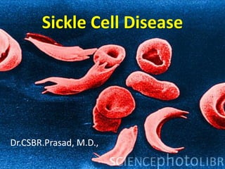
3. Red Blood Cells from Sickle Cell Anemia
• Deoxygenation of SS erythrocytes leads to intracellular
hemoglobin polymerization, loss of deformability and
changes in cell morphology.
OXY-STATE DEOXY-STATE
4.
5. Sickle cell disease Point Mutation
DNA mRNA AMINOACID
C
T
C G
A
G
G A G
C
A
C G
T
G G U G
6.
7. Deoxyhemoglobin S Polymer Structure
A) Deoxyhemoglobin S B) Paired strands of C) Hydrophobic pocket
14-stranded polymer deoxyhemoglobin S for 6b Val
(electron micrograph) (crystal structure)
D) Charge and size prevent
6b Glu from binding.
Dykes, Nature 1978; JMB 1979
Crepeau, PNAS 1981 Wishner, JMB 1975
8. Determinants of Hemoglobin S Polymerization
SS erythrocytes
MCHC ~ 32 g/dl
DeoxyHbS solubility
~16 g/dl
• Intracellular hemoglobin composition
• Intracellular hemoglobin concentration
• Oxygen saturation
9. Basic defect in HGB
Polymerization gives abnormal physiochemical
properties to HGB - sickle hemoglobin (HbS) -
that are responsible for the disease
11. Photomicrograph of red blood cells, showing
abnormal shape characteristic of sickle cell anemia
12. Pathogenesis
The major pathologic manifestations:
1. Chronic hemolysis
2. Microvascular occlusions, and
3. Tissue damage
Several variables affect the rate and degree of
sickling
13. Pathogenesis
Several variables affect the rate and degree of
sickling:
1. Interaction of HbS with the other types of
hemoglobin in the cell
2. Mean cell hemoglobin concentration (MCHC)
3. Intracellular pH
4. Transit time of red cells through microvascular
beds
14. Mechanisms of RBC damage
• Severe derangement in membrane structure
due to distorsion of membrane by needles of
HBS crytals
– influx of Ca++
– efflux of K+ and H2O
• Irreversibly sickled cells
15. Pathophysiology
HgbS fibers are rigid
Hgb S fibers deform
RBC membranes
Membrane disruption
exposes transmembrane
proteins and lipids that
are pro-inflammatory
Progressive sickling
makes cells dense and
inflexible
Frenette et al., Journal of Clinical Investigation 117(4): 850-858, 2007
24. SCD usually manifests early in childhood
– For the first 6 months of life, infants are protected
largely by elevated levels of Hb F; soon thereafter,
the condition becomes evident.
– The following 3 prognostic factors have been
identified as predictors of an adverse outcome: (1)
dactylitis in infants younger than 1 year, (2) Hb
level of less than 7 g/dL, and (3) leukocytosis in
the absence of infection.
25. Painful Crisis
• The most common clinical picture during adult life is
vasoocclusive crisis.
– The crisis begins suddenly, sometimes as a consequence of infection
or temperature change, such as an air-conditioned environment
during a hot summer day. However, often, no precipitating cause can
be identified.
– Severe deep pain is present in the extremities, involving long bones.
The abdomen is affected with severe pain resembling acute abdomen.
The face also may be involved. Pain may be accompanied by fever,
malaise, and leukocytosis. The person in crisis is in extreme
discomfort.
– The crisis may last several hours to several days and terminate as
abruptly as it began.
27. Anemia
• Anemia is universally present.
– It is chronic and hemolytic in nature and usually very well
tolerated.
– While patients with an Hb level of 6-7 g/dL who are able to
participate in the activities of daily life in a normal fashion
are not uncommon, their tolerance for exercise and
exertion tends to be very limited.
– Anemia may be complicated with megaloblastic changes
secondary to folate deficiency. These result from increased
RBC turnover and folate utilization. Periodic bouts of
hyperhemolysis may occur.
28. Aplastic Crisis
• This is caused by infection with the Parvovirus B-19
(B19V). The virus infects RBC progenitors in bone
marrow, resulting in cessation of erythropoiesis.
• Coupled with greatly shortened RBC lifespan, usually
10-20 days, a very rapid drop in Hb occurs.
• The condition is self-limited, with bone marrow
recovery occurring in 7-10 days, followed by brisk
reticulocytosis.
29. Hand-Foot Syndrome
• Problem occurring in infancy is hand-foot
syndrome.
– This is a dactylitis presenting as painful swelling of
the dorsum of the hand and foot.
– Cortical thinning and destruction of the
metacarpal and metatarsal bones appear on
radiographs 3-5 weeks after the swelling begins.
– Leukocytosis or erythema does not accompany
the swelling.
31. Spleen
• The spleen enlarges in the latter part of the first year
of life.
• Splenic sequestration crisis occurs due to a sudden
very painful enlargement of the spleen due to
pooling of large numbers of sickled cells..
• The spleen undergoes repeated infarction
• Over time, the spleen becomes fibrotic and shrinks.
This is, in fact, an autosplenectomy.
• Spleen is nonfunctional. Failure of opsonization
cause inability to deal with infective encapsulated
microorganisms, particularly Streptococcus
pneumoniae.
32. Clinical intraoperative photograph showing massive
splenic infarction in a child with sickle cell anemia. Note
the omental adhesions adherent to site of infarction
37. Infections
• Pneumococcal infections are common in
childhood.
• During adult life, infections with gram-
negative organisms, especially Salmonella,
predominate.
• Of special concern is the frequent occurrence
of Salmonella osteomyelitis in areas of bone
weakened by infarction.
38. Acute Chest Syndrome
• Acute chest syndrome
• Acute chest syndrome (ACS) refers to the combination of
respiratory symptoms, new lung infiltrates, and fever.
• Infection may set off a wave of local ischemia that produces
focal sickling, deoxygenation, and additional sickling.
• Treatment include
– oxygen therapy, empiric antibiotic, analgesics.
– Careful hydration that avoids volume overload.
– Simple transfusion administered early may halt progressive respiratory
deterioration.
– Progress symptoms and failure to improve requires
erythrocytapheresis (exchange transfusion).
39. Acute chest syndrome
Photograph of the coronally sectioned lungs shows dark peripheral areas in both lungs, findings
consistent with infarct, and central, red areas of recent pulmonary hemorrhage
40. CNS
• Central nervous system involvement is one of the
most devastating aspects of SCD.
– It is most prevalent in childhood and adolescence.
– The most severe manifestation is stroke, resulting in
varying degrees of neurological deficit (silent strokes)
– The stroke is mostly thrombotic, but it may also be
hemorrhagic.
– All children with SCD should be screened with transcranial
Doppler.
41. Heart
• The heart is involved due to chronic anemia
and microinfarcts.
– Hemolysis and blood transfusion lead to
hemosiderin deposition in the myocardium.
– Both ventricles and the left atrium are all dilated.
– Usually, a systolic murmur is present, with wide
radiation over the precordium.
42. Liver
• Chronic hemolysis with hyperbilirubinemia is
associated with the formation of bile stones.
• Cholelithiasis may be asymptomatic or result
in acute cholecystitis, requiring surgical
intervention.
• In addition, a hepatopathy may be present.
43. Lungs
• Blood in the pulmonary circulation is deoxygenated,
resulting in a high degree of polymer formation.
• The lungs develop areas of microinfarction. The
resulting areas that lack oxygenation aggravate the
sickling process.
• Pulmonary hypertension may develop. This may be
due in part to the depletion of nitric oxide. Various
studies have found that more than 40% of adults
with SCD have pulmonary hypertension that worsens
with age.
44. Kidneys
• The kidneys lose concentrating capacity.
• Isosthenuria results in a large loss of water,
further contributing to dehydration in these
patients. Renal failure may ensue, usually
preceded by proteinuria.
• Nephrotic syndrome is uncommon but may
occur.
45. Defective Urine Concentrating Ability
in Sickle Cell Trait
• High osmolality and low O2 sat of the renal medulla are conditions that favor polymerization.
• Hemoglobin polymerization correlates inversely with urine concentrating ability.
• a-Thalassemia reduces %HbS, and polymerization potential.
(-a/-a) (-a/aa) (aa/aa)
1000
Urinary Osmolality
(mOsm/kg H2O)
900
800
700
600
500
400
300
20 35 50
Percent Hemoglobin S
Gupta, Kirchner, Nicholson, Adams, Schechter, Noguchi, Steinberg, JCI 1991
46. Extremities
• Leg ulcers are a chronic painful problem. They result
from minor injury to the area around the malleoli.
Because of relatively poor circulation, compounded
by sickling and microinfarcts, healing is delayed and
infection is established.
• Repeated infarction of joints, bones, and growth
plates leads to aseptic necrosis, especially in
weightbearing areas such as the femur. This
complication is associated with chronic pain and
disability and may require changes in employment
and lifestyle.
48. Others
• Priapism is a serious complication and tends to occur
repeatedly. When it is prolonged, it may lead to
impotence.
• Pregnancy represents a special area of concern. The
high rate of fetal loss is due to spontaneous abortion.
Placenta previa and abruption are common due to
hypoxia and placental infarction. At birth, the infant
often is premature or has low birth weight.
49. Clinical Syndromes
Disease Severity is Genotype –Dependent
Genotype
Worsening Disease Severity
Hgb SS
Hgb S / β0 thalassemia
Hgb SC
Hgb S / α thalassemia
Hgb S / D
Hgb S / A
Hgb S / E Asymptomatic
Hgb S / β+ thalassemia + / - Mild Anemia
Hgb S / HPFH
50. Diagnosis
Hemoglobin Electrophoresis
Cellulose acetate, pH 8.4
Embury SH et al. Diagnosis of sickle cell syndromes. In: UpToDate, Rose, BD(Ed),
UpToDate, Waltham, MA, 2008.
51.
52. History of Sickle Cell Disease
• 1927 Hahn and Gillespie: ↓ SaO2 and ↓ pH = ↑ sickling
• 1949 Linus Pauling: Applied Gel Electrophoresis to separate Hgb
A, Hgb AS, Hgb SS, and Hgb F
Disease Molecular Defect *
1st-ever demonstration
• 1956-1959 Hunt and Ingram identified B6 GluVal
• 1950s Xray diffraction (Perutz and Mitchison) and 1960s electron
microscopy (Dobler and Bertles):
deoxygenation hemoglobin Hgb S polymerization sickling
61. Immunohemolytic anemia
• HA due to extra corpuscular defect
• Based on nature of Antibodies involved
1. Warm antibody type
2. Cold agglutinin type
3. Cold hemolysins (paroxysmal cold
hemoglobinuria)
62. Immune Hemolytic Anemias:
• Immunoglobulin ( IgG or IgM ) or
Complement mediated haemolysis,
• Coombs test -- positive
• Haemolysis - extravascular or
intravascular;
66. Immunohemolytic anemia
- Warm antibody type
• Common type
– 50% cases are idiopathic
– 50% -secondary
• Autoantibodies are IgG, sometime IgA
• Antibodies are active at 370C
• Many cases Ab are directed against Rh blood
group antigen
• Moderate splenomegaly
70. Drug induced immune hemolytic
anemia -- :
• Autoantibody induction
• Common with alpha-methyldopa,
• Drug intiates Antibody production
against native erythrocyte antigens
(Rh blood group antigens)
• Only 1% develop Significant hemolysis
71. Cold agglutinin type
• Ab are IgM & are most active in vitro at 0-
4oC
• Agglutination of red cells & fixation of
complement occur at distal body part –
Temp.300C –intravascular hemolysis
• Vascular obstruction-Pallor, cyanosis of body
part exposed to cold temperature (Raynaud
phenomenon)
74. Cold hemolysins (paroxysmal cold
hemoglobinuria)
• Acute intermittent massive hemolysis after
exposure to cold
• Hemolysis is complement dependent
• Autoantibodies are IgG & directed against
P blood group antigen (Donath – Landsteiner
Ab)
75. Cold hemolysins (paroxysmal cold
hemoglobinuria)
• They bound to red cells & complement at low
temp. when temp. is elevated complement
cascade is activated
• Follows Mycoplasmal pneumonia, measles,
mumps, viral/ flu syndrome
• Mechanism of autoAntibody production is
unknown
78. Sickle Cell Disease
• Hereditary Hemoglobinopathy
• Structurally abnormal Hb
• Hb – Tetramer of 4 globin chains / two pairs of
similar globin chains
• Adult :
– 96 % Hb A (α2 β2)
– 3 % Hb A2 (α2 δ2)
– 1 % Hb F (α2 γ2)
79. Sickle cell disease Point Mutation
DNA mRNA AMINOACID
C
T
C G
A
G
G A G
C
A
C G
T
G G U G
80. Sickle Cell Disease
• Point mutation – substitution of valine for
glutamic acid at 6th position of globin chain –
HbS
• Homozygous for sickle mutation – all Hb is
HbS
• Heterozygous for sickle mutation – only 40%
of Hb is HbS
81. Sickle Cell Disease
HbS – Different physiochemical properties
• On deoxygenation HbS molecule undergo
aggregation & polymerization – HbS fibre
formation – Sickle shape
– Intially – sickling is reversible
– Later – irreversibly sickled
82.
83. Sickle Cell Disease
• Defect in membrane phosphorylation
• Detachment of cell membrane from
underlying membrane skeleton
• Cells – dehydrated and dense
84. Sickle Cell Disease
• Sickling depends on
1. Amount of HbS and its interaction with other Hb
chains
– Heterozygotes – less tendency for sickling – No
hemolysis/ anemia (sickle cell trait)
– Homozygotes – Full blown SCD
85. Sickle Cell Disease
• HbF – Inhibits polymerization of HbS
• New born do not manifest disease till 6
months
• HbC has greater tendency to aggregate with
HbS
86. Sickle Cell Disease
• Rate of HbS polymerization affected by Hb
concentration per cell
– Dehydration -↑ MCHC - ↑sickling
– Thalasemia - ↓MCHC – milder disease
• Fall in PH - ↑ sickling
87. Sickle Cell Disease
Consequences of HbS
• Chronic hemolytic anemia
– Splenic sequestration – life span 20 days
• Occlusion of small blood vessel
– ↑ expression of adhesion molecule promote adhesiveness
of red cells to endothelial cell – narrowing of blood vessel
– Ischemia and Infarction
88.
89. Morphology
• BM hyperplasia
– Expansion of BM cavity
– Resorption of bone with secondary new bone formation
– X- ray of skull – crew hair cut appearnce
• Extra medullary hematopoiesis
• ↑ release of Hb & formation of Bilirubin
– Jaundice, pigment gall stone
90.
91. Sickle Cell Disease
• Capillary stasis & thrombosis
– Early stages – splenomegaly
– Erythrostasis leads to thrombosis and infarction -
Autosplenectomy
• Infarction due to vascular occlusion
– Bones, brain, kidney , liver and retina
– Leg ulcers