Neonatal jaundice
•Descargar como PPTX, PDF•
3 recomendaciones•2,698 vistas

Jaundice in newbon, causes and management discussed

Recomendados
Más contenido relacionado
La actualidad más candente
La actualidad más candente (20)
Similar a Neonatal jaundice
Similar a Neonatal jaundice (20)
Más de CSN Vittal
Más de CSN Vittal (20)
Último
Último (20)
Neonatal jaundice
- 2. • Definition of jaundice • Metabolism of bilirubin • Pathophysiology of neonatal jaundice • Types of neonatal jaundice • Complications of jaundice • Management of neonatal jaundice
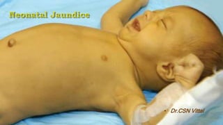
- 3. • Jaundice is yellow color of the skin and sclerae caused by deposits of bilirubin. • When is it visible? – Adult sclera > 2 mg / dL – Newborn skin > 5 mg / dL
- 4. • In term newborns: 60% • In preterms: 80%
- 6. Heme Biliverdin Bilirubin Reticuloendothelialcell Heme oxygenase Biliverdin reductase Bilirubin + albumin Unconjugated Bili Conjugated Bili UDPGA Urobilinogen (stercobilinogen) Stercobilin Enterohepatic circulation (via portal sysem) Moicrobial breakdown Hb = Heme + Globin Yligand 1g of Hb = 34 mg of bilirubin Non heme source 1 mg/kg
- 7. • Appears after 24 hours • Total bilirubin rises by less than 5 mg/dl pe day • Maximum intensity by 4th-5th day in term & • 7th day in preterm • Serum level less than 15 mg / dl • Clinically not detectable after 14 days Physiological • Appears age Appears within 24 hours of age • Increase of bilirubin > 5 mg / dl / day • Serum bilirubin > 15 mg / dl • Jaundice persisting after 14 days • Direct bilirubin > 2 mg / dl • Child sick Pathological
- 8. • Serum TB level of most newborn infants rises to > 2 mg/dL in the first week after birth. 1. Increased RBC volume per kilogram and decreased RBC survival in infants compared to adults 2. Increased ineffective erythropoiesis and 3. increased turnover of nonhemoglobin heme proteins 1. Decreased ligandin 2. Decrease in binding of ligandin by other anions 1. Decreased activity of the gene UGT1A1. (UDP-Glucuronyl Transferase 1A1) 1. Decreased intestinal bacteria, and 2. Decreased gut motility with poor evacuation of bilirubin-laden meconium A. Increased Bilirubin production B. Defective uptake of bilirubin from plasm C. Decreased clearance D. Decreased hepatic excretion
- 10. • Breastfeeding jaundice is the result of the baby not receiving enough milk to lower their bilirubin levels. • This causes the bilirubin to be reabsorbed into the intestines and keep the levels elevated which triggers jaundice. • This usually occurs in the first week of life while the baby and mother are in the early stages of learning how to breastfeed.
- 11. • It develops after the first 5 to 7 days of life and peaks at about 2 – 3 wks up to 10 mg/dL • It is linked to some type of substance in the breast milk that inhibits the liver's ability to break down and process bilirubin leading to increased concentration of beta- glucuronidase in breast milk, causing an increase in the deconjugation and reabsorption of bilirubin. • Mothers should be advised to continue breastfeeding at frequent intervals and TSB levels usually decline over a period of time.
- 12. • Jaundice appears – in the first 24 h, – after the first week of life, or – lasts > 2 wk • Total serum bilirubin rises by > 5 mg/dL/day (> 86 mMol/L/day) • Total serum bilirubin is > 18 mg/dL (> 308 mMol/L/day) • Infant shows symptoms or signs of a serious illness • Rate of rise of total serum bilirubin > 0.2 mg/dL/h (> 3.4 mMol/L/h) or > 5 mg/dL/day (> 86 mMol/L/day) • Conjugated bilirubin concentration > 1 mg/dL (> 17 mMol/L) if TSB is < 5 mg/dL (< 86 mMol/L) or > 20% of TSB (suggests neonatal cholestasis)
- 13. • When the plasma concentration of unconjugated bilirubin exceeds that which can be tightly bound by albumin (20-25 mg/dL), bilirubin can penetrate the blood-brain barrier. • If left untreated, the resulting – hyperbilirubinemic toxic encephalopathy, or kernicterus, – can result in mental retardation
- 14. 1. Hemolytic • Rh , ABO and other blood group incompatibilities • spherocytosis , elliptocytosis, Alpha thalassemia • Sepsis, DIC • Hematomas • Polycythaemia 2. Non hemolytic • Breast milk jaundice • Crigler-Najjar syndrome, types I and II • Gilbert syndrome
- 15. • It is an isoimmune hemolysis associated with – Rh incompatibility or – ABO An Rh+ man and an Rh – woman coul dhave an Rh + baby` 1st Pregnancy: At birth some of the Rh+ blood of fetus may enter mother’s circulation`` After delivery: The mother forms anti-Rh antibodies over next few months 2nd pregnancy with Rh+ fetus: Anti-Rh antibodies pass into the fetus causing its blood cells to lyse
- 17. • Hemolytic disease of newborn: Rh or ABO incompatibility • Intrauterine infections: TORCH, cong malaria, G6PD deficiency • Sepsis • Polycythemia • Cephalhematoma, subarachnoid hemorrhage • Hemolytic disorders, • Crigler-Najjar syndrome • Breast milk jaundicie • Metabolic disorders • Neonatal hepatitis • Extrahepatic biliary atresia • Conj Hyperttophic Pyloric stenosis • Physiological 1st 24 hours After 3rd wk
- 18. SRB ( mg/dL) 5 10 12 15 > 15
- 19. • First Line – Total & direct bilirubin – Blood group and Rh for mother and baby* – peripheral smear – e/o of hemolysis • Second Line – Hematocrit, – Retic count - increased in hemolysis – Direct Coomb’s test – to detect immune hemolysis – G6PD assay – Sepsis screen – Liver and thyroid function • Third Line – TORCH titers – Liver scan when conjugated hyperbilirubinemia – Ultrasonography of the liver and bile ducts in cholestatsis
- 20. • Measurement of bilirubin by jaundice meter – – Trans-cutaneous bilirubin levels
- 22. • Catabolism of heme derived from red cell hemoglobin produces equimolar amounts of CO and bilirubin • An ETCOc cutoff of more than 2.5 parts per million to predict the presence of clinically significant hemolysis
- 23. Score Interpretation 1–3 Subtle signs of acute bilirubin encephalopathy in infants with hyperbilirubinemia 4–6 Moderate acute bilirubin encephalopathy and are likely reversible with urgent and prompt bilirubin reduction strategies. 7–9 Advanced acute bilirubin encephalopathy; urgent, prompt, and individualized intervention are recommended to prevent further brain damage, minimize severity of sequelae, and possibly reverse acute damage
- 24. • Phototherapy • IVIG • Exchange transfusion • Medications
- 25. • Configurational Isomerization – Z isomers are converted to E isomers • Structural Isomerization – Bilirubin converted to lumirubin • Photo oxidation – This is a minor reaction, where photo-products are excreted in urine.
- 26. • Increased insensible water loss • Loose stools • Skin rash • Bronze baby syndrome • Hyperthermia • May result in hypocalcemia
- 27. Indications • Double volume exchange transfusion – Cord bilirubin 5 mg/dL – Cord Hb is 10 g / dL or less – Hydrops fetalis at birth • Partial Exchange Transfusion – CHF in presence of low PCV (< 35%)
- 28. • Occurs when unconjugated hyperbirubinemia as unconjugated bilirubin can cross across the blood brain barriers • When plasma levels exceed that which can be tightly bound albumin at 25 mg/dL
- 29. • Identification of Rh incompatibility antenatally and administration of Anti D (RhoGam) to mother • Ensuring adequate breast feeding • Follow up of high risk newborn • Explaining red flag signs to parents
- 30. Conjugated hyperbilirubinemia is defined by a direct or conjugated bilirubin level >1 mg/dL or >15% of the TB level. May be caused by – defects in intrahepatic bile production, – defects in transmembrane transport of bile, or – mechanical obstruction to flow
- 31. • Associated with hepatomegaly, splenomegaly, pale stools, and dark urine Normal Stool Acholic Stool
- 32. • Obstructive bile duct disorders: Biliary atresia, Choledochal duct cysts • Infectious causes: sepsis and urinary tract infections • Metabolic disorders: α1-antitrypsin deficiency, cystic fibrosis, tyrosinemia, galactosemia, storage diseases • Immunologic disorders: alloimmune liver disease and neonatal lupus erythematosus • Endocrine disorders: Hypothyroidism and panhypopituitarism. • Toxic disorders: total parenteral nutrition (TPN) including lipid
- 33. • History: Acholic stools and dark urine • Laboratory studies: total and direct or conjugated bilirubin, serum alanine aminotransferase (ALT), aspartate aminotransferase (AST), gamma-glutamyl transpeptidase (GGT), alkaline phosphatase, and coagulation studies • Abdominal ultrasonography • Hepatobiliary scintigraphy with technetium-labelled iminodiacetic acid • Percutaneous liver biopsy • intraoperative cholangiography
- 34. • Enteral feedings, even at minimal volumes of 10 mL/kg/day, are initiated as soon as possible. • Discontinue intralipid administration and substitute parenteral fish oil or fish oil emulsion • Surgery: for Congenital biliary atresia – hepaoportoenterostoly (Kasai Procedure)
- 35. • Dr.CSN Vittal