04 Neurologic
•Descargar como PPT, PDF•
7 recomendaciones•2,878 vistas

This document provides an overview of central nervous system infections, focusing on acute bacterial meningitis. It describes the typical causes, pathogenesis, clinical manifestations, diagnosis, complications and treatment of bacterial meningitis. Key points include that the most common causes are Neisseria meningitidis, Streptococcus pneumoniae and Haemophilus influenzae. Bacteria reach the subarachnoid space via the bloodstream or direct invasion. Typical symptoms are fever, headache, vomiting and signs of meningeal irritation. Diagnosis involves CSF analysis showing cloudy appearance, high pressure, neutrophil pleocytosis, elevated proteins and low glucose. Complications can include subdural effusions, hydrocephalus and brain damage.

Recomendados
Más contenido relacionado
La actualidad más candente
La actualidad más candente (20)
Destacado
Destacado (20)
Similar a 04 Neurologic
Similar a 04 Neurologic (20)
Más de Deep Deep
Más de Deep Deep (20)
Último
Último (20)
04 Neurologic
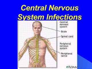
- 1. Central Nervous System Infections
- 2. Cerebrum Frontal lobe Parietal lobe Temporal lobe Occipital lobe Cerebellum Brain Stem
- 3. Pia mater, Arachnoid, Dura mater Subdural space, Subarachnoid space
- 4. The meninges of the spine cord is the continuation of the meninges of the brain
- 5. Acute Bacterial Meningitis Viral Meningoencephalitis General Introduction
- 8. 4. The anatomic distribution of infection may be diffuse or focal Meningitis: primary involvement of meninges Encephalitis: brain parenchymal involvement Meningoencephalitis: involvement of both Diffuse Focal Brain abscess: The neurologic expression of this infection is determined by the site and extent of the abscess(es)
- 9. 5. The diagnosis of diffuse CNS infections depends on careful examination of cerebrospinal fluid(CSF) obtained by lumbar puncture(LP). Production, circulation and function of CSF: Produced by the choroid plexus in the cerebral ventricles Circulates in the subarachnoid space surrounding the spinal cord and brain, protect them from injury
- 10. Acute Bacterial Meningitis Most common CNS infection in children One of the most potentially serious infections associated with a high rate of acute complications and risk of chronic morbidity Prompt and reasonable treatment may improve the prognosis largely
- 11. Etiology Pathogenesis Pathology Clinical manifestations Laboratory examinations Diagnosis Complications Treatment Differential diagnosis Epidemiolgy Purulent meningitis Acute Bacterial Meningitis
- 16. Commonest Meningitis-Causing Bacteria according to Patient Age N. meningitidis, S. pneumoniae over 12 years H. influenzae, S.pneumoniae, N. meningitidis 2 months to 12 years Group B streptococcus,gram-negative enteric bacilli,Listeria monocytogenes Birth to two month of age Common bacteria causing meningitis Age
- 17. Epidemiology Meningitis can occur at any age, but the risk is greatest among infants between 1 and 12 mo of age ; 95% of cases occur between 1 mo and 5 yr of age The lack of immunity to specific pathogens associated with young age. The mode of transmission is probably person to person contact through respiratory tract secretions or droplets. The predisposing season is the winter or spring
- 20. Large occipital encephalocele Small encephalocele high in the occipital region Posterior parietal encephalocele Lumbar myelomenigocele
- 48. Normal papilla of optic nerve Papilledema
- 58. Changes in the appearance, composition or the pressure of the CSF indicates diseases of CNS First drop appearance First tube culture Second biochemistry test Third tube cell count, Gram stain Skin subcutaneous tissue yellow ligament(first feel of reduced resistance suddenly) dural mater and arachnoid(second feel)
- 59. Indication of LP: LP should be performed when diffuse infection of CNS is suspected Contraindications for an immediate LP : cardiorespiratory compromise increased intracranial pressure (ICP) infection in the area of needle insertion bleeding diathesis (low platelet, hemophilia,DIC)
- 60. Features of normal CSF: A clear colourless fluid, contains little protein(<40mg/dl), glucose(2.8~4.4mmol/L or 50~80mg/dl) and some other components (chlorides:118~128mmol/L), and few cells(0-8/mm 3 in infants and 0-5/mm 3 in adults, 0-30 mm 3 in neonates; lymphocytic or monocytic predominance) Pressure of CSF is 25-70mmH 2 O in infants, 65-195mmH 2 O in adults
- 61. Typical CSF features of bacterial meningitis: Appearance: cloudy (turbid), puric Pressure: >200-300mmH 2 O Cells: pleocytosis 1000- 10,000 WBC/mm 3 Neutrophilic predominance 75-95% Protein: Elevated protein concentration ( 100-1000mg/dL) Glucose: Hypoglycorrhachia (<1.1mmol/L) Glucose level Ratio of CSF to serum<0.4 (Continued)
- 62. Gram stain Bacterial cultrue Define the specific pathogen Special test for CSF: Countercurrent immunoelectroporesis Latex particle agglutination antigens Determination of CSF C-reactive protein and tumor necrosis factor levels
- 63. Other investigations : Peripheral WBC: leukocytosis(20000-40000/mm 3 ), neutrophilic predominant( >80%) But in very severe cases, WBC may be low. Blood culture: Culture and staining of petechial lesions: meningcoccal meningitis. Head CT scan: brain abscesses subdural effusions or empyemas ventriculitis hydrocephalus
- 68. *: Concurrently decreased glucose and chloride in CSF is a hallmark of TB-M CSF features in various infectious meningitis Fungal + Ink+ lymphocytes ~ Fungal Viral + – normal lymphocytes Viral M.tuber-culosis+ Acid-fast+ * lymphocytes Tuberculous bacterial + Gram + neutrophils Bacterial predominance number culture stain glucose protein Pleocytosis
- 79. Viral Meningoencephalitis Relatively common An acute inflammatory process involving the meninges and brain tissue Caused by a number of different viral agents Most cases are mild and self-limited, but in severe and progressive cases death or severe sequence may occur.
- 80. Etiology Enteroviruses 80% Arboviruses Herpesviruses An important cause of severe encephalitis with a high mortality and severe sequence. Mumps
- 81. Pathology Meningeal congestion, Mononuclear (lymphocytic) infiltration Vascular and perivascular inflammatory changes Demyelination and damaged neurons. The involvement of brain tissues, spinal cord, nerve roots, and peripheral nerves is quite variable the clinical neurologic presentations are often variable.
- 82. Synapse Dendrites Axon(with myelin) Soma The structure of a neuron
- 83. Pathogenesis Virus lymphatic system(first multiplication) bloodstream (extraneural phase, systemic symptoms) CNS(secondary multiplication, neurologic disease) Neurologic damage is caused by: A direct invasion and destruction of neural tissues by actively multiplying viruses Neuronal destruction A reaction of the patient’s nervous tissue to antigens of the virus. Demyelination and vascular and perivascular destruction .
- 86. Normal conduction along a myelinated axon Effects of demyelination on impulse conduction
- 87. Clinical Manifestations A wide range of severity, several patterns of onset: Non-specific initial manifestations :fever, headache, nasopharyngitis, abdominal pain, nausea and vomiting, or in infants, screaming spells Mildly affected initially, only to lapse into coma and die suddenly. Ushered in by high fever, violent convulsions, bizarre movement, and hallucinations Signs of meningeal irritation, increased ICP, local neurologic signs Specific forms: acute transverse myelitis, Guillain-Barre syndrome, acute hemiplegia , and acute cerebellar ataxia.
- 88. Laboratory Findings 1. Analysis of CSF: Pressure: normal~ elevated. Appearance: generally clear Leukocyte count: none~ several hundred/mm 3 , mononuclear or lymphocytic predominance(but during the first 8-12 hr of the onset may be polymorphonuclear predominance). Protein concentration: normal~ slightly elevated( but may be very high if brain destruction is extensive HSV encephalitis. Glucose level : usually normal(but may be depressed in mumps infection Gram stain or bacterial culture: no bacteria recovered.
- 89. 2. Virological examinations 3. Electroencephalogram(EEG): typically shows diffuse slow wave activity 4. Neuroimaging studies(CT or MRI): may show swelling of the brain parenchyma or focal lesions.
- 90. Diagnosis 1. Clinical manifestations 2. Features of CSF 3. Virological findings 4. EEG 5. CT or MRI appearance
- 91. Differential Diagnosis which sometimes present acute onset, and has the similar CSF changes to that in viral meningoencephalitis. Cytological examination of CSF and neurological roentgenogram should be done . 1. Partial treated pyogenic meningitis The special antigen identification and gram stain of CSF may help the diagnose . 2. Other nonbacterial CNS infections Fungi, rickettsiae, Mycoplasma, protozoa and other parasites 3. Primary or secondary brain tumor,or brain abscess
- 92. Treatment 1.Antiviral agents: Acyclovir for herpes simplex Gancrclovir for CMV, EBV Virazole(ribavirin), interferon 2. Antibiotics: When bacterial cause cannot be excluded definitely. 3. General treament: Controlling fever, reducing intracranial pressure, maintenance of fluid and electrolyte balance, improving the cadiopulmonary function,etc.Supportive and rehabilitative efforts are very important after the patient recovers.
- 93. Prognosis Depends on the severity of clinical illness, the specific cause, and the age of the child. Most patients completely recover Various, degree of neurologic sequence may be left in some cases( especially in those with infection caused by Herpes simplex virus