Im409 22 24 Haenssler
•
1 recomendación•594 vistas

Implants, International Magazine of Oral Implantology

Recomendados
Más contenido relacionado
La actualidad más candente
La actualidad más candente (20)
Similar a Im409 22 24 Haenssler
Similar a Im409 22 24 Haenssler (20)
Más de Virgil Koszegi
Último
Último (20)
Im409 22 24 Haenssler
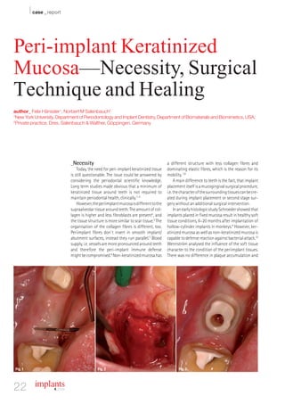
- 1. I case _ report Peri-implant Keratinized Mucosa—Necessity, Surgical Technique and Healing author_ Felix Hänssler1, Norbert M Salenbauch2 1 New York University, Department of Periodontology and Implant Dentistry, Department of Biomaterials and Biomimetics, USA; 2 Private practice, Dres. Salenbauch & Walther, Göppingen, Germany _Necessity a different structure with less collagen fibres and Today, the need for peri-implant keratinized tissue dominating elastic fibres, which is the reason for its is still questionable. The issue could be answered by mobility. 7,8 considering the periodontal scientific knowledge. A main difference to teeth is the fact, that implant Long term studies made obvious that a minimum of placement itself is a mucogingival surgical procedure, keratinized tissue around teeth is not required to i.e. the character of the surrounding tissues can be cre- maintain periodontal health, clinically.1–4 ated during implant placement or second stage sur- However, the periimplant mucosa is different to the gery without an additional surgical intervention. supraalveolar tissue around teeth. The amount of col- In an early histologic study Schroeder showed that lagen is higher and less fibroblasts are present5, and implants placed in fixed mucosa result in healthy soft the tissue structure is more similar to scar tissue.5 The tissue conditions, 6–20 months after implantation of organisation of the collagen fibres is different, too. hollow-cylinder implants in monkeys.9 However, ker- Periimplant fibres don`t insert in smooth implant/ atinized mucosa as well as non-keratinized mucosa is abutment surfaces, instead they run parallel.5 Blood capable to defense reaction against bacterial attack.10 supply, i.e. vessels are more pronounced around teeth Wennström analysed the influence of the soft tissue and therefore the peri-implant immune defense character to the condition of the periimplant tissues. might be compromised.6 Non-keratinized mucosa has There was no difference in plaque accumulation and Fig. 1 Fig. 2 Fig. 3 22 implants 4_ 2009
- 2. case _ report I peri-implant health between a keratinized mucosa without keratinized tissue. However, a long lasting width of < 2mm and > 2mm.11 Several other authors ideal oral hygiene can not be garantueed for every pa- report from healthy situations withouth keratinized tient and maybe compromised due to general diseases mucosa around implants, too.12,13 later. Therefore it is recommended to create kera- However, Chung and coworkers showed statistical tinized tissue of at least 2 mm. Beside the functional significant differences in plaque accumulation and aspect, keratinized tissue is esthetically more apeal- gingival inflammation when comparing implants with ing.32 < 2mm keratinized mucosa and implants surrounded with > 2mm keratinized mucosa. The annual bone loss _Surgical techniques was more pronounced in the group with less than Basically, two different approaches are used to cre- 2mm keratinized mucosa.14 A correlation between ker- ate keratinized mucosa around implants.33-35 Depend- atinized mucosa and implant loss was established, too. ing on the amount of missing keratinized tissue, api- In a study of 443 implants, 2.9% implant loss (97.1% cally repositioned flaps (Figs. 1–3) or free gingival success rate) occured in the group of implants sur- grafts (Figs. 4–6) are the procedures of choice. Consid- rounded with keratinized mucosa, while 29.5% im- ering both techniques, free gingival grafts are more plant loss (70.5% success rate) occured in the group of predictable. However, healing of the donor site is often implants without keratinized mucosa.15 uncomfortable for the patient. Besides, color match of Studies are confirming significant more bone loss tissue taken from the palate and the recipient bed is and inflammation16 as well as a higher susceptebility critical. A combination of an apical repositioned flap to inflammation without keratinized mucosa.17–21 This with a free gingival graft is also shown.34 position is supported by several authors.11, 20, 22–31 Summerized, there is a higher risk for plaque accu- _Healing mulation, inflammation, and periimplantitis without The healing of free gingival grafts was discovered keratinized tissue. If a perfect oral hygiene is estab- in histologic and clinical observations. After fixation of lished, non-keratinized mucosa might be maintained the graft on the recipient bed it is solely dependent healthy.25 This explains the good results in studies upon diffusion from its host bed, this occurs most ef- mentioned above, concluding periimplant stability ficiently through the fibrin clot.36 At the first day, cap- AD maxon dental Ceramic Drills for Implantology. When patients insist on metal-free treatment, maxon's ceramic drills can be applied. The maxon ceramic drill is based on three-lip drill geometry, which means that the implantologist can guide the drill securely and quietly. The ZrO ² ceramic cutting edges are exceptionally sharp and undergo almost no wear and tear. www.maxondental.com maxon motor driven by precision
- 3. I case _ report Fig. 4 Fig. 5 Fig. 6 illary proliferation begins. Between second and third Healing of the donor site is characterized by day, some capillaries have extended into the graft.36 An epithelialisation and regeneration of the connective adequate blood supply appears to be present about the tissue. Farnoush wrote that the healing process pro- eighth day.37 A connective tissue union between the ceeds by secondary intention and that reepitheliali- graft and its bed begins around the fourth day and is sation takes about 2–4 weeks, depending on the complete by the tenth day.38 If the graft is placed on wound size and surgical technique.44 The application a strong bleeding recipient bed or no adequate appli- of hemostatic agents can accelerate wound healing. cation of pressure is allowed after surgery, a The placement of oxidized regenerated cellulose over hematoma will form. This will seperate the graft from the wound exhibited complete healing after 21 its bed and the risk for necrosis will increase, since days.42 Healing and regeneration of the underlying neither rapid capillary penetration nor nutriment connective tissue will take at least 9 weeks.45,46 diffusion can occur through the hematoma.39 There- fore, pressure against the graft for five minutes is _Conclusion recommended after surgery.38 When transplanted, a Due to a possible decrease in oral hygiene and the diffusion system will maintain the graft for approxi- fact, that implant placement itself includes peri-im- mately three days until circulation is restored.36 The plant mucogingival surgery keratinized tissue must thinner graft can be easily maintained by diffusion be created around implants. The appropriate surgical and is easier to vascularize. The thicker graft shows technique depends on the residual amount of kera- more desquamation, its vascularization is delayed tinized mucosa. Small-sized dimensions can be gen- and necrosis occurs. A graft properly immobilized on erated by an apical repositioned flap while a large di- the nonbleeding, rigid recipient bed will undergo mension will be established with a free gingival graft. rapid vascularization.38 In contrast, if the graft is mo- A complete healing of a free gingival graft can be ex- bile, the ingrowing capillaries will be torn. This tear- pected after approximately 12 weeks._ ing results in bleeding and hematoma formation.40 To support adaptation and immobilization a crossing suture over the entire transplant must be placed. In suturing, the graft is stretched to conform to the re- cipient bed. This tension counteracts primary con- _contact implants traction and aids vascularization by reopening the grafts collapsed vessels.37 It appears that ten weeks is Felix Hänssler, Dr med dent a sufficient time for complete histological healing of Department of Periodontology and a graft of intermediate thickness (0.75 mm), but 16 Implant Dentistry weeks may not be long enough for complete healing Department of Biomaterials and Biomimetics of a thick graft (1.75 mm).41 The shrinkage of the free New York University College of Dentistry gingival graft is about 24 %.42 Rateitschak et al ob- 345 East 24th Street served patients over 4 years, finding that graft New York, NY 10010, USA shrinkage occured during the first 30 days and then E-Mail: haenssler@nyu.edu remained constant.43 24 I implants 4_ 2009