Cataract surgery
•Descargar como PPT, PDF•
114 recomendaciones•60,012 vistas

Intra capsular cataract extraction (ICCE) was an early surgical technique for removing cataracts but had high complication rates. Extra capsular cataract extraction (ECCE) was developed to address these issues by leaving the posterior capsule intact. ECCE became the standard technique with improvements in microscopes, irrigation/aspiration systems, and intraocular lenses. Phacoemulsification, an ECCE variant using ultrasonic fragmentation, further reduced complications through smaller incisions allowing faster recovery.

Recomendados
Más contenido relacionado
La actualidad más candente
La actualidad más candente (20)
Similar a Cataract surgery
Similar a Cataract surgery (20)
Más de Suleman Muhammad
Más de Suleman Muhammad (16)
Último
Último (20)
Cataract surgery
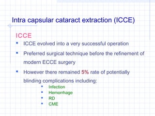
- 1. Intra capsular cataract extraction (ICCE) ICCE ICCE evolved into a very successful operation Preferred surgical technique before the refinement of modern ECCE surgery However there remained 5% rate of potentially blinding complications including: Infection Hemorrhage RD CME
- 2. Intra capsular cataract extraction (ICCE) ECCE has replaced ICCE, almost entirely in most parts of the world: 1. Better operating microscopes 2. More sophisticated surgical aspiration systems 3. More sophisticated IOL implants
- 3. Techniques (ICCE) Smith’s method Arruga’s method Erysiphakes Cryo surgery Chemical dissolution of zonular fibers
- 4. Smith’s technique Smith used external pressure with muscle hook to mechanically break the inferior zonules Expelled the lens through the limbal incision The lens would “Tumble”, I.e. the inferior pole would exit the eye before the superior pole
- 5. Arruga’s method Toothless forceps (Arruga’s) used to grasp the lens capsule and then gently pulled from the eye using side-to-side motion that broke the zonules
- 7. Erysiphakes technique Suction cuplike devices were used to remove the lens with traction
- 8. Cryo surgery Cryprobe: Hollow metal-tipped probe, cooled by liquid nitrogen, that is touched to the lens surface As the temperature of the probe tip falls below freezing, an ice ball forms and the lens adheres to it This instrument forms an ice ball, fusing the lens capsule, cortex, and nucleus Lessening the risk of capsular rupture as the cataract is removed
- 9. Chemical dissolution of zonular fibers The enzyme is irrigated into posterior chamber to dissolve the zonular fibers in order to facilitate ICCE surgery Enzyme alpha-chymotrypsin enhances the safety of ICCE by increasing the ease of lens removal
- 10. Extra capsular cataract extraction (ECCE) Shift from ICCE to modern ECCE To decrease the rate of potentially blinding: Complications To facilitate the placement of PC IOLs By leaving the PC intact, the surgeon could decrease the risk of: Vitreous loss and Complications like RD, CME, and Bullous Keratopathy
- 11. Extra capsular cataract extraction (ECCE) Key to the development of modern ECCE technique were the growing use of: Operating microscopes for increased magnification & Improved methods of cortical removal
- 12. Extra capsular cataract extraction (ECCE) Charles Kelman in 1967 developed phacoemulsification This new type of ECCE: Ultrasonically emulsified the lens nucleus, Allowing the operation to be performed through a small incision This method has continued to grow in popularity as: Techniques & Instrumentation
- 13. Indications of ICCE Operating microscopes not available Unstable / luxated cataracts Week zonular support
- 14. Advantages of ICCE • Entire lens removed with no capsule left behind to: • Opacify or • Require additional surgery • Less sophisticated instrumentation required • Non automated extraction devices: Cryoprobes Capsular forceps Erysiphakes Allow this procedure To be performed Under most conditions
- 15. Disadvantages of ICCE • Large ICCE incision 12 – 14 mm (160° - 180°) Delayed healing Iris incarceration Delayed visual rehabilitation Vitreous incarceration • Postoperative wound leaks with inadvertent filteration • Endothelial cell loss > following ICCE than ECCE • Corneal / endothelial cell trauma from lifting / folding of the cornea (lens delivery / cryprobe) • Cystoid macular edema (transient 50%, persistent 2% - 4%)
- 16. Disadvantages of ICCE (cont’d) Vitreous complications: In young patients PC is firmly adherent to anterior hyaloid; attempted ICCE will usually result in vitreous loss Intact vitreous face may opacify and ↓ vision Adherence to corneal endothelium (corneal edema) Adherence to iris (pupillary block glaucoma) Broken vitreous face may incarcerate in the wound with vitreous traction causing: RD CME Vitreous in AC causing open angle glaucoma
- 17. Disadvantages of ICCE (cont’d) IOL implantation problematic since posterior capsular support missing IOL choices include: ACL /Sutured PC IOL (Iris fixation IOLs no longer available) These significant disadvantages and risks led to loss of popularity of ICCE
- 18. Patient preparation Pharmacologic pupillary dilation with topical mydriatic and cycloplegic agents to facilitate lens removal (iris retractors intraoperatively) Anaesthesia
- 19. Patient preparation (cont’d) Orbital massage / osmotic agents (manitol, glycerine, isosorbide) before surgery 1. Intermittent digital pressure on closed eye lids or 2. Occulopressive device (honann baloon, mercury bag, sponge ball, strap) 3. Massage helps to: Distribute the anaesthetic agent within orbit ↓ Orbital volume ↓ Pressure on the globe ↓ IOP
- 20. Patient preparation (cont’d) Orbital massage (cont’d) 4. Minimizes vitreous prolapse during cataract extraction and facilitates an angle supported IOL 5. Osmotic agents are used less frequently: Volume load in patients with heart and kidney failure Nausea (Occasional) Urinary urgency during surgery
- 21. Patient preparation (cont’d) Procedure Postoperative course VA should be consistent with: 1. Refractive state of the eye 2. Clarity of the cornea 3. Clarity of the media 4. Visual potential of the retina and optic nerve
- 22. Patient preparation (cont’d) ECCE ECCE involves removal of the nucleus and cortex through an opening in the anterior capsule (anterior capsulotomy), leaving the posterior capsule in place.
- 23. Patient preparation (cont’d) ECCE (cont’d) Methods 1. Nucleus expression (manual) 2. Phacoemulsification (Ultrasonic fragmentation)
- 24. Patient preparation (cont’d) ECCE (cont’d) Methods Preferred method of routine cataract surgery Selection of technique for nucleus removal depends upon: Instrumentation available Surgeon’s level of experience with each technique
- 25. Advantages of ECCE surgery (cont’d) Smaller incision Less traumatic to corneal endothelium Eliminates complications (short and long term) associated with vitreous adherent to: Incision wound Iris Cornea
- 26. Advantages of ECCE surgery (cont’d) Intact posterior capsule allows better anatomical position for IOL fixation Intact posterior capsule ↓ incidence of: CME RD Corneal edema
- 27. Advantages of ECCE surgery (cont’d) Intact posterior capsule ↓ ability of bacteria, introduced into eye, to gain access to vitreous cavity and cause endophthalmitis 2ndry IOL implantation Filtration surgery Corneal Transplantation Wound rapair Technically easier and safer when intact PC is present
- 28. Contraindications (ECCE) Zonular weakness ECCE requires zonular integrity for selective removal of nucleus and cortical material Therefore when zonular support appears insufficient to allow safe removal of the cataract through ECCE surgery, ICCE or Pars Plana Lensectomy should be considered
- 29. Instrumentation (ECCE) A wide range of instruments is available for each step of ECCE: Opening the anterior capsule Dissecting and removing the nucleus Removing the lens cortex Polishing PC
- 30. Cystotome Used for anterior capsulotomy (opening in the anterior of the lens) Fashioned from 25 gauge needles by bending at its hub and beveled tip Prefabricated cystotomes also commercially available The needle tip is used to puncture and tear the anterior capsule
- 31. Irrigation and aspiration system coaxial, double-lumen blunt cannulas One lumen irrigates BSS into the AC Second lumen aspirates lens material out of the AC Irrigation is gravity fed from a solution bottle Fluid flow is regulated with adjustment of bottle height The flow may be constant, or the surgeon can employ a foot control connected to a pinch valve
- 32. Irrigation and aspiration system coaxial, double-lumen blunt cannulas (cont’d) Aspiration: Syringe connected to the cannula Elaborate pump system controlled by a foot switch
- 33. Lens nucleus Removed by a variety of techniques, each with its own set of instruments: Lens expressor Lens loop Spoon, Vectis
- 34. Procedure ECCE Pupillary dilation Critical to the success of ECCE esp. phacoemulsification Cycloplegic / mydriatic drops NSAID (topical/oral) these agents help to maintain dialation during surgery
- 35. Procedure ECCE (cont’d) Incision Incision: Mid limbal, chord length 8 – 12 mm, which is smaller than for ICCE The initial incision consists of a limbal groove Some surgeons prefer more posterior incision with anterior dissection creating a flap of tunnel A stab incision is made into AC AC depth stabilized by viscoelastic agents, air bubble, or continuous fluid irrigation Cystotome is inserted for anterior capsulotomy
- 36. Procedure ECCE Capsulotomy Christmas tree Can-opener Capsulorrhexis (cont’d)
- 37. Procedure ECCE (cont’d) Capsulotomy (cont’d) Christmas tree With cystotome anterior capsule punctured inferiorly and The flap of the capsule drawn toward the wound and cut with scissors
- 38. Procedure ECCE (cont’d) Capsulotomy (cont’d) Can-Opener Cystotome used to make a series of connected punctures or small tears in circle
- 39. Procedure ECCE (cont’d) Capsulorrhexis Continuous tear anterior capsulotomy popular in phacoemulsification, can be performed with either: Csytotome or Capsulorrhexis forceps First a small tear is created, The edge this tear is then grasped with cytotome tip/forceps, and A smooth tear is created, removing a circular portion of anterior capsule
- 41. Procedure ECCE (cont’d) Capsulorrhexis (cont’d) This technique provides: Structural integrity for the lens capsule Maintain implant stability Centeration
- 42. Nuclear expression Manual 1. Whole (Lens loop, spoon, vectis, irrigation) 2. Fragmentation with forceps/nuclear splitter) Ultrasonic fragmentation
- 43. Lens cortex aspiration 1. Syringe connected to cannula 2. Pump system controlled by foot switch
- 44. Posterior capsular polishing Abrasive tipped irrigation cannula / low vacuum clean using low aspiration remove epithelial and cortical particles from the capsular surface
- 45. IOL implantation AC filled with viscoelastic / BBS / air Viscoelastic most reliable AC maintainer It also protects corneal endothelial IOL inserted in the ciliary sulcus / capsular bag Sulcus fixation: Requires greater IOL diameter (>12.5 mm) Large diameter optic (6 mm) More forgiving in case of postoperative decentration Bag fixation: IOL diameter <12.5 mm Optic diameter 5.00 mm
- 46. Wound suturing 10/0 Nylon Proper suture tension ↓ postoperative Astigmatism Loose sutures – Against-the-rule Astigmatism Tight sutures – With-the rule Astigmatism
- 47. Postoperative course ECCE As with ICCE, VA on the first postoperative day should be consistent with: Refractive state of the eye Clarity of the cornea Clarity of the media Visual potential of the retina and optic nerve
- 48. Postoperative course ECCE Lid: Mild eye lid edema and erythema may occur Conjunctiva: May be injected and boggy Cornea: Should be clear and free of striate / edema AC: Should be of normal depth and mild cellular reaction typical
- 49. Postoperative course ECCE (cont’d) Posterior capsule: Should be clear and intact Implant: Should be well positioned and stable Red reflex: Should be strong and clear IOP: Elevations may be associated with retained viscoelastic
- 50. Postoperative course ECCE Antibiotics and Corticosteroids: Topical antibiotic and corticosteroids are used for first few weeks Vision: Steady improvement in vision and comfort, as inflammation subsides
- 51. Postoperative course ECCE (Cont’d) Refraction: Refraction stable by 6th – 8th weeks, Glasses may then be prescribed Astigmatism: If significant astigmatism along the axis of incision, selective sutures removed by 6th week, according to keratometry corneal topography
- 52. Phacoemulsification Phacoemulsification is an ECCE technique that differs from “standard ECCE with nuclear expression” by the: 1. Size of incision required 2. Method of nucleus removal This technique uses ultrasonically driven needle (phaco tip) to fragment the nucleus and aspirate the lens substance through a needle port
- 53. Phacoemulsification (cont’d) Advantages Lower incidence of wound related complications Faster healing Rapid visual rehabilitation AC depth controlled during surgery and providing safeguards against positive vitreous pressure and choroidal haemorrhage (closed system)
- 54. Phacoemulsification (cont’d) Instrumentation Ultrasound Irrigation system Aspiration system
- 55. Phacoemulsification (cont’d) Ultrasound The phacoemulsification hand piece contains a piezoelectic crystal that vibrates at frequency of 24000 – 56000 Hz The vibration is transmitted to the head which is attached to the phaco tip
- 56. Phacoemulsification (cont’d) Aspiration The aspiration system of phacoemulsification machine varies according to the pump design: 1. Peristaltic Pump 2. Diaphragm Pump 3. Venture Pump
- 57. Phacoemulsification (cont’d) Aspiration (cont’d) Peristaltic Pump Consists of set of rollers that move along a flexible tubing, forcing fluid through the tubing and creating a relative vacuum at the aspiration port of phacoemulsification needle
- 58. Phacoemulsification (cont’d) Aspiration (cont’d) Diaphragm Pump Flexible diaphragm overlying a fluid chamber with one-way valves at the inlet and outlet
- 59. Phacoemulsification (cont’d) Aspiration (cont’d) Venturi Pump Creates a vacuum based on the venturi principle:- That a flow of gas across a port creates a vacuum proportional to the rate of the gas
- 60. Phacoemulsification Irrigation Fluid dynamics of phacoemulsification requires constant irrigation through the irrigation sleeve around the ultrasound tip Constant irrigation: Maintains AC depth Cools the phacoemulsification probe Prevents heat buildup and adjacent tissue damage