Acute coronary syndrome
•Descargar como DOCX, PDF•
4 recomendaciones•697 vistas

Acute coronary syndrome

Recomendados
Más contenido relacionado
La actualidad más candente
La actualidad más candente (20)
Destacado
Destacado (15)
Similar a Acute coronary syndrome
Similar a Acute coronary syndrome (20)
Más de Medvizz institute of medical education
Más de Medvizz institute of medical education (17)
Último
Último (20)
Acute coronary syndrome
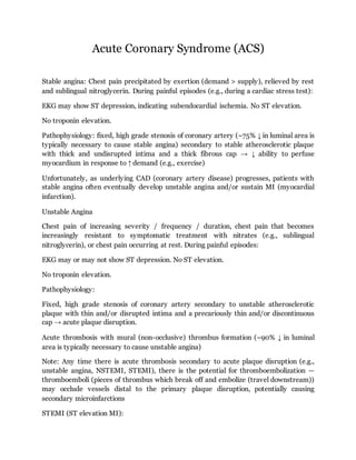
- 1. Acute Coronary Syndrome (ACS) Stable angina: Chest pain precipitated by exertion (demand > supply), relieved by rest and sublingual nitroglycerin. During painful episodes (e.g., during a cardiac stress test): EKG may show ST depression, indicating subendocardial ischemia. No ST elevation. No troponin elevation. Pathophysiology: fixed, high grade stenosis of coronary artery (~75% ↓ in luminal area is typically necessary to cause stable angina) secondary to stable atherosclerotic plaque with thick and undisrupted intima and a thick fibrous cap → ↓ ability to perfuse myocardium in response to ↑ demand (e.g., exercise) Unfortunately, as underlying CAD (coronary artery disease) progresses, patients with stable angina often eventually develop unstable angina and/or sustain MI (myocardial infarction). Unstable Angina Chest pain of increasing severity / frequency / duration, chest pain that becomes increasingly resistant to symptomatic treatment with nitrates (e.g., sublingual nitroglycerin), or chest pain occurring at rest. During painful episodes: EKG may or may not show ST depression. No ST elevation. No troponin elevation. Pathophysiology: Fixed, high grade stenosis of coronary artery secondary to unstable atherosclerotic plaque with thin and/or disrupted intima and a precariously thin and/or discontinuous cap → acute plaque disruption. Acute thrombosis with mural (non-occlusive) thrombus formation (~90% ↓ in luminal area is typically necessary to cause unstable angina) Note: Any time there is acute thrombosis secondary to acute plaque disruption (e.g., unstable angina, NSTEMI, STEMI), there is the potential for thromboembolization — thromboemboli (pieces of thrombus which break off and embolize (travel downstream)) may occlude vessels distal to the primary plaque disruption, potentially causing secondary microinfarctions STEMI (ST elevation MI):
- 2. Sx consistent with ACS Specific EKG findings: persistent ST elevation ≥1mm in two contiguous limb leads or persistent ST elevation ≥2mm in two contiguous chest leads or new LBBB (left bundle branch block)). ↑ troponins (however, elevated cardiac biomarkers are not technically required for diagnosis of STEMI if a patient presents with ACS Sx and persistent ST elevation) Note: STEMI → “Q-wave MI” (old terminology) EKG is crucial to the differential diagnosis in patients presenting with ACS (acute coronary syndrome): UA (unstable angina): Sx consistent with ACS No persistent ST elevation — EKG may be normal or only show minor changes in ~50% of UA patients, however when abnormal, EKG may show new T-wave inversion ≥2mm or dynamic/transient ST deviation (i.e., elevation or depression) ≥0.5mm Normal serum levels of troponins (indicating no infarction) within 24 hours of Sx onset The EKG is the most important test in the evaluation of acute chest pain, and should be done immediately after stabilizing the patient and taking vitals. In this setting, characteristic EKG changes are highly specific for myocardial ischemia. A patient presenting with acute chest pain and a normal EKG has <10% chance of having an acute MI. ST depression → subendocardial infarct ST elevation → transmural infarct Q waves → old transmural infarct Troponin: protein that helps control interaction between actin and myosin in skeletal and cardiac muscle. Relative to other biomarkers (eg, CK-MB, LDH), cardiac forms of troponin I (cTnI) and troponin T (cTnT) have ↑ sensitivity for myocardial damage, and are ∴ currently the preferred biomarkers for detecting myocardial necrosis Begins to rise 3-4 hours after onset, peaks between 18-36 hours and remains elevated for 7-10 days, returns to normal within 10-14 days
- 3. CK-MB: enzyme that reversibly transfers a phosphate group from creatine phosphate to ADP. Mostly found in cardiac muscle but can also be found in skeletal muscle ∴ not as specific as troponins in diagnosing acute MI Begins to rise 3-8 hours after onset, peaks at 24 hours, and returns to normal within 2-3 days Re-elevation of CK-MB can diagnose a re-infarction between 3 and 10 days post myocardial infarction, however troponin cannot diagnose re-infarction between days 3 and 10 because troponin is still elevated from the first infarction! Cardiac Catheterization and Angiography is the gold standard for localizing and quantifying CAD In a diagnostic catheterization, the coronary vasculature is accessed via the coronary ostium. Contraindicated with severe renal failure (due to contrast agent toxicity), MUST check renal status Acute Management of NSTEMI/Unstable Angina – BEMOAN B-Blocker, Enoxaparin, Morphine, O2, ASA, Nitrates The Thrombolysis In Myocardial Infarction (TIMI) Risk Score allows for grading risk thrombosis in UA. If TIMI risk score ≥ 3, consider early LMWH and angiography. Age >65 Three or more risk factors for CAD (Fam history, DM, HTN, Smoking, HLD) Aspirin use within the past 7 days Coronary Artery stenosis >50% Severe angina (>2 events over the past 24 hours ST segment changes on ECG Elevated troponins or other cardiac enzymes Studies illustrate that Clopidogrel and other glycoprotein IIB/IIIA inhibitors may be used during angina and MI as well as following PTCA or thrombolysis for their ability to lower thrombosis risk. Acute Management of STEMI: After diagnosis is made, do not wait for results of further investigations before implementing reperfusion therapy
- 4. Goal is to re-perfuse artery: Thrombolysis (EMS-to-needle) within 30 minutes Primary PCI (EMS-to-balloon) within 90 minutes PCI is the method of choice for re-perfusion in experienced centers Treatment of Chronic Stable Angina Goals: To reduce myocardial oxygen demand and/or increase oxygen supply through lifestyle modification and treatment of risk factors Lifestyle modification through diet and exercise Diet and exercise reduce the risk and morbidity of subsequent coronary events as well as decreases whole-body inflammatory process associated with plaque rupture as measured through acute phase reactants such as CRP. Elevated CRP levels correlate with increased risk of CAD, MI, CV, as well as PVD. Anti-platelet therapy (first-line treatment): ASA and Clopidegrel when ASA absolutely contraindicated B-Blockers (first-line therapy and decreases overall mortality): Cardioselective agents preferred, e.g. metoprolol and atenolol). In patients undergoing successful direct PCI for AMI, treatment with carvedilol, in contrast to metoprolol, was associated with a significant decrease in QT-RR slopes, suggesting greater cardiac electrical stability. Nitrates (symptomatic control, no clear impact on survival) Calcium Channel Blockers (Second-line or in combination) – caution: verapamil/diltiazem combined with BB may cause symptomatic sinus bradycardia or AV block ACE Inhibitors (ACEIs, not used to treat symptomatic angina): especially in patients with concomitant DM, renal dysfunction, or LV systolic dysfunction. ARB when ACEIs contraindicated Statins – Statin therapy in appropriately-selected individuals can not only decrease mortality, but also angina episodes. Invasive strategies – Revascularization
- 5. Assessing Risk Using Non-invasive Stress Testing: Exercise test results stratify patients into risk groups: Low-risk patients can be treated medically without invasive testing Intermediate risk patients may need additional testing in the form of exercise imaging studies or cardiac catherization High risk patients should be referred for cardiac catherization Treadmill Stress Testing follows the Bruce Protocol, a multistage test consisting of progressively greater workloads. This allows for detection and signs of angina and impending ACS. Indications for terminating Exercise Stress Test: Drop in SBP of >10mmHg from baseline despite an increase in workload Moderate to severe angina ST elevation (>1mm) in leads without diagnostic Q-waves (other than V1 or aVR) Increasing nervous symptoms (eg ataxia, dizziness, or near syncope) Signs of poor perfusion (cyanosis or pallor) Sustained Ventricular Tachycardia Patient’s desire to stop Nuclear Cardiology is myocardial perfusion imaging with ECG-gated single photon emission computed tomography (SPECT), using a radiolabeled tracer. They are commonly used in patients who can not tolerate a treadmill stress test. Role in evaluating myocardial viability, detecting ischemia, and simultaneously assessing perfusion and left ventricular function May be used in conjunction to treadmill or vasodilator stress with IV drugs such as dipyridamole and adenosine that act to increase coronary flow. Images of the heart are obtained during stress and at rest 3-4 hours later and classify defects as fixed (impaired perfusion at rest and during stress) or reversible defect (impaired perfusion only during stress) Stress Testing in Women – exercise treadmill stress testing in women has a higher false- positive rate. This is multifactorial, given lower prevalence of CHD in women, older age
- 6. of presentation and thus lower capacity for exercise, even hormonal medication and autonomic influences. Long-Term Management of ACS including a pre-discharge ECG and ECHO and continuation of medications required during hospitalization. Anti-platelet therapy (first-line treatment): ASA and Clopidegrel when ASA absolutely contraindicated B-Blockers (first-line therapy and decreases overall mortality): Cardioselective agents preferred, e.g. metoprolol and atenolol) Nitrates (symptomatic control, no clear impact on survival) Calcium Channel Blockers, specifically verapamil or diltiazem (Second-line or in combination) – caution: verapamil/diltiazem combined with BB may cause symptomatic sinus bradycardia or AV block ACE Inhibitors (ACEIs, not used to treat symptomatic angina): especially in patients with concomitant DM, renal dysfunction, or LV systolic dysfunction. ARB when ACEIs contraindicated +/- Aldosterone antagonists if on ACEI and BB and LVEF<40% and CHF or DM. Significant mortality benefit shown with eplerenone by 30days. Eplerenone: contraindicated in the presence of acvanced real function (GFR < 30) or significant hyperkalemia. Creatinine and potassium need to be serially followed (especially given the person is likely to be on a concomitant ACE inhibitor or ARB) Statins – early, intensive, irrespective of cholesterol level