Retinal Ganglion Cells and Visual Pathways
•Descargar como PPT, PDF•
2 recomendaciones•1,667 vistas

This document summarizes research on the neuronal architecture and functional organization of the mammalian visual system. It describes how photoreceptors in the retina respond to light and dark stimuli and how this information is transmitted through bipolar and ganglion cells which have receptive fields. It discusses how information from the two eyes is projected from the retina to the lateral geniculate nucleus and then to the primary visual cortex. Different types of cells in visual cortex, like simple and complex cells, respond to visual features like orientation and motion.

Recomendados
Más contenido relacionado
La actualidad más candente
La actualidad más candente (16)
Similar a Retinal Ganglion Cells and Visual Pathways
Similar a Retinal Ganglion Cells and Visual Pathways (20)
Más de PS Deb
Más de PS Deb (20)
Último
Último (20)
Retinal Ganglion Cells and Visual Pathways
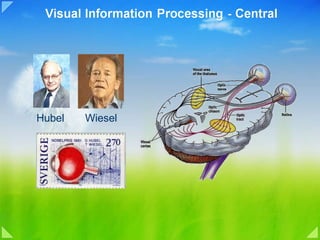
- 1. Hubel Wiesel
- 2. Neuronal architecture of mammalian visual system
- 5. Photoreceptors Rod and Cone
- 6. Anatomical Distribution of Rods and Cones
- 9. Ganglion Cell : Contrast discrimination
- 10. Receptive Field Receptive fields of photoreceptors and their connections. (A) The receptive field center provides a direct input from the photoreceptors to the bipolar cell, and the receptive field surround provides indirect input from the photoreceptor to the bipolar cells via horizontal cells. (B) 1: Photoreceptor cell; 2: on-center bipolar cell; 3: off-center bipolar cell; 4: on-center ganglion cell; 5: off-center ganglion cell.
- 11. Receptive field of bipolar cells
- 12. Receptive field Responses of retinal bipolar and ganglion cells to darkness and illumination in the receptive field center.A) Changes in the electrical activity of the photoreceptor and on- and off-center bipolar and ganglion cells when the photoreceptor receptive field center is in the dark. (B) Changes in the electrical activity of the photoreceptor and on- and off-center bipolar and ganglion cells when the photoreceptor receptive field center is illuminated
- 13. Lateral inhibition mechanism Responses of retinal bipolar and ganglion cells to darkness and illumination in the receptive field surround. (A) Changes in the electrical activity of the photoreceptor and on- and off-center bipolar and ganglion cells when the photoreceptor receptive field surround is in the dark. (B) Changes in the electrical activity of the photoreceptor and on- and off-center bipolar and ganglion cells when the photoreceptor receptive field surround is illuminated.
- 14. Receptive field of two ganglion cells overlap Two neighboring retinal ganglion cells receive input over the direct path from two overlapping groups of receptors. The areas of retina occupied by these receptors make up their receptive-field centers, shown face on by the large overlapping circles.
- 15. Ganglion Cells Characteristics M Cells P Cells Color No Yes Contrast High Low Spatial Low High Temporal High Low Population 5% 90% Action Potential Phasic, fast Tonic, slow Function Movement Shape Receptive field Large Small Retinal mapping Periphery Fovea
- 17. Central Projections of Retinal Ganglion Cells
- 19. Retinal projection to Lateral Geniculate Nucleus
- 21. LGN Projection to Occipital Cortex
- 22. Occipital lobe
- 23. LGN to Visual Cortex projection
- 24. Receptive field of a simple cell in the primary visual cortex
- 25. Simple cell of visual cortex
- 26. Complex Cell
- 27. What Primary Visual Cortex do?
- 29. Complex Cells
- 30. Retinal image of an object
- 31. Significance of Movement Cells
- 32. A rough indication of physiological cell types found in the different layers of the striate cortex.
- 33. The ocular dominance columns
- 34. Ocular Dominance Columns Ocular dominance remains constant in vertical microelectrode penetrations through the striate cortex. Penetrations parallel to the surface show alternation from left eye to right eye and back, roughly one cycle every millimeter.
- 36. Ocular Dominance Column input from LGN R L
- 37. The overlap and blurring of ocular-dominance columns beyond layer 4 is due to horizontal or diagonal connections.
- 38. Orientation column of visual cortex: Optical imaging
- 40. Primary Visual Cortex Architecture
- 41. Hypercomplex Cells End Stopping cells
- 42. V2-3: Assembling simple features into objects. V1 V2
- 43. Binding Problem
- 44. Binding Problem
- 45. Illusory contour
- 46. What we perceive depends not only on the visual image but also on our interpretation of what we see Interpretation based on our memories modifies what we see. For example if we expect to see the letter m in “exanple” we may not notice that is has been misspelled.
- 47. Visual Area Beyond V2
- 48. Evidence for dorsal and ventral pathway
- 49. Inferior Temporal neuron response to Form
- 50. Face and Complex Form Recognition ITC
Notas del editor
- Overview The human visual system is extraordinary in the quantity and quality of information it supplies about the world. A glance is sufficient to describe the location, size, shape, color, and texture of objects and, if the objects are moving, their direction and speed. Equally remarkable is the fact that visual information can be discerned over a wide range of stimulus intensities, from the faint light of stars at night to bright sunlight. The next two chapters describe the molecular, cellular, and higher-order mechanisms that allow us to see. The first steps in the process of seeing are determined by the optics of the eye, the molecular mechanisms by which light energy is transduced into electrical signals in the retina, and the retinal circuitry that determines the information relayed from the eye to the lateral geniculate nucleus of the thalamus, and ultimately to the primary visual cortex in the occipital lobe In the early 1960s, David Hubel and Torsten Wiesel (who won the Nobel Prize for Medicine in 1981) were the first to use microelectrodes to explore the receptive fields of the neurons in the lateral geniculate nucleus and the visual cortex. First, Hubel and Wiesel showed that the neurons of the lateral geniculate nucleus behave practically the same way as the ganglion cells in the retina. Then the scientists discovered the existence of three relatively independent pathways in the processing of visual information, each of which takes care of a different aspect of vision.
- The initial stages of the mammalian visual system have the platelike organization often found in the central nervous system. The first three stages are housed in the retina; the remainder are in the brain: in the lateral geniculate bodies and the stages beyond in the cortex
- Kandel Figure 26-1 Photoreceptors are located in the retina. The location of the retina within the eye is shown at left. Detail of the retina at the fovea is shown on the right (the diagram has been simplified by eliminating lateral connections mediated by interneurons; see Figure 26-6). In most of the retina light must pass through layers of nerve cells and their processes before it reaches the photoreceptors. In the center of the fovea, or foveola, these proximal neurons are shifted to the side so that light has a direct pathway to the photoreceptors. As a result, the visual image received at the foveola is the least distorted. The Retina Contains the Eye's Receptor Sheet The eye is designed to focus the visual image on the retina with minimal optical distortion. Light is focused by the cornea and the lens, then traverses the vitreous humor that fills the eye cavity before reaching photoreceptors in the retina (Figure 26-1). The retina lies in front of the pigment epithelium that lines the back of the eye. Cells in the pigment epithelium are filled with the black pigment melanin, which absorbs any light not captured by the retina. This prevents light from being reflected off the back of the eye to the retina again (which would degrade the visual image). Because the photoreceptors lie in the back of the eye, immediately in front of the pigment epithelium, all other retinal cells lie in front of the photoreceptors, closer to the lens. Therefore, light must travel through layers of other retinal neurons before striking the photoreceptors. To allow light to reach the photoreceptors without being absorbed or greatly scattered (which would distort the visual image), the axons of neurons in the proximal layers of the retina are unmyelinated so that these layers of cells are relatively transparent. Moreover, in one region of the retina, the fovea , the cell bodies of the proximal retinal neurons are shifted to the side, enabling the photoreceptors there to receive the visual image in its least distorted form (Figure 26-1). This shifting is most pronounced at the center of the fovea, the foveola. Humans therefore constantly move their eyes so that scenes of interest are projected onto the fovea. The retina also contains a region called the optic disc, where the optic nerve fibers leave the retina. This region has no photoreceptors and therefore is a blind spot in the visual field (see Figure 27-2). The projection of the visual field onto the two retinas is described in Chapter 27.
- Retinal cell types Neural Circuitry of Retina Hyperpolarization Neurotransmitters Electrical synapse with graded conduction Lateral inhibition Bipolar cell depolarization/ hyperpolarization Neural Function of the Retina Neural Circuitry of the Retina Figure 50–1 shows the tremendous complexity of neural organization in the retina. To simplify this, Figure 50–11 presents the essentials of the retina’s neural connections, showing at the left the circuit in the peripheral retina and at the right the circuit in the foveal retina. The different neuronal cell types are as follows: 1. The photoreceptors themselves—the rods and cones —which transmit signals to the outer plexiform layer, where they synapse with bipolar cells and horizontal cells 2. The horizontal cells, which transmit signals horizontally in the outer plexiform layer from the rods and cones to bipolar cells 3. The bipolar cells, which transmit signals vertically from the rods, cones, and horizontal cells to the inner plexiform layer, where they synapse with ganglion cells and amacrine cells 4. The amacrine cells, which transmit signals in two directions, either directly from bipolar cells to ganglion cells or horizontally within the inner plexiform layer from axons of the bipolar cells to dendrites of the ganglion cells or to other amacrine cells 5. The ganglion cells, which transmit output signals from the retina through the optic nerve into the brain A sixth type of neuronal cell in the retina, not very prominent and not shown in the figure, is the interplexiform cell. This cell transmits signals in the retrograde direction from the inner plexiform layer to the outer plexiform layer. These signals are inhibitory and are believed to control lateral spread of visual signals by the horizontal cells in the outer plexiform layer. Their role may be to help control the degree of contrast in the visual image. The Visual Pathway from the Cones to the Ganglion Cells Functions Differently from the Rod Pathway. As is true for many of our other sensory systems, the retina has both an old type of vision based on rod vision and a new type of vision based on cone vision. The neurons and nerve fibers that conduct the visual signals for cone vision are considerably larger than those that conduct the visual signals for rod vision, and the signals are conducted to the brain two to five times as rapidly. Also, the circuitry for the two systems is slightly different, as follows. To the right in Figure 50–11 is the visual pathway from the foveal portion of the retina, representing the new, fast cone system. This shows three neurons in the direct pathway: (1) cones, (2) bipolar cells, and (3) ganglion cells. In addition, horizontal cells transmit inhibitory signals laterally in the outer plexiform layer, and amacrine cells transmit signals laterally in the inner plexiform layer. To the left in Figure 50–11 are the neural connections for the peripheral retina, where both rods and cones are present. Three bipolar cells are shown; the middle of these connects only to rods, representing the type of visual system present in many lower animals. The output from the bipolar cell passes only to amacrine cells, which relay the signals to the ganglion cells. Thus, for pure rod vision, there are four neurons in the direct visual pathway: (1) rods, (2) bipolar cells, (3) amacrine cells, and (4) ganglion cells. Also, horizontal and amacrine cells provide lateral connectivity. The other two bipolar cells shown in the peripheral retinal circuitry of Figure 50–11 connect with both rods and cones; the outputs of these bipolar cells pass both directly to ganglion cells and by way of amacrine cells. Neurotransmitters Released by Retinal Neurons. Not all the neurotransmitter chemical substances used for synaptic transmission in the retina have been entirely delineated. However, both the rods and the cones release glutamate at their synapses with the bipolar cells. Histological and pharmacological studies have shown there to be many types of amacrine cells secreting at least eight types of transmitter substances, including gamma-aminobutyric acid, glycine, dopamine, acetylcholine, and indolamine, all of which normally function as inhibitory transmitters. The transmitters of the bipolar, horizontal, and interplexiform cells are unclear, but at least some of the horizontal cells release inhibitory transmitters. Transmission of Most Signals Occurs in the Retinal Neurons by Electrotonic Conduction, Not by Action Potentials. The only retinal neurons that always transmit visual signals by means of action potentials are the ganglion cells, and they send their signals all the way to the brain through the optic nerve. Occasionally, action potentials have also been recorded in amacrine cells, although the importance of these action potentials is questionable. Otherwise, all the retinal neurons conduct their visual signals by electrotonic conduction, which can be explained as follows. Electrotonic conduction means direct flow of electric current, not action potentials, in the neuronal cytoplasm and nerve axons from the point of excitation all the way to the output synapses. Even in the rods and cones, conduction from their outer segments, where the visual signals are generated, to the synaptic bodies is by electrotonic conduction. That is, when hyperpolarization occurs in response to light in the outer segment of a rod or a cone, almost the same degree of hyperpolarization is conducted by direct electric current flow in the cytoplasm all the way to the synaptic body, and no action potential is required. Then, when the transmitter from a rod or cone stimulates a bipolar cell or horizontal cell, once again the signal is transmitted from the input to the output by direct electric current flow, not by action potentials. The importance of electrotonic conduction is that it allows graded conduction of signal strength. Thus, for the rods and cones, the strength of the hyperpolarizing output signal is directly related to the intensity of illumination; the signal is not all or none, as would be the case for each action potential. Lateral Inhibition to Enhance Visual Contrast— Function of the Horizontal Cells The horizontal cells, shown in Figure 50–11, connect laterally between the synaptic bodies of the rods and cones, as well as connecting with the dendrites of the bipolar cells. The outputs of the horizontal cells are always inhibitory. Therefore, this lateral connection provides the same phenomenon of lateral inhibition that is important in all other sensory systems—that is, helping to ensure transmission of visual patterns with proper visual contrast. This phenomenon is demonstrated in Figure 50–12, which shows a minute spot of light focused on the retina. The visual pathway from the centralmost area where the light strikes is excited, whereas an area to the side is inhibited. In other words, instead of the excitatory signal spreading widely in the retina because of spreading dendritic and axonal trees in the plexiform layers, transmission through the horizontal cells puts a stop to this by providing lateral inhibition in the surrounding areas. This is essential to allow high visual accuracy in transmitting contrast borders in the visual image. Some of the amacrine cells probably provide additional lateral inhibition and further enhancement of visual contrast in the inner plexiform layer of the retina as well.
- Figure 11.8. Structural differences between rods and cones. Although generally similar in structure, rods (A) and cones (B) differ in their size and shape, as well as in the arrangement of the membranous disks in their outer segments. Functional Specialization of the Rod and Cone Systems Figure 11.9. The range of luminance values over which the visual system operates. At the lowest levels of illumination, only rods are activated. Cones begin to contribute to perception at about the level of starlight and are the only receptors that function under relatively bright conditions. The two types of photoreceptors, rods and cones, are distinguished by shape (from which they derive their names), the type of photopigment they contain, distribution across the retina, and pattern of synaptic connections ( Figure 11.8 ). These properties reflect the fact that the rod and cone systems (the receptors and their connections within the retina) are specialized for different aspects of vision. The rod system has very low spatial resolution but is extremely sensitive to light; it is therefore specialized for sensitivity at the expense of resolution. Conversely, the cone system has very high spatial resolution but is relatively insensitive to light; it is therefore specialized for acuity at the expense of sensitivity. The properties of the cone system also allow us to see color. The range of illumination over which the rods and cones operate is shown in Figure 11.9 . At the lowest levels of light, only the rods are activated. Such rod-mediated perception is called scotopic vision . The difficulty of making visual discriminations under very low light conditions where only the rod system is active is obvious. The problem is primarily the poor resolution of the rod system (and, to a lesser degree, the fact that there is no perception of color in dim light because the cones are not involved to a significant degree). Although cones begin to contribute to visual perception at about the level of starlight, spatial discrimination is still very poor. As illumination increases, cones become more and more dominant in determining what is seen, and they are the major determinant of perception under relatively bright conditions such as normal indoor lighting or sunlight. The contributions of rods to vision drops out nearly entirely in so-called photopic vision because their response to light saturates—that is, the membrane potential of individual rods no longer varies as a function of illumination because all of the membrane channels are closed (see Figure 11.5 ). Mesopic vision occurs in levels of light at which both rods and cones contribute—at twilight, for example. From these considerations it should be clear that most of what we think of as “seeing” is mediated by the cone system, and that loss of cone function is devastating, as occurs in elderly individuals suffering from macular degeneration ( Box C ). Individuals who have lost cone function are legally blind, whereas those who have lost rod function only experience difficulty seeing at low levels of illumination (night blindness; see Box B ). Differences in the transduction mechanisms of the two receptor types also contribute to the ability of rods and cones to respond to different ranges of light intensity. For example, rods produce a reliable response to a single photon of light, whereas more than 100 photons are required to produce a comparable response in a cone. It is not, however, that cones fail to effectively capture photons. Rather, the change in current produced by single photon capture in cones is comparatively small and difficult to distinguish from noise. Another difference is that the response of an individual cone does not saturate at high levels of steady illumination, as does the rod response. Although both rods and cones adapt to operate over a range of luminance values, the adaptation mechanisms of the cones are more effective. This difference in adaptation is apparent in the time course of the response of rods and cones to light flashes. The response of a cone, even to a bright light flash that produces the maximum change in photoreceptor current, recovers in about 200 milliseconds, more than four times faster than rod recovery. The arrangement of the circuits that transmit rod and cone information to retinal ganglion cells also contributes to the different characteristics of scotopic and photopic vision. In most parts of the retina, rod and cone signals converge on the same ganglion cells; i.e., individual ganglion cells respond to both rod and cone inputs, depending on the level of illumination. The early stages of the pathways that link rods and cones to ganglion cells, however, are largely independent. For example, the pathway from rods to ganglion cells involves a distinct class of bipolar cell (called rod bipolar) that, unlike cone bipolar cells, does not contact retinal ganglion cells. Instead, rod bipolar cells synapse with the dendritic processes of a specific class of amacrine cell that makes gap junctions and chemical synapses with the terminals of cone bipolars; these processes, in turn, make synaptic contacts on the dendrites of ganglion cells in the inner plexiform layer. Finally, the rod and cone systems differ dramatically in their degree of convergence, a factor that contributes greatly to their distinct properties. Each rod bipolar cell is contacted by a number of rods, and many rod bipolar cells contact a given amacrine cell. In contrast, the cone system is much less convergent. Thus, each retinal ganglion cell that dominates central vision (called midget ganglion cells) receives input from only one cone bipolar cell, which, in turn, is contacted by a single cone. Convergence makes the rod system a better detector of light, because small signals from many rods are pooled to generate a large response in the bipolar cell. At the same time, convergence reduces the spatial resolution of the rod system, since the source of a signal in a rod bipolar cell or retinal ganglion cell could have come from anywhere within a relatively large area of the retinal surface. The one-to-one relationship of cones to bipolar and ganglion cells is, of course, just what is required to maximize acuity. Rods Detect Dim Light Rods contain more photosensitive visual pigment than cones, enabling them to capture more light. Even more important, rods amplify light signals more than cones do. A single photon can evoke a detectable electrical response in a rod; in contrast, tens or hundreds of photons must be absorbed by a cone to evoke a similar response. In addition, the rod system is highly convergent: Many rods have synapses on the same target interneuron, known as the bipolar cell (see below). Thus, signals from the rods are pooled in the bipolar cell and reinforce one another, strengthening the signals evoked by light in individual receptors and increasing the ability of the brain to detect dim lights. In contrast, fewer cones converge on each bipolar cell. In fact, cones in the foveola have small diameters, are closely spaced, and do not converge at all; each bipolar cell receives input from a single cone.
- Figure 11.10. Distribution of rods and cones in the human retina. Graph illustrates that cones are present at a low density throughout the retina, with a sharp peak in the center of the fovea. Conversely, rods are present at high density throughout most of the retina, with a sharp decline in the fovea. Boxes at top illustrate the appearance of cross sections through the outer segments of the photoreceptors at different eccentricities. The increased density of cones in the fovea is accompanied by a striking reduction in the diameter of their outer segments. Anatomical Distribution of Rods and Cones The distribution of rods and cones across the surface of the retina also has important consequences for vision ( Figure 11.10 ). Despite the fact that perception in typical daytime light levels is dominated by cone-mediated vision, the total number of rods in the human retina (91 million) far exceeds the number of cones (roughly 4.5 million). As a result, the density of rods is much greater than cones throughout most of the retina. However, this relationship changes dramatically in the fovea , a highly specialized region of the central retina that measures about 1.2 millimeters in diameter ( Figure 11.11 ). In the fovea, cone density increases almost 200-fold, reaching, at its center, the highest receptor packing density anywhere in the retina. This high density is achieved by decreasing the diameter of the cone outer segments such that foveal cones resemble rods in their appearance. The increased density of cones in the fovea is accompanied by a sharp decline in the density of rods. In fact, the central 300 µm of the fovea, called the foveola , is totally rod-free. The extremely high density of cone receptors in the fovea, and the one-to- one relationship with bipolar cells and retinal ganglion cells (see earlier), endows this region (and the cone system generally) with the capacity to mediate high visual acuity. As cone density declines with eccentricity and the degree of convergence onto retinal ganglion cells increases, acuity is markedly reduced. Just 6° eccentric to the line of sight, acuity is reduced by 75%, a fact that can be readily appreciated by trying to read the words on any line of this page beyond the word being fixated on. The restriction of highest acuity vision to such a small region of the retina is the main reason humans spend so much time moving their eyes (and heads) around—in effect directing the foveas of the two eyes to objects of interest (see Chapter 20 ). It is also the reason why disorders that affect the functioning of the fovea have such devastating effects on sight (see Box C ). Conversely, the exclusion of rods from the fovea, and their presence in high density away from the fovea, explain why the threshold for detecting a light stimulus is lower outside the region of central vision. It is easier to see a dim object (such as a faint star) by looking away from it, so that the stimulus falls on the region of the retina that is richest in rods (see Figure 11.10 ). Another anatomical feature of the fovea (which literally means “pit”) that contributes to the superior acuity of the cone system is that the layers of cell bodies and processes that overlie the photoreceptors in other areas of the retina are displaced around the fovea, and especially the foveola (see Figure 11.11 ). As a result, light rays are subjected to a minimum of scattering before they strike the photoreceptors. Finally, another potential source of optical distortion that lies in the light path to the receptors—the retinal blood vessels—are diverted away from the foveola. This central region of the fovea is therefore dependent on the underlying choroid and pigment epithelium for oxygenation and metabolic sustenance
- Figure 11.6. Cyclic GMP-gated channels in the outer segment membrane are responsible for the light-induced changes in the electrical activity of photoreceptors (a rod is shown here, but the same scheme applies to cones). In the dark, cGMP levels in the outer segment are high; this molecule binds to the Na+-permeable channels in the membrane, keeping them open and allowing sodium (and other cations) to enter, thus depolarizing the cell. Exposure to light leads to a decrease in cGMP levels, a closing of the channels, and receptor hyperpolarization. Phototransduction In most sensory systems, activation of a receptor by the appropriate stimulus causes the cell membrane to depolarize, ultimately stimulating an action potential and transmitter release onto the neurons it contacts. In the retina, however, photoreceptors do not exhibit action potentials; rather, light activation causes a graded change in membrane potential and a corresponding change in the rate of transmitter release onto postsynaptic neurons. Indeed, much of the processing within the retina is mediated by graded potentials, largely because action potentials are not required to transmit information over the relatively short distances involved. Perhaps even more surprising is that shining light on a photoreceptor, either a rod or a cone, leads to membrane hyperpolarization rather than depolarization ( Figure 11.5 ). In the dark, the receptor is in a depolarized state, with a membrane potential of roughly -40 mV (including those portions of the cell that release transmitters). Progressive increases in the intensity of illumination cause the potential across the receptor membrane to become more negative, a response that saturates when the membrane potential reaches about -65 mV. Although the sign of the potential change may seem odd, the only logical requirement for subsequent visual processing is a consistent relationship between luminance changes and the rate of transmitter release from the photoreceptor terminals. As in other nerve cells, transmitter release from the synaptic terminals of the photoreceptor is dependent on voltage-sensitive Ca2+ channels in the terminal membrane. Thus, in the dark, when photoreceptors are relatively depolarized, the number of open Ca2+ channels in the synaptic terminal is high, and the rate of transmitter release is correspondingly great; in the light, when receptors are hyperpolarized, the number of open Ca2+ channels is reduced, and the rate of transmitter release is also reduced. The reason for this unusual arrangement compared to other sensory receptor cells is not known. The relatively depolarized state of photoreceptors in the dark depends on the presence of ion channels in the outer segment membrane that permit Na+ and Ca2+ ions to flow into the cell, thus reducing the degree of inside negativity ( Figure 11.6 ). The probability of these channels in the outer segment being open or closed is regulated in turn by the levels of the nucleotide cyclic guanosine monophosphate (cGMP) (as in many other second messenger systems; see Chapter 8 ). In darkness, high levels of cGMP in the outer segment keep the channels open. In the light, however, cGMP levels drop and some of the channels close, leading to hyperpolarization of the outer segment membrane, and ultimately the reduction of transmitter release at the photoreceptor synapse. The series of biochemical changes that ultimately leads to a reduction in cGMP levels begins when a photon is absorbed by the photopigment in the receptor disks. The photopigment contains a light-absorbing chromophore ( retinal , an aldehyde of vitamin A) coupled to one of several possible proteins called opsins that tune the molecule's absorption of light to a particular region of the spectrum. Indeed, it is the different protein component of the photopigment in rods and cones that contributes to the functional specialization of these two receptor types. Most of what is known about the molecular events of phototransduction has been gleaned from experiments in rods, in which the photopigment is rhodopsin ( Figure 11.7A ). When the retinal moiety in the rhodopsin molecule absorbs a photon, its configuration changes from the 11- cis isomer to all- trans retinal; this change then triggers a series of alterations in the protein component of the molecule ( Figure 11.7B ). The changes lead, in turn, to the activation of an intracellular messenger called transducin , which activates a phosphodiesterase that hydrolyzes cGMP. All of these events take place within the disk membrane. The hydrolysis by phosphodiesterase at the disk membrane lowers the concentration of cGMP throughout the outer segment, and thus reduces the number of cGMP molecules that are available for binding to the channels in the surface of the outer segment membrane, leading to channel closure. One of the important features of this complex biochemical cascade initiated by photon capture is that it provides enormous signal amplification. It has been estimated that a single light-activated rhodopsin molecule can activate 800 transducin molecules, roughly eight percent of the molecules on the disk surface. Although each transducin molecule activates only one phosphodiesterase molecule, each of these is in turn capable of catalyzing the breakdown of as many as six cGMP molecules. As a result, the absorption of a single photon by a rhodopsin molecule results in the closure of approximately 200 ion channels, or about 2% of the number of channels in each rod that are open in the dark. This number of channel closures causes a net change in the membrane potential of about 1 mV. Equally important is the fact that the magnitude of this amplification varies with the prevailing levels of illumination, a phenomenon known as light adaptation . At low levels of illumination, photoreceptors are the most sensitive to light. As levels of illumination increase, sensitivity decreases, preventing the receptors from saturating and thereby greatly extending the range of light intensities over which they operate. The concentration of Ca2+ in the outer segment appears to play a key role in the light-induced modulation of photoreceptor sensitivity. The cGMP-gated channels in the outer segment are permeable to both Na+ and Ca2+; thus, light-induced closure of these channels leads to a net decrease in the internal Ca2+ concentration. This decrease triggers a number of changes in the phototransduction cascade, all of which tend to reduce the sensitivity of the receptor to light. For example, the decrease in Ca2+ increases the activity of guanylate cyclase, the cGMP synthesizing enzyme, leading to an increase in cGMP levels. Likewise, the decrease in Ca2+ increases the affinity of the cGMP-gated channels for cGMP, reducing the impact of the light-induced reduction of cGMP levels. The regulatory effects of Ca2+ on the phototransduction cascade are only one part of the mechanism that adapts retinal sensitivity to background levels of illumination; another important contribution comes from neural interactions between horizontal cells and photoreceptor terminals. Once initiated, additional mechanisms limit the duration of this amplifying cascade and restore the various molecules to their inactivated states. The protein arrestin , for instance, blocks the ability of activated rhodopsin to activate transducin, and facilitates the breakdown of activated rhodopsin. The all- trans retinal then dissociates from the opsin, diffuses into the cytosol of the outer segment, and is transported out of the outer segment and into the pigment epithelium, where appropriate enzymes ultimately convert it to 11- cis retinal. After it is transported back into the outer segment, the 11- cis retinal recombines with opsin in the receptor disks. The recycling of rhodopsin is critically important for maintaining the light sensitivity of photoreceptors. Even under intense levels of illumination, the rate of regeneration is sufficient to maintain a significant number of active photopigment molecules.
- Figure 26-7 Retinal ganglion cells respond optimally to contrast in their receptive fields. Ganglion cells have circular receptive fields, with specialized center ( pink ) and surround ( gray ) regions. On-center cells are excited when stimulated by light in the center and inhibited when stimulated in the surround; off-center cells have the opposite responses. The figure shows the responses of both types of cells to five different light stimuli (the stimulated portion of the receptive field is shown in yellow ). The pattern of action potentials fired by the ganglion cell in response to each stimulus is also shown in extracellular recordings. Duration of illumination is indicated by a bar above each record. (Adapted from Kuffler 1953.) A. On-center cells respond best when the entire central part of the receptive field is stimulated ( 3 ). These cells also respond well, but less vigorously, when only a portion of the central field is stimulated by a spot of light ( 1 ). Illumination of the surround with a spot of light ( 2 ) or ring of light ( 4 ) reduces or suppresses the cell firing, which resumes more vigorously for a short period after the light is turned off. Diffuse illumination of the entire receptive field ( 5 ) elicits a relatively weak discharge because the center and surround oppose each other's effects. B. The spontaneous firing of off-center cells is suppressed when the central area of the receptive field is illuminated ( 1, 3 ) but accelerates for a short period after the stimulus is turned off. Light shone onto the surround of the receptive field excites the cell ( 2, 4 ). THE CONCEPT OF A RECEPTIVE FIELD Narrowly defined, the term receptive field refers simply to the specific receptors that feed into a given cell in the nervous system , with one or more synapses intervening. In this narrower sense, and for vision, it thus refers simply to a region on the retina, but since Kuffler's time and because of his work the term has gradually come to be used in a far broader way. Retinal ganglion cells were historically the first example of cells whose receptive fields had a substructure: stimulating different parts of the receptive fields gave qualitatively different responses, and stimulating a large area resulted in cancellation of the effects of the subdivisions rather than addition. As presently used, receptive field tends to include a description of the substructure, or if you prefer, an account of how you have to stimulate an area to make the cell respond. When we speak of "mapping out a cell's receptive field", we often mean not simply demarcating its boundaries on the retina or the screen the animal is looking at, but also describing the substructure. As we get deeper into the central nervous system, where receptive fields tend to become more and more complex, we will find that their descriptions become increasingly elaborate. Receptive-field maps are especially useful because they allow us to predict the behavior of a cell. In the case of retinal ganglion cells, for example, suppose we stimulate an on-center cell with a long, narrow rectangle of light, just wide enough to span the receptive-field center, and long enough to go beyond the whole field, center plus surround. We would predict from the on-center map on the previous page that such a stimulus would evoke a strong response, since it covers all the center and only a small fraction of the antagonistic surround. Furthermore, from the radial symmetry of the map we can predict that the magnitude of the cell's response will be independent of the slit's orientation. Both predictions are confirmed experimentally. The receptive fields of two neighboring retinal ganglion cells will usually overlap. The smallest spot of light we can shine on the retina is likely to influence hundreds of ganglion cells, some off center and some on center. The spot will fall on the centers of some receptive fields and on the surrounds of others. My second comment concerns the important question of what a population of cells such as the output cells of the retina, are doing in response to light. To understand what ganglion cells, or any other cells in a sensory system are doing, we have to go at the problem in two ways. By mapping the receptive field, we ask how we need to stimulate to make one cell respond. But we also want to know how some particular retinal stimulus affects the entire population of ganglion cells. To answer the second question we need to begin by asking what two neighboring ganglion cells, sitting side by side in the retina, have in common. The description I have given so far of ganglion-cell receptive fields could mislead you into thinking of them as forming a mosaic of nonoverlapping little circles on the retina, like the tiles on a bathroom floor. Neighboring retinal ganglion cells in fact receive their inputs from richly overlapping and usually only slightly different arrays of receptors, as shown in the diagram on this page. This is the equivalent of saying that the receptive fields almost completely overlap You can see why by glancing at the simplified circuit in the diagram above: the cell colored purple and the one colored blue receive inputs from the overlapping regions, shown in cross section, of the appropriate colors. Because of divergence, in which one cell makes synapses with many others at each stage, one receptor can influence hundreds or thousands of ganglion cells. It will contribute to the receptive-field centers of some cells and to the surrounds of others. It will excite some cells, through their centers if they are on-center cells and through their surrounds if they are off-center cells; and it will similarly inhibit others, through their centers or surrounds. Thus a small spot shining on the retina can stir up a lot of activity, in many cells. This region was the ganglion cell's receptive field. As you might expect, the receptive field was generally centered at or very near the tip of the electrode. It soon became clear that ganglion cells were of two types, and for reasons that I will soon explain, he called them on-center cells and off-center cells . An on-center cell discharged at a markedly increased rate when a small spot was turned on anywhere within a well-defined area in or near the center of the receptive field. If you listen to the discharges of such a cell over a loudspeaker, you will first hear spontaneous firing, perhaps an occasional click, and then, when the light goes on, you will hear a barrage of impulses that sounds like a machine gun firing. We call this form of response an on response. When Kuffler moved the spot of light a small distance away from the center of the receptive field, he discovered that the light suppressed the spontaneous firing of the cell, and that when he turned off the light the cell gave a brisk burst of impulses, lasting about i second. We call this entire sequence—suppression during light and discharge following light—an off response . Exploration of the receptive field soon showed that it was cleanly subdivided into a circular on region surrounded by a much larger ring-shaped off region. The more of a given region, on or off, the stimulus filled, the greater was the response, so that maximal on responses were obtained to just the right size circular spot, and maximal off responses to a ring of just the right dimensions (inner and outer diameters). Typical recordings of responses to such stimuli are shown on this page. The center and surround regions interacted in an antagonistic way: the effect of a spot in the center was diminished by shining a second spot in the surround – as f you were telling the cell to fire faster and slower at the same time. The most impressive demonstration of this interaction between center and surround occurred when a large spot covered the entire receptive field of ganglion cell. This evoked a response that was much weaker than the response to a spot just filling the center; indeed, for some cells the effects of center to the same set stimulating the two regions cancelled each other completely. An off-center cell had just the opposite behavior. Its receptive field consisted of a small center from which off responses were obtained, and a surround that gave on responses. The two kinds of cells were intermixed and seemed to be equally common. An off-center cell discharges at its highest rate in response to a black spot on a white background, because we are now illuminating only the surround of its receptive field. In nature, dark objects are probably just as common as light ones, which may help explain why information from the retina is in the form of both on-center cells and off-center cells. If you make a spot progressively larger, the response improves until the receptive-field center is filled, then it starts to decline as more and more of the surround is included, as you can see from the graph on the next page. With a spot covering the entire field, the center either just barely wins out over the surround, or the result is a draw. This effect explains why neurophysiologists before Kuffler had such lack of success: they had recorded from these cells but had generally used diffuse light – clearly from the ideal stimulus. THE RECEPTIVE FIELDS OF RETINAL GANGLION CELLS: THE OUTPUT OF THE EYE In studying the retina we are confronted with two main problems: First, how do the rods and cones translate the light they receive into electrical, and then chemical, signals? Second, how do the subsequent cells in the next two layers—the bipolar, horizontal, amacrine, and ganglion cells—interpret this information? Before discussing the physiology of the receptors and inter-mediate cells, I want to jump ahead to describe the output of the retina—represented by the activity of the ganglion cells. The map of the receptive field of a cell is a powerful and convenient shorthand description of the cell's behavior, and thus of its output. Understanding it can help us to understand why the cells in the intermediate stages are wired up as they are, and will help explain the purpose of the direct and indirect paths. If we know what ganglion cells are telling the brain, we will have gone far toward understanding the entire retina. Around 1950, Stephen Kuffler became the first to record the responses of retinal ganglion cells to spots of light in a mammal, the cat. He was then working at the Wilmer Institute of Ophthalmology at the Johns Hopkins Hospital. In retrospect, his choice of animals was lucky because the cat's retina seems to have neither the complexity of movement responses we find in the frog or rabbit retina nor the color complications we find in the retinas of fish, birds, or monkeys. Kuffler used an optical stimulator designed by Samuel Talbot. This optical device, a modified eye doctor's ophthalmoscope, made it possible to flood the retina with steady, weak, uniform background light and also to project small, more intense stimulus spots, while directly observing both the stimulus and the electrode tip. The background light made it possible to stimulate either rods or cones or both, because only the cones work when the prevailing illumination is very bright, and only the rods work in very dim light. Kuffler recorded extracellularly from electrodes inserted through the sclera (white of the eye) directly into the retina from the front. He had little difficulty finding retinal ganglion cells, which are just under the surface and are fairly large. With a steady, diffuse background light, or even in utter darkness, most retinal ganglion cells kept up a steady, somewhat irregular firing of impulses, at rates of from 1to 2 up to about 20 impulses per second. Because one might have expected the cells to be silent in complete darkness, this firing itself came as a surprise. By searching with a small spot of light, Kuffler was able to find a region in the retina through which he could influence—increase or suppress—the retinal ganglion cell's firing. Two classes of ganglion cells can be distinguished by their responses to a small spot of light applied to the center of their receptive field (Figure 26-7). On-center ganglion cells are excited when light is directed to the center of their receptive field. Light applied to the surround inhibits the cell; the most effective inhibitory stimulus is a ring of light on the entire surround. Offcenter ganglion cells are inhibited by light applied to the center of their receptive field. However, their firing rate increases for a short period of time after the light is removed; that is, they are excited when the spot of light on the center is turned off. Light excites an offcenter ganglion cell when it is directed to the surround of the receptive field. In both types of cells the response evoked by a ring of light on the entire surround cancels the response evoked by light directed to the center almost completely. For this reason, diffuse illumination of the entire receptive field evokes only a small response in either type of cell (Figure 26-7). Not all ganglion cells have a center-surround receptive field organization. For example, a few ganglion cells respond to changes in the overall luminance of the visual field and are important in controlling pupillary reflexes (see Chapter 27). On-center and off-center ganglion cells are present in roughly equal numbers, and every photoreceptor sends output to both types. Thus, ganglion cells provide two parallel pathways for the processing of visual information. In addition, their receptive fields vary in size across the retina. In the foveal region of the primate retina, where visual acuity is greatest, the receptive fields are small, with centers that are only a few minutes of arc (60 min = 1 degree). At the periphery of the retina, where acuity is low, the fields are larger, with centers of 3°-5° (1° on the retina is equal to about 0.25 mm).
- Like bipolar cells, ganglion cells have circular receptive fields, with centre-surround opposition. In addition, the ON or OFF characteristic of a bipolar cell is passed on to the ganglion cell to which it is connected. Most ganglion cells are not very sensitive to light stimuli that strike both the centre and the surround of their receptive fields. Hence, in total darkness or uniform light, they emit few action potentials. However, these cells are highly sensitive to differences in illumination at different points in their receptive fields, such as when an area of light or darkness sweeps across one side of a receptive field but not the other. The information conveyed by the action potentials from ganglion cells thus has more to do with the contrasts in illumination between light and dark areas than with the absolute degree of luminosity. The perception of light and darkness therefore is not absolute, but relative. THE SIGNIFICANCE OF CENTER-SURROUND FIELDS Why should evolution go to the trouble of building up such curious entities as center-surround receptive fields? This is the same as asking what use they are to the animal. Answering such a deep question is always difficult, but we can make some reasonable guesses. The messages that the eye sends to the brain can have little to do with the absolute intensity of light shining on the retina, because the retinal ganglion cells do not respond well to changes in diffuse light. What the cell does signal is the result of a comparison of the amount of light hitting a certain spot on the retina with the average amount falling on the immediate surround. We can illustrate this comparison by the following experiment. We first find an on-center cell and map out its receptive field. Then, beginning with the screen uniformly and dimly lit by a steady background light, we begin turning on and off a spot that just fills the field center, starting with the light so dim we cannot see it and gradually turning up the intensity. At a certain brightness, we begin to detect a response, and we notice that this is also the brightness at which we just begin to see the spot. If we measure both the background and the spot with a light meter, we find that the spot is about 2 percent brighter than the background. Now we repeat the procedure, but we start with the background light on the screen five times as bright. We gradually raise the intensity of the stimulating light. 21 Again at some point we begin to detect responses, and once again, this is the brightness at which we can just see the spot of light against the new background. When we measure the stimulating light, we find that it, too, is five times as bright as previously, that is, the spot is again 2 percent brighter than the background. The conclusion is that both for us and for the cell, what counts is the relative illumination of the spot and its surround. The cell's failure to respond well to anything but local intensity differences may seem strange, because when we look at a large, uniformly lit spot, the interior seems as vivid to us as the borders. Given its physiology, the ganglion cell reports information only from the borders of the spot; we see the interior as uniform because no ganglion cells with fields in the interior are reporting local intensity differences. The argument seems convincing enough, and yet we feel uncomfortable because, argument or no argument, the interior still looks vivid! As we encounter the same problem again and again in later chapters, we have to conclude that the nervous system often works in counterintuitive ways. Rationally, however, we must concede that seeing the large spot by using only cells whose fields are confined to the borders—instead of tying up the entire population whose centers are distributed throughout the entire spot, borders plus interior—is the more efficient system: if you were an engineer that is probably exactly how you would design a machine. I suppose that if you did design it that way, the machine, too, would think the spot was uniformly lit. In one way, the cell's weak responses or failure to respond to diffuse light should not come as a surprise. Anyone who has tried to take photographs without a light meter knows how bad we are at judging absolute light intensity. We are lucky if we can judge our camera setting to the nearest f-stop, a factor of two; to do even that we have to use our experience, noting that the day is cloudy-bright and that we are in the open shade an hour before sunset, for example, rather than just looking. But like the ganglion cell, we are very good at spatial comparisons—judging which of two neighboring regions is brighter or darker. As we have seen, we can make this comparison when the difference is only 2 percent, just as a monkey's most sensitive retinal ganglion cells can. This system carries another major advantage in addition to efficiency. We see most objects by reflected light, from sources such as the sun or a light bulb. Despite changes in the intensity of these sources, our visual system preserves to a remarkable degree the appearance of objects. The retinal ganglion cell works to make this possible. Consider the following example: a newspaper looks roughly the same—white paper, black letters—whether we view it in a dimly lit room or out on a beach on a sunny day. Suppose, in each of these two situations, we measure the light coming to our eyes from the white paper and from one of the black letters of the headline. In the following table you can read the figures I got by going from my office out into the sun in the Harvard Medical School quadrangle: Outdoors Room 22 ____________________________ White paper 120 6.0 Black letter 12 0.6 The figures by themselves are perfectly plausible. The light outside is evidently twenty times as bright as the light in the room, and the black letters reflect about one-tenth the light that white paper does. But the figures, the first time you see them, are nevertheless amazing, for they tell us that the black letter outdoors sends twice as much light to our eyes as white paper under room lights. Clearly, the appearance of black and white is not a function of the amount of light an object reflects. The important thing is the amount of light relative to the amount reflected by surrounding objects. A black-and-white television set, turned off, in a normally lit room, is white or greyish white. The engineer supplies electronic mechanisms for making the screen brighter but not for making it darker, and regardless of how it looks when turned off, no part of it will ever send less light when it is turned on. We nevertheless know very well that it is capable of giving us nice rich blacks. The blackest part of a television picture is sending to our eyes at least the same amount of light as it sends when the set is turned off. The conclusion from all this is that "black" and "white" are more than physical concepts; they are biological terms, the result of a computation done by our retina and brain on the visual scene. As we will see in Chapter 8, the entire argument I have made here concerning black and white applies also to color. The color of an object is determined Black letter 12 0.6 not just by the light coming from it, but also—and to just as important a degree as in the case of black and white—by the light coming from the rest of the scene. As a result, what we see becomes independent not only of the intensity of the light source, but also of its exact wavelength composition. And again, this is done in the interests of preserving the appearance of a scene despite marked changes in the intensity or spectral composition of the light source.
- RECEPTIVE FIELDS, FROM THE RETINA TO THE CORTEX Each of the neurons in the various layers of the retina "covers" an area in your field of vision. This area in space where the presence of an appropriate stimulus will modify the activity of this neuron is called the receptive field of this neuron. The receptive field of a single photoreceptor cell, for example, can be said to be limited to the tiny spot of light, within your field of vision, that corresponds to this photoreceptor's precise location on your retina. But in each succeeding layer of the retina, the receptive fields become increasingly complex, and they become even more complex when it comes to the neurons of the visual cortex.
- Kandel Figure 26-9 On-center and off-center bipolar cells establish parallel pathways for the signal of a single cone. Each bipolar cell makes an excitatory connection with a ganglion cell of the same type. When the cone is hyperpolarized by light, the on-center bipolar cell is excited and the off-center bipolar cell is inhibited. These opposite and simultaneous actions are initiated by the transmitter glutamate. In the dark the cone releases large amounts of transmitter because it is depolarized. Light, by hyperpolarizing the cone, causes a reduction in transmitter release. The same transmitter has different actions because the two types of bipolar cells have different postsynaptic receptors that gate different types of ion channels. The responses of the ganglion cells are largely determined by the inputs from the bipolar cells. The on-center bipolar cell, which becomes depolarized by illumination of its receptive field center, will depolarize the on-center ganglion cells; the off-center cell shows the opposite response.
- See text for details Figure 26-10 Signals from cones in the surround of a bipolar cell's receptive field are mediated by horizontal cells. Center-surround antagonism is illustrated here for an on center bipolar cell. The horizontal cell receives input from a cone in the surround of the on center bipolar cell and also has a connection with a postsynaptic cone in the center of the bipolar cell's receptive field. In the dark, horizontal cells release an inhibitory transmitter that maintains postsynaptic cones in the receptive field center in a slightly hyperpolarized state. Illumination of cones in the bipolar cell's surround hyperpolarizes those cones, which in turn hyperpolarize the postsynaptic horizontal cell. (In the dark the cones in the surround are maintained in a depolarized state and thus excite those horizontal cells.) This hyperpolarization of the horizontal cell reduces the amount of inhibitory transmitter released by the horizontal cell onto postsynaptic cones in the receptive field center, and as a result these cones become depolarized (the opposite effect of light absorption by these cones). This in turn allows the on-center bipolar cell to become hyperpolarized, the opposite effect of illumination in the receptive field center. Excitation of Some Bipolar Cells and Inhibition of Others—The Depolarizing and Hyperpolarizing Bipolar Cells Two types of bipolar cells provide opposing excitatory and inhibitory signals in the visual pathway: (1) the depolarizing bipolar cell and (2) the hyperpolarizing bipolar cell. That is, some bipolar cells depolarize when the rods and cones are excited, and others hyperpolarize. There are two possible explanations for this difference. One explanation is that the two bipolar cells are of entirely different types—one responding by depolarizing in response to the glutamate neurotransmitter released by the rods and cones, and the other responding by hyperpolarizing. The other possibility is that one of the bipolar cells receives direct excitation from the rods and cones, whereas the other receives its signal indirectly through a horizontal cell. Because the horizontal cell is an inhibitory cell, this would reverse the polarity of the electrical response. Regardless of the mechanism for the two types of bipolar responses, the importance of this phenomenon is that it allows half the bipolar cells to transmit positive signals and the other half to transmit negative signals. We shall see later that both positive and negative signals are used in transmitting visual information to the brain. Another important aspect of this reciprocal relation between depolarizing and hyperpolarizing bipolar cells is that it provides a second mechanism for lateral inhibition, in addition to the horizontal cell mechanism. Because depolarizing and hyperpolarizing bipolar cells lie immediately against each other, this provides a mechanism for separating contrast borders in the visual image, even when the border lies exactly between two adjacent photoreceptors. In contrast, the horizontal cell mechanism for lateral inhibition operates over a much greater distance. Two classes of ganglion cells can be distinguished by their responses to a small spot of light applied to the center of their receptive field (Figure 26-7). On-center ganglion cells are excited when light is directed to the center of their receptive field. Light applied to the surround inhibits the cell; the most effective inhibitory stimulus is a ring of light on the entire surround. Offcenter ganglion cells are inhibited by light applied to the center of their receptive field. However, their firing rate increases for a short period of time after the light is removed; that is, they are excited when the spot of light on the center is turned off. Light excites an off center ganglion cell when it is directed to the surround of the receptive field. In both types of cells the response evoked by a ring of light on the entire surround cancels the response evoked by light directed to the center almost completely. For this reason, diffuse illumination of the entire receptive field evokes only a small response in either type of cell (Figure 26-7). Not all ganglion cells have a center-surround receptive field organization. For example, a few ganglion cells respond to changes in the overall luminance of the visual field and are important in controlling pupillary reflexes (see Chapter 27). On-center and off-center ganglion cells are present in roughly equal numbers, and every photoreceptor sends output to both types. Thus, ganglion cells provide two parallel pathways for the processing of visual information. In addition, their receptive fields vary in size across the retina. In the foveal region of the primate retina, where visual acuity is greatest, the receptive fields are small, with centers that are only a few minutes of arc (60 min = 1 degree). At the periphery of the retina, where acuity is low, the fields are larger, with centers of 3°-5° (1° on the retina is equal to about 0.25 mm). Bipolar Cells Convey Cone Signals to Ganglion Cells Through Direct or Indirect Pathways Visual information is transferred from cones to ganglion cells along two types of pathways in the retina. Cones in the center of a ganglion cell's receptive field make direct synaptic contact with bipolar cells that in turn directly contact the ganglion cells; these connections are known as direct or vertical pathways. Signals from cones in the surround of the ganglion cell's receptive field are also conveyed to the ganglion cell through bipolar cells but only indirectly by means of horizontal and some amacrine cells; these indirect connections are called lateral pathways. Horizontal cells, which have large dendritic trees, transfer information from distant cones to bipolar cells. (Horizontal cells are also electrically coupled to each other by gap junctions and thus are able to respond to inputs from even more distant cones that contact neighboring horizontal cells.) Curiously, the horizontal cells do not appear to convey information to the bipolar cells directly, but rather by feeding back onto cones in the center of the bipolar cell's receptive field (see Figure 26-10). Some types of amacrine cells transfer information from distant bipolar cells to ganglion cells (see Figure 26-6). Most synaptic contacts in the retina are grouped in two plexiform (network-like) layers. The outer plexiform layer contains the processes of receptor, bipolar, and horizontal cells, while the inner plexiform layer contains the processes of bipolar, amacrine, and ganglion cells (see Figure 26-6). Thus the bipolar cells bridge the two plexiform layers by having processes in both. We have seen that photoreceptors respond to light with graded changes in membrane potential rather than by firing action potentials. The same is true of horizontal and bipolar cells. These cells lack voltage-gated Na+ channels capable of generating action potentials; instead they transmit signals passively (see Chapter 8). Because these cells are small and have short processes, the signals spread to their synaptic terminals without significant reduction. (Passive signal spread in cells with short processes occurs in many different parts of the brain.) In contrast, the axons of ganglion cells project considerable distances to their targets in the brain and transfer information in the form of trains of action potentials. Many types of amacrine cells also fire action potentials. Amacrine Cells and Their Functions About 30 types of amacrine cells have been identified by morphological or histochemical means. The functions of about half a dozen types of amacrine cells have been characterized, and all of them are different. One type of amacrine cell is part of the direct pathway for rod vision—that is, from rod to bipolar cells to amacrine cells to ganglion cells. Another type of amacrine cell responds strongly at the onset of a continuing visual signal, but the response dies rapidly. Other amacrine cells respond strongly at the offset of visual signals, but again, the response dies quickly. Still other amacrine cells respond when a light is turned either on or off, signaling simply a change in illumination, irrespective of direction. Still another type of amacrine cell responds to movement of a spot across the retina in a specific direction; therefore, these amacrine cells are said to be directional sensitive. In a sense, then, many or most amacrine cells are interneurons that help analyze visual signals before they ever leave the retina. Ganglion Cells and Optic Nerve Fibers Each retina contains about 100 million rods and 3 million cones; yet the number of ganglion cells is only about 1.6 million. Thus, an average of 60 rods and 2 cones converge on each ganglion cell and the optic nerve fiber leading from the ganglion cell to the brain. However, major differences exist between the peripheral retina and the central retina. As one approaches the fovea, fewer rods and cones converge on each optic fiber, and the rods and cones also become more slender. These effects progressively increase the acuity of vision in the central retina. In the center, in the central fovea, there are only slender cones—about 35,000 of them—and no rods. Also, the number of optic nerve fibers leading from this part of the retina is almost exactly equal to the number of cones, as shown to the right in Figure 50–11. This explains the high degree of visual acuity in the central retina in comparison with the much poorer acuity peripherally. Light beam Neither excited nor inhibited Excited area Inhibited area Three Types of Retinal Ganglion Cells and Their Respective Fields There are three distinct types of ganglion cells, designated W, X, and Y cells. Each of these serves a different function. Transmission of Rod Vision by the W Cells. The W cells, constituting about 40 per cent of all the ganglion cells, are small, having a diameter less than 10 micrometers, and they transmit signals in their optic nerve fibers at the slow velocity of only 8 m/sec. These ganglion cells receive most of their excitation from rods, transmitted by way of small bipolar cells and amacrine cells. They have broad fields in the peripheral retina because the dendrites of the ganglion cells spread widely in the inner plexiform layer, receiving signals from broad areas. On the basis of histology as well as physiologic experiments, the W cells seem to be especially sensitive for detecting directional movement in the field of vision, and they are probably important for much of our crude rod vision under dark conditions. Transmission of the Visual Image and Color by the X Cells. The most numerous of the ganglion cells are the X cells, representing 55 per cent of the total. They are of medium diameter, between 10 and 15 micrometers, and transmit signals in their optic nerve fibers at about 14 m/sec. The X cells have small fields because their dendrites do not spread widely in the retina. Because of this, their signals represent discrete retinal locations. Therefore, it is mainly through the X cells that the fine details of the visual image are transmitted. Also, because every X cell receives input from at least one cone, X cell transmission is probably responsible for all color vision. Function of the Y Cells to Transmit Instantaneous Changes in the Visual Image. The Y cells are the largest of all, up to 35 micrometers in diameter, and they transmit their signals to the brain at 50 m/sec or faster. They are the least numerous of all the ganglion cells, representing only 5 per cent of the total. Also, they have broad dendritic fields, so that signals are picked up by these cells from widespread retinal areas. The Y ganglion cells respond, like many of the amacrine cells, to rapid changes in the visual image— either rapid movement or rapid change in light intensity— sending bursts of signals for only small fractions of a second. These ganglion cells presumably apprise the central nervous system almost instantaneously when a new visual event occurs anywhere in the visual field, but without specifying with great accuracy the location of the event, other than to give appropriate clues that make the eyes move toward the exciting vision. Excitation of the Ganglion Cells Spontaneous, Continuous Action Potentials in the Ganglion Cells. It is from the ganglion cells that the long fibers of the optic nerve lead into the brain. Because of the distance involved, the electrotonic method of conduction employed in the rods, cones, and bipolar cells within the retina is no longer appropriate; therefore, ganglion cells transmit their signals by means of repetitive action potentials instead. Furthermore, even when unstimulated, they still transmit continuous impulses at rates varying between 5 and 40 per second. The visual signals, in turn, are superimposed onto this background ganglion cell firing. Ganglion Cells Are Specialized for the Detection of Contrasts and Rapid Changes in the Visual Image Why do ganglion cells have a center-surround receptive field organization, and why are there parallel on-center and off-center pathways? As we have just seen, ganglion cells respond only weakly to uniform illumination because of the center-surround structure of their receptive fields. They respond best when the light intensities in the center and surround are quite different. They therefore report principally the contrasts in light, rather than its absolute intensity. Most of the useful information in a visual scene is, however, contained in the pattern of contrasts. The absolute amount of light reflected by objects is relatively uninformative because it is largely determined by the intensity of the light source. Doubling the ambient light intensity will double the amount of light reflected by objects but does not alter contrasts between the objects. The center-surround organization of the receptive field of ganglion cells is therefore an adaptation for detecting useful information in the visual scene. As we shall see in Chapters 28 and 29, perception of the brightness and color of objects relies mainly on information about contrast rather than the absolute amount of light and can therefore be influenced by the contrast between an object and its surroundings. For example, the same gray ring looks much lighter against a black background than against a white one (Figure 26-8). Why does the detection of contrast start in the retina? In principle the information from photoreceptors could be sent directly to higher centers for this processing. However, signals transmitted through several relay steps to the cortex inevitably become slightly distorted. One way of minimizing the effect of transmission errors is for the retina itself to measure the difference and to transmit that information. This, in effect, is what the ganglion cell does. The firing rate of a ganglion cell provides a measure of the difference in the intensities of light illuminating the center and surround. In this way information about small differences in intensities is directly transmitted to higher centers. Parallel on-center and off-center pathways also enhance the performance of the visual system because each type of ganglion cell responds best to either rapid increases or decreases in illumination. On-center ganglion cells have a low rate of firing under dim illumination; rapid increases in firing thus signal rapid increases in light intensity in their receptive field center. In contrast, off-center ganglion cells discharge at a low rate in the light; rapid increases in firing in these cells therefore signal rapid decreases in light intensity in their receptive field center. This specialization has been demonstrated by experiments in which the function of on-center ganglion cells in awake monkeys was blocked using a pharmacological agent, aminophosphorobutyrate (APB), which selectively antagonizes transmission from photoreceptors to on-center bipolar cells. Detection of rapid increases, but not decreases, in illumination was severely impaired in these animals.
- THE CONCEPT OF A RECEPTIVE FIELD Narrowly defined, the term receptive field refers simply to the specific receptors that feed into a given cell in the nervous system, with one or more synapses intervening. In this narrower sense, and for vision, it thus refers simply to a region on the retina, but since Kuffler's time and because of his work the term has gradually come to be used in a far broader way. Retinal ganglion cells were historically the first example of cells whose receptive fields had a substructure: stimulating different parts of the receptive fields gave qualitatively different responses, and stimulating a large area resulted in cancellation of the effects of the subdivisions rather than addition. As presently used, receptive field tends to include a description of the substructure, or if you prefer, an account of how you have to stimulate an area to make the cell respond. When we speak of "mapping out a cell's receptive field", we often mean not simply demarcating its boundaries on the retina or the screen the animal is looking at, but also describing the substructure. As we get deeper into the central nervous system, where receptive fields tend to become more and more complex, we will find that their descriptions become increasingly elaborate. Receptive-field maps are especially useful because they allow us to predict the behavior of a cell. In the case of retinal ganglion cells, for example, suppose we stimulate an on-center cell with a long, narrow rectangle of light, just wide enough to span the receptive-field center, and long enough to go beyond the whole field, center plus surround. We would predict from the on-center map on the previous page that such a stimulus would evoke a strong response, since it covers all the center and only a small fraction of the antagonistic 10 surround. Furthermore, from the radial symmetry of the map we can predict that the magnitude of the cell's response will be independent of the slit's orientation. Both predictions are confirmed experimentally. surround. Furthermore, from the radial symmetry of the map we can predict that the magnitude of the cell's response will be independent of the slit's orientation. Both predictions are confirmed experimentally. The receptive fields of two neighboring retinal ganglion cells will usually overlap. The smallest spot of light we can shine on the retina is likely to influence hundreds of ganglion cells, some off center and some on center. The spot will fall on the centers of some receptive fields and on the surrounds of others. Two neighboring retinal ganglion cells receive input over the direct path from two overlapping groups of receptors. The areas of retina occupied by these receptors make up their receptive-field centers, shown face on by the large overlapping circles. My second comment concerns the important question of what a population of cells such as the output cells of the retina, are doing in response to light. To understand what ganglion cells, or any other cells in a sensory system are doing, we have to go at the problem in two ways. By mapping the receptive field, we ask how we need to stimulate to make one cell respond. But we also want to know how some particular retinal stimulus affects the entire population of ganglion cells. To answer the second question we need to begin by asking 11 what two neighboring ganglion cells, sitting side by side in the retina, have in common. The description I have given so far of ganglion-cell receptive fields could mislead you into thinking of them as forming a mosaic of nonoverlapping little circles on the retina, like the tiles on a bathroom floor. Neighboring retinal ganglion cells in fact receive their inputs from richly overlapping and usually only slightly different arrays of receptors, as shown in the diagram on this page. This is the equivalent of saying that the receptive fields almost completely overlap You can see why by glancing at the simplified circuit in the diagram above: the cell colored purple and the one colored blue receive inputs from the overlapping regions, shown in cross section, of the appropriate colors. Because of divergence, in which one cell makes synapses with many others at each stage, one receptor can influence hundreds or thousands of ganglion cells. It will contribute to the receptive-field centers of some cells and to the surrounds of others. It will excite some cells, through their centers if they are on-center cells and through their surrounds if they are off-center cells; and it will similarly inhibit others, through their centers or surrounds. Thus a small spot shining on the retina can stir up a lot of activity, in many cells.
- Just like bipolar cells, ganglion cells have concentric receptive fields with a centre-surround antagonism. But contrary to the two types of bipolar cells, ON-centre ganglion cells and OFF-centre ganglion cells do not respond by depolarizing or hyperpolarizing, but rather by increasing or decreasing the frequency with which they discharge action potentials . Conversely, if light were shined on the surround of the receptive field of this same ON-centre bipolar cell, it would become hyperpolarized. In contrast, another kind of bipolar cell becomes depolarized when an area of darkness strikes the centre of its receptive field, and hyperpolarized when it strikes the surround. Bipolar cells of this kind are called OFF-centre cells. Human vision depends in large part on our ability to discern contrasts between objects and the backgrounds behind them. The establishment of parallel pathways for the processing of visual information starting in the retina is one of the mechanisms that makes this discrimination possible. In addition to the simple cells found mainly in layer IV of the visual cortex, there are other cells, outside of layer IV, that respond to a light stimulus only if it has a particular orientation and is moving. This centre-surround structure of the receptive fields of bipolar cells is transmitted to the ganglion cells via synapses located in the inner plexiform layer . Thus, some synapses connect ON-centre bipolar cells to ON-centre ganglion cells, while others connect OFF-centre bipolar cells to OFF-centre ganglion cells. The accentuation of contrasts by the centre-surround receptive fields of the bipolar cells is thereby preserved and passed on to the ganglion cells, and ultimately to the visual cortex. That said, the response to the stimulation of the centre of the receptive field is always inhibited by the stimulation of the surround. While the other neurons in the retina emit only graduated electrical potentials, the ganglion cells are the only ones that send out neural signals in the form of action potentials . When you consider that it is the ganglion cells' axons that form the optic nerve and thereby transmit information from the retina over large distances, the significance of the generation of action potentials in these cells becomes apparent. Note that these potentials are generated spontaneously; it is the frequency at which they are discharged that is increased or decreased by the appearance of light in these cells' receptive fields. Though most ganglion cells have either ON-centre OFF-surround receptive fields or the reverse , there are other criteria that define other categories. On the basis of overall appearance, neural connections, and electrophysiological traits, at least three such categories of ganglion cells have been distinguished in the retinas of macaques (short-tailed monkeys whose retinas are very similar to our own). The small parvocellular (or "type P") ganglion cells (from the Latin parvus , meaning "small") represent about 90% of the total population of ganglion cells. Large magnocellular (or "type M") ganglion cells (from the Latin magnus , meaning "large") account for about 5%. Non-M, non-P ganglion cells, which have not yet been well characterized, account for the remaining 5%. In addition to being larger themselves, type M ganglion cells have larger receptive fields, propagate action potentials more quickly in the optic nerve, and are more sensitive to low-contrast stimuli. In addition, the positive response of an M cell to a stimulus consists of a brief salvo of action potentials, whereas the response of P cells is more tonic, continuing as long as the stimulus is active. The most commonly accepted theory is that M cells are particularly involved in detecting movement in a stimulus, whereas P cells, with their small receptive fields, would be more sensitive to its shape and details.
- RECEPTIVE FIELDS, FROM THE RETINA TO THE CORTEX Bipolar cells have centre-surround receptive fields . The centre of each such field receives direct connections from a small number of photoreceptors, while the surrounding area (called the "surround") receives inputs from a larger set of photoreceptors whose activity is relayed by the horizontal cells . Light shining on the centre of a bipolar cell's receptive field and light shining on the surround produce opposite changes in the cell's membrane potential. The diagram here uses an ON-centre bipolar cell as an example. If light is shined on the centre of this cell's receptive field, the first change is a hyperpolarization of the photoreceptor cell, causing depolarization of the bipolar cell, because of the inhibitory nature of the synapse between them . This depolarization in turn excites the following cell, a ganglion cell, causing it to emit action potentials at a higher frequency. Source: Adapted from J.E. Dowling Conversely, if light were shined on the surround of the receptive field of this same ON-centre bipolar cell, it would become hyperpolarized. In contrast, another kind of bipolar cell becomes depolarized when an area of darkness strikes the centre of its receptive field, and hyperpolarized when it strikes the surround. Bipolar cells of this kind are called OFF-centre cells.
- Figure 12.1. The retinal surface of the right eye, viewed with an ophthalmoscope. The optic disk is the region where the ganglion cell axons leave the retina to form the optic nerve; it is also characterized by the entrance and exit, respectively, of the ophthalmic arteries and veins that supply the retina. The macula lutea can be seen as a distinct area at the center of the optical axis (the optic disk lies nasally); the macula is the region of the retina that has the highest visual acuity. The fovea is a depression or pit about 1.5 mm in diameter that lies at the center of the macula (see Chapter 11 ). Figure 12.2. Central projections of retinal ganglion cells. Ganglion cell axons terminate in the lateral geniculate nucleus of the thalamus, the superior colliculus, the pretectum, and the hypothalamus. For clarity, only the crossing axons of the right eye are shown. Figure 12.3. The circuitry responsible for the pupillary light reflex. This pathway includes bilateral projections from the retina to the pretectum and projections from the pretectum to the Edinger-Westphal nucleus. Neurons in the Edinger-Westphal nucleus terminate in the ciliary ganglion, and neurons in the ciliary ganglion innervate the pupillary constrictor muscles. Notice that the afferent axons activate both Edinger-Westphal nuclei via the neurons in the pretectum. 12. Central Visual Pathways Overview Information supplied by the retina initiates interactions between multiple subdivisions of the brain that eventually lead to conscious perception of the visual scene, at the same time stimulating more conventional reflexes such as adjusting the size of the pupil, directing the eyes to targets of interest, and regulating homeostatic behaviors that are tied to the day/night cycle. The pathways and structures that mediate this broad range of functions are necessarily diverse. Of these, the primary visual pathway from the retina to the dorsal lateral geniculate nucleus in the thalamus and on to the primary visual cortex is the most important and certainly the most thoroughly studied component of the visual system. Different classes of neurons within this pathway encode the varieties of visual information—luminance, spectral differences, orientation, and motion—that we ultimately see. The parallel processing of different categories of visual information continues in cortical pathways that extend beyond primary visual cortex, supplying a variety of visual areas in the occipital, parietal, and temporal lobes. Visual areas in the temporal lobe are primarily involved in object recognition, whereas those in the parietal lobe are concerned with motion. Normal vision depends on the integration of information in all these cortical areas. The processes underlying visual perception are not understood and remain one of the central challenges of modern neuroscience Central Projections of Retinal Ganglion Cells Ganglion cell axons exit the retina through a circular region in its nasal part called the optic disk (or optic papilla), where they bundle together to form the optic nerve . This region of the retina contains no photoreceptors and, because it is insensitive to light, produces the perceptual phenomenon known as the blind spot ( Box A ). The optic disk is easily identified as a whitish circular area when the retina is examined with an ophthalmoscope; it also is recognized as the site from which the ophthalmic artery and veins enter (or leave) the eye ( Figure 12.1 ). In addition to being a conspicuous retinal landmark, the appearance of the optic disk is a useful gauge of intracranial pressure. The subarachnoid space surrounding the optic nerve is continuous with that of the brain; as a result, increases in intracranial pressure—a sign of serious neurological problems such as a space-occupying lesion—can be detected as a swelling of the optic disk (called papilledema). Axons in the optic nerve run a straight course to the optic chiasm at the base of the diencephalon. In humans, about 60% of these fibers cross in the chiasm, while the other 40% continue toward the thalamus and midbrain targets on the same side. Once past the chiasm, the ganglion cell axons on each side form the optic tract . Thus, the optic tract, unlike the optic nerve, contains fibers from both eyes. The partial crossing (or decussation) of ganglion cell axons at the optic chiasm allows information from corresponding points on the two retinas to be processed by approximately the same cortical site in each hemisphere, an important issue that is considered in the next section. The ganglion cell axons in the optic tract reach a number of structures in the diencephalon and midbrain ( Figure 12.2 ). The major target in the diencephalon is the dorsal lateral geniculate nucleus of the thalamus. Neurons in the lateral geniculate nucleus, like their counterparts in the thalamic relays of other sensory systems, send their axons to the cerebral cortex via the internal capsule. These axons pass through a portion of the internal capsule called the optic radiation and terminate in the primary visual (or striate) cortex (also referred to as Brodmann's area 17 or V1), which lies largely along and within the calcarine fissure in the occipital lobe. The retinogeniculostriate pathway, or primary visual pathway , conveys information that is essential for most of what is thought of as seeing. Thus, damage anywhere along this route results in serious visual impairment. A second major target of the ganglion cell axons is a collection of neurons that lies between the thalamus and the midbrain in a region known as the pretectum . Although small in size compared to the lateral geniculate nucleus, the pretectum is particularly important as the coordinating center for the pupillary light reflex (i.e., the reduction in the diameter of the pupil that occurs when sufficient light falls on the retina) ( Figure 12.3 ). The initial component of the pupillary light reflex pathway is a bilateral projection from the retina to the pretectum. Pretectal neurons, in turn, project to the Edinger-Westphal nucleus , a small group of nerve cells that lies close to the nucleus of the oculomotor nerve (cranial nerve III) in the midbrain. The Edinger-Westphal nucleus contains the preganglionic parasympathetic neurons that send their axons via the oculomotor nerve to terminate on neurons in the ciliary ganglion (see Chapter 20 ). Neurons in the ciliary ganglion innervate the constrictor muscle in the iris, which decreases the diameter of the pupil when activated. Shining light in the eye thus leads to an increase in the activity of pretectal neurons, which stimulates the Edinger-Westphal neurons and the ciliary ganglion neurons they innervate, thus constricting the pupil. In addition to its normal role in regulating the amount of light that enters the eye, the pupillary reflex provides an important diagnostic tool that allows the physician to test the integrity of the visual sensory apparatus, the motor outflow to the pupillary muscles, and the central pathways that mediate the reflex. Under normal conditions, the pupils of both eyes respond identically, regardless of which eye is stimulated; that is, light in one eye produces constriction of both the stimulated eye (the direct response) and the unstimulated eye (the consensual response; see Figure 12.3 ). Comparing the response in the two eyes is often helpful in localizing a lesion. For example, a direct response in the left eye without a consensual response in the right eye suggests a problem with the visceral motor outflow to the right eye, possibly damage to the oculomotor nerve or Edinger-Westphal nucleus in the brainstem. Failure to elicit a response (either direct or indirect) to stimulation of the left eye if both eyes respond normally to stimulation of the right eye suggests damage to the sensory input from the left eye, possibly to the left retina or optic nerve. There are two other important targets of retinal ganglion cell axons. One is the suprachiasmatic nucleus of the hypothalamus, a small group of neurons at the base of the diencephalon (see Figure 28.4 ). The retinohypothalamic pathway is the route by which variation in light levels influences the broad spectrum of visceral functions that are entrained to the day/night cycle (see Chapters 21 and 28 ). The other target is the superior colliculus , a prominent structure visible on the dorsal surface of the midbrain (see Figure 1.14 ). The superior colliculus coordinates head and eye movements; its functions are considered in Chapter 20 . Figure 27-4 A simplified diagram of the projections from the retina to the visual areas of the thalamus (lateral geniculate nucleus) and midbrain (pretectum and superior colliculus). The retinal projection to the pretectal area is important for pupillary reflexes, and the projection to the superior colliculus contributes to visually guided eye movements. The projection to the lateral geniculate nucleus, and from there to the visual cortex, processes visual information for perception. The Superior Colliculus Controls Saccadic Eye Movements The superior colliculus is a structure of alternating gray cellular and white (axonal) layers lying on the roof of the midbrain. Retinal ganglion cells project directly to the superficial layers and form a map of the contralateral visual field. Cells in the superficial layers in turn project through the pulvinar nucleus of the thalamus to a broad area of the cerebral cortex, thus forming an indirect pathway from the retina to the cerebral cortex. The superior colliculus also receives extensive cortical inputs. The superficial layers receive input from the visual cortex, while deeper layers receive projections from many other areas of the cerebral cortex. These deep layers have the same map of the visual field found in the superficial layers, but the cells also respond to auditory and somatosensory stimuli as well. The locations in space represented by these multisensory inputs are aligned with one another. For example, neurons that respond to a bird flying within the contralateral visual field also will respond to its singing when it is in that same part of the field. In this way, different types of sensory information about an object are conveyed to a common region of the superior colliculus. The auditory and somatosensory inputs are adjusted to fit with the visual map in situations where the maps of these other modalities might diverge. An example of such divergence occurs when our eyes are directed to one side but our head is directed straight ahead (with respect to the body); a bird sitting where we are looking will fall in the center of the visual field but its song will locate it to one side of the auditory field. Many cells lying in the deeper layers of the colliculus also discharge vigorously before the onset of saccadic eye movements, those movements that shift the gaze rapidly from one point in the visual scene to another. These cells form a movement map in the intermediate layers of the colliculus, and this map is in register with the visual map: Cells responding to stimuli in the left visual field will discharge vigorously before a leftward-directed saccade. Although the superior colliculus receives direct retinal input, the control of these saccadic eye movements is thought to be determined more by the inputs from the cerebral cortex that reach the intermediate layers. The organization within the brain of this system for generating saccadic eye movements is considered in
- LAYERING OF THE LATERAL GENICULATE Each lateral geniculate body is composed of six layers of cells stacked one on the other like a club sandwich. Each layer is made up of cells piled four to ten or more deep. The whole sandwich is folded along a fore-and-aft axis, giving the cross-sectional appearance shown in the illustration on the top of the next page. The six cell layers show clearly in the left lateral geniculate body of a macaque monkey, seen in a section cut parallel to the face. The section is stained to show cell bodies, each of which appears as a dot. The stacked-plate organization is preserved in going from retina to geniculate, except that the fibers from the retinas are bundled into a cable and splayed out again, in an orderly way, at their geniculate destination. In the scheme in which one plate projects to the next, an important complication arises in the transition from retina to geniculate; here the two eyes join up, with the two separate plates of retinal ganglion cells projecting to the sextuple geniculate plate. A single cell in the lateral geniculate body does not receive convergent input from the two eyes: a cell is a right-eye cell or a left-eye cell. These two sets of cells are segregated into separate layers, so that all the cells in any one layer get input from one eye only. The layers are stacked in such a way that the eyes alternate. In the left lateral geniculate body, the sequence in going from layer to layer, from above downwards, is right, left, right, left, left, right. It is not at all clear why the sequence reverses between the fourth and fifth layers—sometimes I think it is just to make it harder to remember. We really have no good idea why there is a sequence at all. As a whole, the sextuple-plate structure has just one topography. Thus the two left half-retinal surfaces project to one sextuple plate, the left lateral geniculate (see the bottom figure on the previous page). Similarly, the right half-retinas project to the right geniculate. Any single point in one layer corresponds to a point in the layer implies movement in the visual field along some path dictated by the visual-field-to-geniculate map. If we move instead in a direction perpendicular to the layers—for example, along the radial line in the figure on the top of the previous page—as the electrode passes from one layer to the next, the receptive fields stay in the same part of the visual field but the eyes switch—except, of course, where the sequence reverses. The half visual field maps onto each geniculate six times, three for each eye, with the maps in precise register. The lateral geniculate body seems to be two organs in one. With some justification we can consider the ventral, or bottom, two layers {ventral means "belly") as an entity because the cells they contain are different from the cells in the other four layers: they are bigger and respond differently to visual stimuli. We should also consider the four dorsal , or upper, layers {dorsal means "back" as opposed to "belly") as a separate structure because they are histologically and physiologically so similar to each other. Because of the different sizes of their cells, these two sets of layers are called magnocellular (ventral) and parvocellular (dorsal). Fibers from the six layers combine in a broad band called the optic radiations , which ascends to the primary visual cortex (see the illustration on page 2.) There, the fibers fan out in a regular way and distribute themselves so as to make a single orderly map, just as the optic nerve did on reaching the geniculate. This brings us, finally, to the cortex.
- P (small) ganglion cells, primarily from the fovea, project to a part of the thalamus called the lateral geniculate nucleus (LGN). M (large) ganglion cells, primarily from the peripheral retina, code where objects are & project both to LGN and several structures in the brainstem, including the superior colliculus (SC). The SC causes the eye and head to turn to an interesting visual object: the “visual grasp reflex”. This points the fovea at the object. Now foveal P cells can inform the cortex about the object’s details. In the LGN, the two eyes maintain their own separate representations in different layers. Ganglion cell axons grow to specific locations within each layer. Neighbouring cells grow to neighbouring locations in the LGN. Step 1: The eye develops a chemical gradient of some substance based on its location in the head (eg nasal vs temporal) Step 2: Ganglion cells are given a location identity by the position specific chemical gradient in the eye. Step 3: A similar gradient is set up in the LGN Step 4: Axons from the medial (nasal) retina are guided by this “scent” to the correct location in the contralateral LGN. Step 5: Some time later, axons from the lateral (temporal) retina are guided to the ipsilateral LGN.
- Figure 27-9 Each half of the visual field is represented in the contralateral primary visual cortex. In humans the primary visual cortex is located at the posterior pole of the cerebral hemisphere and lies almost exclusively on the medial surface. (In some individuals it is shifted so that part of it extends onto the lateral surface.) Areas in the primary visual cortex are devoted to specific parts of the visual field, as indicated by the corresponding numbers. The upper fields are mapped below the calcarine fissure, and the lower fields above it. The striking aspect of this map is that about half of the neural mass is devoted to representation of the fovea and the region just around it. This area has the greatest visual acuity. The Primary Visual Cortex Organizes Simple Retinal Inputs Into the Building Blocks of Visual Images The first point in the visual pathway where the receptive fields of cells are significantly different from those of cells in the retina is the primary visual cortex, also called visual area 1 (abbreviated V1). This region of cortex, Brodmann's area 17, is also called the striate cortex because it contains a prominent stripe of white matter in layer 4, the stria of Gennari , consisting of myelinated axons. Like the lateral geniculate nucleus and superior colliculus, the primary visual cortex in each cerebral hemisphere receives information exclusively from the contralateral half of the visual field (Figure 27-9). Organization and Function of the Visual Cortex Figures 51–2 and 51–3 show the visual cortex located primarily on the medial aspect of the occipital lobes. Like the cortical representations of the other sensory systems, the visual cortex is divided into a primary visual cortex and secondary visual areas. Primary Visual Cortex. The primary visual cortex (see Figure 51–2) lies in the calcarine fissure area, extending forward from the occipital pole on the medial aspect of each occipital cortex. This area is the terminus of direct visual signals from the eyes. Signals from the macular area of the retina terminate near the occipital pole, as shown in Figure 51–2, while signals from the more peripheral retina terminate at or in concentric half circles anterior to the pole but still along the calcarine fissure on the medial occipital lobe. The upper portion of the retina is represented superiorly and the lower portion inferiorly. Note in the figure the especially large area that represents the macula. It is to this region that the retinal fovea transmits its signals. The fovea is responsible for the highest degree of visual acuity. Based on retinal area, the fovea has several hundred times as much representation in the primary visual cortex as do the most peripheral portions of the retina. The primary visual cortex is also called visual area I . Still another name is the striate cortex because this area has a grossly striated appearance. Secondary Visual Areas of the Cortex. The secondary visual areas, also called visual association areas, lie lateral, anterior, superior, and inferior to the primary visual cortex. Most of these areas also fold outward over the lateral surfaces of the occipital and parietal cortex, as shown in Figure 51–3. Secondary signals are transmitted to these areas for analysis of visual meanings. For instance, on all sides of the primary visual cortex is Brodmann’s area 18 (see Figure 51–3), which is where virtually all signals from the primary visual cortex pass next. Therefore, Brodmann’s area 18 is called visual area II, or simply V-2.The other, more distant secondary visual areas have specific designations—V-3, V-4, and so forth—up to more than a dozen areas. The importance of all these areas is that various aspects of the visual image are progressively dissected and analyzed. Two Major Pathways for Analysis of Visual Information— (1) The Fast “Position” and “Motion” Pathway; (2) The Accurate Color Pathway Figure 51–3 shows that after leaving the primary visual cortex, the visual information is analyzed in two major pathways in the secondary visual areas. 1. Analysis of Third-Dimensional Position, Gross Form, and Motion of Objects. One of the analytical pathways, demonstrated in Figure 51–3 by the black arrows, analyzes the third-dimensional positions of visual objects in the space around the body. This pathway also analyzes the gross physical form of the visual scene as well as motion in the scene. In other words, this pathway tells where every object is during each instant and whether it is moving. After leaving the primary visual cortex, the signals flow generally into the posterior midtemporal area and upward into the broad occipitoparietal cortex. At the anterior border of the parietal cortex, the signals overlap with signals from the posterior somatic association areas that analyze threedimensional aspects of somatosensory signals. The signals transmitted in this position-form-motion pathway are mainly from the large Y optic nerve fibers of the retinal Y ganglion cells, transmitting rapid signals but depicting only black and white with no color. 2. Analysis of Visual Detail and Color. The red arrows in Figure 51–3, passing from the primary visual cortex into secondary visual areas of the inferior, ventral, and medial regions of the occipital and temporal cortex, show the principal pathway for analysis of visual detail. Separate portions of this pathway specifically dissect out color as well. Therefore, this pathway is concerned with such visual feats as recognizing letters, reading, determining the texture of surfaces, determining detailed colors of objects, and deciphering from all this information what the object is and what it means.
- 1. Why there are 6 layers, not 4. Two of the p layers appear to be redundant. 2. 80 to 90% of the input is not from the retina but from the reticular formation and the primary visual cortex. What does this input do? 3. The receptive fields of LGN neurons are the same as those of ganglion cells. Thus the LGN does not appear to further process visual information. If so, why have the synapse? Why not send information from the ganglion cells directly to the cortex? Understanding the LGN is important because the LGN is part of the thalamus. All the other sensory modalities also synapse in the thalamus prior to projecting to the cortex. What the thalamus does is also largely a mystery. To where do LGN neurons project? To primary visual cortex located at the back of the head mostly on the medial (inside) side. Primary visual cortex has many names: V1, area 17, & striate cortex. V1, like every cortical area, is made up of a thin sheet of grey matter near the surface. To pack lots of grey matter into a skull, this sheet is folded. Having lots of grey matter is good because this is where the cells and connections are. Below the grey matter lies the white matter. White matter contains the nerve fibers that interconnect the cells in the grey matter. The gray matter has 6 layers. V1 is also called the striate cortex because of a very thick layer 4. This is where massive input from the LGN ends.
- The main connections made by axons from the lateral geniculate body to the striate cortex and from the striate cortex to other brain regions. To the right, the shading indicates the relative density of Nissl staining, for comparison with the illustration on page 5. The fibers coming to the cortex from the lateral geniculate body enter from the white matter. Running diagonally, most make their way up to layer 4C, branching again and again, and finally terminate by making synapses with the stellate cells that populate that layer. Axons originating from the two ventral (magnocellular) geniculate layers end in the upper half of 4C, called 4C alpha; those from the four dorsal (parvocellular) geniculate layers end in the lower half of 4C (4C Bata). As you can see from the diagram on this page, these subdivisions of layer 4C have different projections to the upper layers: 4C alpha sends its output to 4.B; 4Q Bata, to the deepest part of 3. And those layers in turn differ in their projections. Seeing these differences in the pathways stemming from the two sets of geniculate layers is one of many reasons to think that they represent two different systems. Most pyramidal cells in layers 2, 3, 4A, 5, and 6 send axons out of the cortex, but side-branches, called "collaterals", of these same descending axons connect locally and help to distribute the information through the full cortical thickness. The layers of the cortex differ not only in their inputs and their local interconnections but also in the more distant structures to which they project. All layers except 1, 4A, and 4C send fibers out of the cortex. Layers 2 and 3 and layer 4B project mainly to other cortical regions, whereas the deep layers project down to subcortical structures: layer 5 projects to the superior colliculus in the midbrain, and layer 6 projects mainly back to the lateral geniculate body. Although we have known for almost a century that the inputs from the geniculate go mostly to layer 4, we did not know the differences in outputs of the different cortical layers until 1969, when Japanese scientist Keisuke Toyama first discovered them with physiological techniques; they have been confirmed anatomically many times since.
- THE CELLULAR STRUCTURE OF THE VISUAL CORTEX The primary visual cortex, like all the other parts of the neocortex , has a stratified cellular structure. Layers I to VI, originally described by Brodmann, had to be further subdivided as more was learned about the input and output pathways of the visual cortex. First, layer IV was divided into three sublayers designated IV A, IV B, and IV C. Then layer IV C was itself subdivided into IV Ca and IV Cb when a difference was found between the connectivities of the cells of the upper and lower parts of this sublayer. The axons of the cells of the lateral geniculate nucleus transmit information from the eye along various pathways that project mainly into layer IV C. In addition, the neighbouring cells in this layer receive receive information from neighbouring areas of the retina, thus preserving a retinotopic structure. We also know that the information flows emerging from the lateral geniculate nucleus use separate channels arising from its internal structure . In layer IV C, these information streams are received by the stellate cells , whose axons pass them on to the dendrites of the pyramidal cells in layers IV B and III. These pyramidal cells then project their axons to other areas of the cortex. As for the other output pathways from the primary visual cortex, we know that the pyramidal cells in layer V project to the superior colliculus and the pons at the subcortical level, and that the axons from layer VI return massively to the lateral geniculate nucleus, thus exerting a feedback effect on this structure. This stratification of the visual cortex into horizontal layers can be readily revealed through simple staining of its neurons. But the visual cortex is also organized into vertical columns, which were not detected until electrophysiological recordings were made of these neurons. David Hubel and Torsten Wiesel were the first scientists to propose this columnar structure superimposed on the horizontal layers. Using microelectrodes to explore the receptive fields of the neurons of the visual cortex, they showed that this cortex can be regarded as a collection of essentially identical columns. The difference from one column to the next comes simply from the portion of the visual field that is assigned to each of them. The succession of functions of the various layers from the top of the column to the bottom remains the same, but each column processes a characteristic (contrast, colour, orientation, movement, etc.) of a different part of the visual field . Figure 27-10 The primary visual cortex has distinct anatomical layers, each with characteristic synaptic connections. (Adapted from Lund 1988.) A. Most afferent fibers from the lateral geniculate nucleus terminate in layer 4. The axons of cells in the parvocellular layers (P) terminate primarily in layer 4Cb, with minor inputs to 4A and 1, while the axons of cells in the magnocellular layers (M) terminate primarily in layer 4Ca. Collaterals of both types of cells also terminate in layer 6. Cells of the intralaminar regions (I) of the lateral geniculate nucleus terminate in the blob regions of layers 2 and 3. B. Several types of neurons make up the primary visual cortex. Spiny stellate and pyramidal cells, both of which have spiny dendrites, are excitatory. Smooth stellate cells are inhibitory. Pyramidal cells project out of the cortex, whereas both types of stellate cells are local neurons. C. Conception of information flow based on anatomical connections. (LGN = lateral geniculate nucleus; MT = middle temporal area.) Inputs. Axons from M and P cells in the lateral geniculate nucleus end on spiny stellate cells in the sublayers of 4C, and these cells project axons to layer 4B or the upper layers 2 and 3. Axons from cells in the intralaminar zones of the lateral geniculate nucleus project directly to layers 2 and 3. Intracortical connections. Axon collaterals of pyramidal cells in layers 2 and 3 project to layer 5 pyramidal cells, whose axon collaterals project both to layer 6 pyramidal cells and back to cells in layers 2 and 3. Axon collaterals of layer 6 pyramidal cells then make a loop back to layer 4C onto smooth stellate cells. Output. Each layer, except for 4C, has outputs for V1 and each is different. The cells in layers 2, 3, and 4B project to extrastriate visual cortical areas. Cells in layer 5 project to the superior colliculus, the pons, and the pulvinar. Cells in layer 6 project back to the lateral geniculate nucleus and the claustrum.
- Figure 27-11 Receptive field of a simple cell in the primary visual cortex. The receptive field of a cell in the visual system is determined by recording activity in the cell while spots and bars of light are projected onto the visual field at an appropriate distance from the fovea. The records shown here are for a single cell. Duration of illumination is indicated by a line above each record of action potentials. (Adapted from Hubel and Wiesel 1959 and Zeki 1993.) 1. The cell's response to a bar of light is strongest if the bar of light is vertically oriented in the center of its receptive field. 2. Spots of light consistently elicit weak responses or no response. A small spot in the excitatory center of the field elicits only a weak excitatory response ( a ). A small spot in the inhibitory area elicits a weak inhibitory response ( b ). Diffuse light produces no response ( c ). 3. By using spots of light, the excitatory or “on” areas (+) and inhibitory or “off” areas (-) can be mapped. The map of the responses reveals an elongated “on” area and a surrounding “off” area, consistent with the optimal response of the cell to a vertical bar of light. Simple and Complex Cells Decompose the Outlines of a Visual Image Into Short Line Segments of Various Orientations How is the complexity of the circuitry in the cerebral cortex reflected in the response properties of cortical cells? Hubel, Wiesel, and their colleagues found that most cells above and below layer 4 respond optimally to stimuli that are substantially more complex than those that excite cells in the retina and lateral geniculate nucleus. Their most unexpected finding was that small spots of light—which are so effective in the retina, lateral geniculate nucleus, and in the input layer of the cortex 4C—are much less effective in all other layers of the visual cortex except possibly the blob regions in the superficial layers. Instead, cells respond best to stimuli that have linear properties, such as a line or bar. These cells belong to two major groups, simple and complex. Figure 12.9. Neurons in the primary visual cortex respond selectively to oriented edges. (A) An anesthetized animal is fitted with contact lenses to focus the eyes on a screen, where images can be projected; an extracellualr electrode records the neuronal responses. (B) Neurons in visual cortex typically respond vigorously to a bar of light oriented at a particular angle and weakly—or not at all—to other orientations. The Functional Organization of the Striate Cortex Much in the same way that Stephen Kuffler explored the response properties of individual retinal ganglion cells (see Chapter 11 ), David Hubel and Torsten Wiesel used microelectrode recordings to examine the properties of neurons in more central visual structures. The responses of neurons in the lateral geniculate nucleus were found to be remarkably similar to those in the retina, with a center-surround receptive field organization and selectivity for luminance increases or decreases. However, the small spots of light that were so effective stimulating neurons in the retina and lateral geniculate nucleus were largely ineffective in visual cortex. Instead, most cortical neurons in cats and monkeys were found to respond vigorously to light-dark bars or edges, and only if the bars were presented at a particular range of orientations within the cell's receptive field ( Figure 12.9 ). The responses of cortical neurons are thus tuned to the orientation of edges, much like cone receptors are tuned to the wavelength of light; the peak in the tuning curve (the orientation to which a cell is most responsive) is referred to as the neuron's preferred orientation. By sampling the responses of a large number of single cells, Hubel and Weisel demonstrated that all edge orientations were roughly equally represented in visual cortex. As a result, a given orientation in a visual scene appears to be “encoded” in the activity of a distinct population of orientation-selective neurons . Hubel and Wiesel also found that there are subtly different subtypes within a class of neurons that preferred the same orientation. For example, the receptive fields of some cortical cells, which they called simple cells , were composed of spatially separate “on” and “off” response zones, as if the “on” and “off” centers of lateral geniculate cells that supplied these neurons were arrayed in separate parallel bands. Other neurons, referred to as complex cells , exhibited mixed “on” and “off” responses throughout their receptive field, as if they received their inputs from a number of simple cells. Further analysis uncovered cortical neurons sensitive to the length of the bar of light that was moved across their receptive field, decreasing their rate of response when the bar exceeded a certain length. Still other cells responded selectively to the direction in which an edge moved across their receptive field. Although the mechanisms responsible for generating these selective responses are still not well understood, there is little doubt that the specificity of the receptive field properties of neurons in the striate cortex (and beyond) plays an important role in determining the basic attributes of visual scenes. Another feature that distinguishes the responses of neurons in the striate cortex from those at earlier stages in the primary visual pathway is binocularity . Although the lateral geniculate nucleus receives inputs from both eyes, the axons terminate in separate layers, so that individual geniculate neurons are monocular, driven by either the left or right eye but not by both ( Figure 12.10 ; see also Figure 12.14 ). In some species, including most (but not all) primates, inputs from the left and right eyes remain segregated to some degree even beyond the geniculate because the axons of geniculate neurons terminate in alternating eye-specific columns within cortical layer IV—the so-called ocular dominance columns (see the next section). Beyond this point, the signals from the two eyes are combined at the cellular level. Thus, most cortical neurons have binocular receptive fields, and these fields are almost identical, having the same size, shape, preferred orientation, and roughly the same position in the visual field of each eye. Bringing together the inputs from the two eyes at the level of the striate cortex provides a basis for stereopsis , the special sensation of depth that arises from viewing nearby objects with two eyes instead of one. Because the two eyes look at the world from slightly different angles, objects that lie in front of or behind the plane of fixation project to noncorresponding points on the two retinas. To convince yourself of this fact, hold your hand at arm's length and fixate on the tip of one finger. Maintain fixation on the finger as you hold a pencil in your other hand about half as far away. At this distance, the image of the pencil falls on noncorresponding points on the two retinas and will therefore be perceived as two separate pencils (a phenomenon called double vision, or diplopia ). If the pencil is now moved toward the finger (the point of fixation), the two images of the pencil fuse and a single pencil is seen in front of the finger. Thus, for a small distance on either side of the plane of fixation, where the disparity between the two views of the world remains modest, a single image is perceived; the disparity between the two eye views of objects nearer or farther than the point of fixation is interpreted as depth ( Figure 12.11 ). Although the neurophysiological basis of stereopsis is not understood, some neurons in the striate cortex and in other visual cortical areas have receptive field properties that make them good candidates for extracting information about binocular disparity. Unlike many binocular cells whose monocular receptive fields sample the same region of visual space, these neurons have monocular fields that are slightly displaced (or perhaps differ in their internal organization) so that the cell is maximally activated by stimuli that fall on noncorresponding parts of the retinas. Some of these neurons (so-called far cells ) discharge to disparities beyond the plane of fixation, while others ( near cells ) respond to disparities in front of the plane of fixation. The pattern of activity in these different classes of neurons seems likely to contribute to sensations of stereoscopic depth ( Box B ). Interestingly, the preservation of the binocular responses of cortical neurons is contingent on the normal activity from the two eyes during early postnatal life. Anything that creates an imbalance in the activity of the two eyes—for example, the clouding of one lens or the abnormal alignment of the eyes during infancy (strabismus)—can permanently reduce the effectiveness of one eye in driving cortical neurons, and thus impair the ability to use binocular information as a cue for depth. Early detection and correction of visual problems is therefore essential for normal visual function in maturity (see Chapter 24 ).
- Figure 27-11 Receptive field of a simple cell in the primary visual cortex. The receptive field of a cell in the visual system is determined by recording activity in the cell while spots and bars of light are projected onto the visual field at an appropriate distance from the fovea. The records shown here are for a single cell. Duration of illumination is indicated by a line above each record of action potentials. (Adapted from Hubel and Wiesel 1959 and Zeki 1993.) 1. The cell's response to a bar of light is strongest if the bar of light is vertically oriented in the center of its receptive field. 2. Spots of light consistently elicit weak responses or no response. A small spot in the excitatory center of the field elicits only a weak excitatory response (a). A small spot in the inhibitory area elicits a weak inhibitory response (b). Diffuse light produces no response (c). 3. By using spots of light, the excitatory or “on” areas (+) and inhibitory or “off” areas (-) can be mapped. The map of the responses reveals an elongated “on” area and a surrounding “off” area, consistent with the optimal response of the cell to a vertical bar of light. Figure 27-12 The receptive fields of simple cells in the primary visual cortex are different and more varied than those of the neurons in the retina and lateral geniculate nucleus. A. Cells of the retina and lateral geniculate nucleus fall into two classes: on-center and offcenter. The receptive fields of these neurons have a center-surround organization due to antagonistic excitatory (+) and inhibitory (-) regions. B. The receptive fields of simple cells in the primary visual cortex have narrow elongated zones with either excitatory (+) or inhibitory (-) flanking areas. Despite the variety, the receptive fields of simple cells share three features: (1) specific retinal position, (2) discrete excitatory and inhibitory zones, and (3) a specific axis of orientation. C. Model of the organization of inputs in the receptive field of simple cells proposed by Hubel and Wiesel. According to this model, a simple cortical neuron in the primary visual cortex receives convergent excitatory connections from three or more on-center cells that together represent light falling along a straight line in the retina. As a result, the receptive field of the simple cortical cell has an elongated excitatory region, indicated by the colored outline in the receptive field diagram. The inhibitory surround of the simple cortical cells is probably provided by off-center cells whose receptive fields (not shown) are adjacent to those of the on-center cells. (Adapted from Hubel and Wiesel 1962). Figure 27-13 The receptive field of a complex cell in the primary visual cortex has no clearly excitatory or inhibitory zones. Orientation of the light stimulus is important, but position within the receptive field is not. (Adapted from Hubel and Wiesel 1962). A. In this example the cell responds best to a vertical edge moving across the receptive field from left to right. This figure shows the patterns of action potentials fired by the cell in response to two types of variation in the stimulus: differences in orientation and differences in position. The line above each record indicates the period of illumination. Different orientations of the light stimulus produce different rates of firing in the cell. A vertical bar of light on the left of the receptive field produces a strong excitatory response (a). Orientations other than vertical are less effective ( b-d ). The position of the border of the light within the receptive field affects the type of response in the cell. If the edge of the light comes from any point on the right within the receptive field, the stimulus produces an excitatory response ( a-d ). If the edge comes from the left, the stimulus produces an inhibitory response ( f-i ). Illumination of the entire receptive field produces no response ( e ). B. According to Hubel and Wiesel, the receptive fields of complex cells are determined by the pattern of inputs. Each complex cell receives convergent excitatory input from several simple cortical cells, each of which has a receptive field with the same organization: a central rectilinear excitation zone (+) and flanking inhibitory regions (-). In this way the receptive field of a complex cell is built up from the individual fields of the presynaptic cells. The simple cells respond best to a bar of light with a specific orientation. For example, a cell that responds best to a vertical bar will not respond, or respond only weakly, to a bar that is horizontal or even oblique (Figure 27-11). Thus, an array of cells in the cortex, all receiving impulses from the same point on the retina but with rectilinear receptive fields with different axes of orientation, is able to represent every axis of rotation for that point on the retina. Simple cells also have excitatory and inhibitory zones in their receptive fields, although these zones are slightly larger than those for lateral geniculate cells (Figure 27-12A, B). For example, a cell may have a rectilinear excitatory zone (with its long axis running from 12 to 6 o'clock such as in Figure 27-12B upper right). For a cell with such a field, an effective stimulus must excite the specific segment of the retina innervated by receptors in the excitatory zone and have the correct linear properties (in this case an edge) and have a specific axis of orientation (in this case vertical, running from 12 to 6 o'clock). Rectilinear receptive fields could be built up from many circular fields if the presynaptic connections from the lateral geniculate nucleus were appropriately arrayed on the simple cell (Figure 27-12C). Indeed, experiments have indicated that the excitatory (“on”) regions in the receptive field of simple cells largely represent the input from on-center lateral geniculate cells while the inhibitory (“off”) regions represent inputs from off- center lateral geniculate cells. The receptive fields of complex cells in the cortex are usually larger than those of simple cells. These fields also have a critical axis of orientation, but the precise position of the stimulus within the receptive field is less crucial because there are no clearly defined on or off zones (Figure 27-13A). Thus, movement across the receptive field is a particularly effective stimulus for certain complex cells. Although some complex cells have direct connections with cells of layer 4C, Hubel and Wiesel proposed that a significant input to complex cells comes from a group of simple cortical cells with the same axis of orientation but with slightly offset receptive field positions (Figure 27-13B). Some Feature Abstraction Is Accomplished by Progressive Convergence The pattern of convergence of inputs throughout the pathway that leads to the complex cells suggests that each complex cell surveys the activity of a group of simple cells, each simple cell surveys the activity of a group of geniculate cells, and each geniculate cell surveys the activity of a group of retinal ganglion cells. The ganglion cells survey the activity of bipolar cells that, in turn, survey an array of receptors. At each level each cell has a greater capacity for abstraction than cells At each level of the afferent pathway the stimulus properties that activate a cell become more specific. Retinal ganglion and geniculate neurons respond primarily to contrast. This elementary information is transformed in the simple and complex cells of the cortex, through the pattern of excitation in their rectilinear fields, into relatively precise line segments and boundaries. Hubel and Wiesel suggest that this processing is an important step in analyzing the contours of objects. In fact, contour information may be sufficient to recognize an object. Monotonous interior or background surfaces contain no critical visual information! David Hubel describes this unexpected feature of perception: Many people, including myself, still have trouble accepting the idea that the interior of a form… does not itself excite cells in our brain,… that our awareness of the interior as black or white. … depends only on cells' sensitivity to the borders. The intellectual argument is that perception of an evenly lit interior depends on the activation of cells having fields at the borders and on the absence of activation of cells whose fields are within the borders, since such activation would indicate that the interior is not evenly lit. So our perception of the interior as black, white, gray or green has nothing to do with cells whose fields are in the interior—hard as that may be to swallow.… What happens at the borders is the only information you need to know: the interior is boring. It is the information carried by edges that allows us to recognize objects in a picture readily even when the objects are sketched only in rough outline (see Figure 25-3). Since simple and complex cells in V1 receive input from both the M and P pathways, both pathways could contribute to what the theoretical biologist David Marr called the primal sketch , the initial two-dimensional approximation of the shape of a stimulus. We will return in Chapter 28 to the fate of the P and M pathways.
- In addition to the simple cells found mainly in layer IV of the visual cortex, there are other cells, outside of layer IV, that respond to a light stimulus only if it has a particular orientation and is moving. These are called complex cells . They detect movement through two mechanisms. First, when the axons of many simple cells with the same orientation and adjacent but not identical receptive fields converge on a complex cell, it can detect movement from the differences between these fields. Second, complex cells can detect movement through the phenomenon of temporal summation: if a cell that has already been excited once is excited again shortly afterward, its membrane is still depolarized enough that a stimulus that would not normally suffice to trigger another action potential can do so. Thus, when a moving light beam activates several simple cells in succession, the temporal summation of the stimuli applied to them causes the complex cell to respond to the movement. Complex cells also frequently display selectivity for direction, responding only when the stimulus is moving in one direction and not in the other. And unlike simple cells, complex cells are not fussy about where the band of light is located in their receptive field. Complex cells represent a further level of visual information processing, but certainly not the ultimate one, because researchers have also discovered the existence of hypercomplex cells Figure 27-13 The receptive field of a complex cell in the primary visual cortex has no clearly excitatory or inhibitory zones. Orientation of the light stimulus is important, but position within the receptive field is not. (Adapted from Hubel and Wiesel 1962). A. In this example the cell responds best to a vertical edge moving across the receptive field from left to right. This figure shows the patterns of action potentials fired by the cell in response to two types of variation in the stimulus: differences in orientation and differences in position. The line above each record indicates the period of illumination. Different orientations of the light stimulus produce different rates of firing in the cell. A vertical bar of light on the left of the receptive field produces a strong excitatory response (a). Orientations other than vertical are less effective ( b-d ). The position of the border of the light within the receptive field affects the type of response in the cell. If the edge of the light comes from any point on the right within the receptive field, the stimulus produces an excitatory response ( a-d ). If the edge comes from the left, the stimulus produces an inhibitory response ( f-i ). Illumination of the entire receptive field produces no response ( e ). B. According to Hubel and Wiesel, the receptive fields of complex cells are determined by the pattern of inputs. Each complex cell receives convergent excitatory input from several simple cortical cells, each of which has a receptive field with the same organization: a central rectilinear excitation zone (+) and flanking inhibitory regions (-). In this way the receptive field of a complex cell is built up from the individual fields of the presynaptic cells. The simple cells respond best to a bar of light with a specific orientation. For example, a cell that responds best to a vertical bar will not respond, or respond only weakly, to a bar that is horizontal or even oblique (Figure 27-11). Thus, an array of cells in the cortex, all receiving impulses from the same point on the retina but with rectilinear receptive fields with different axes of orientation, is able to represent every axis of rotation for that point on the retina. Simple cells also have excitatory and inhibitory zones in their receptive fields, although these zones are slightly larger than those for lateral geniculate cells (Figure 27-12A, B). For example, a cell may have a rectilinear excitatory zone (with its long axis running from 12 to 6 o'clock such as in Figure 27-12B upper right). For a cell with such a field, an effective stimulus must excite the specific segment of the retina innervated by receptors in the excitatory zone and have the correct linear properties (in this case an edge) and have a specific axis of orientation (in this case vertical, running from 12 to 6 o'clock). Rectilinear receptive fields could be built up from many circular fields if the presynaptic connections from the lateral geniculate nucleus were appropriately arrayed on the simple cell (Figure 27-12C). Indeed, experiments have indicated that the excitatory (“on”) regions in the receptive field of simple cells largely represent the input from on-center lateral geniculate cells while the inhibitory (“off”) regions represent inputs from off- center lateral geniculate cells. The receptive fields of complex cells in the cortex are usually larger than those of simple cells. These fields also have a critical axis of orientation, but the precise position of the stimulus within the receptive field is less crucial because there are no clearly defined on or off zones (Figure 27-13A). Thus, movement across the receptive field is a particularly effective stimulus for certain complex cells. Although some complex cells have direct connections with cells of layer 4C, Hubel and Wiesel proposed that a significant input to complex cells comes from a group of simple cortical cells with the same axis of orientation but with slightly offset receptive field positions (Figure 27-13B). Some Feature Abstraction Is Accomplished by Progressive Convergence The pattern of convergence of inputs throughout the pathway that leads to the complex cells suggests that each complex cell surveys the activity of a group of simple cells, each simple cell surveys the activity of a group of geniculate cells, and each geniculate cell surveys the activity of a group of retinal ganglion cells. The ganglion cells survey the activity of bipolar cells that, in turn, survey an array of receptors. At each level each cell has a greater capacity for abstraction than cells At each level of the afferent pathway the stimulus properties that activate a cell become more specific. Retinal ganglion and geniculate neurons respond primarily to contrast. This elementary information is transformed in the simple and complex cells of the cortex, through the pattern of excitation in their rectilinear fields, into relatively precise line segments and boundaries. Hubel and Wiesel suggest that this processing is an important step in analyzing the contours of objects. In fact, contour information may be sufficient to recognize an object. Monotonous interior or background surfaces contain no critical visual information! David Hubel describes this unexpected feature of perception: Many people, including myself, still have trouble accepting the idea that the interior of a form… does not itself excite cells in our brain,… that our awareness of the interior as black or white. … depends only on cells' sensitivity to the borders. The intellectual argument is that perception of an evenly lit interior depends on the activation of cells having fields at the borders and on the absence of activation of cells whose fields are within the borders, since such activation would indicate that the interior is not evenly lit. So our perception of the interior as black, white, gray or green has nothing to do with cells whose fields are in the interior—hard as that may be to swallow.… What happens at the borders is the only information you need to know: the interior is boring. It is the information carried by edges that allows us to recognize objects in a picture readily even when the objects are sketched only in rough outline (see Figure 25-3). Since simple and complex cells in V1 receive input from both the M and P pathways, both pathways could contribute to what the theoretical biologist David Marr called the primal sketch , the initial two-dimensional approximation of the shape of a stimulus. We will return in Chapter 28 to the fate of the P and M pathways.
- Ganglion cells see dots. Simple cells in cortex see lines. By analysing line segments, the visual cortex improves acuity. This is called hyperacuity. Note that the smallest line offset that you can detect is smaller than that of the smallest dot offset. Clinical letter charts test the acuity of both the eye and the hyper-acuity of the visual cortex. A problem with either causes impaired vision.
- Cell A in the LGN shares its information with many simple cells in the visual cortex. By grouping different LGN neurons, sensitivity to a variety of orientations can be achieved using only a small number of LGN neurons. Each LGN neuron, e.g. A, sends a signal to many simple cells, each with different orientations. In this figure, cell A shares its information with 3 simple cells. If there were a simple cell for each 5 deg change in orientation, the same cell A would provide information to 36 simple cells (180 deg/ 5 deg = 36).
- V1 contains several types of cells. 1. Layer 4 cells with receptive fields the same as that of LGN & ganglion cell. 2. Simple cells with elongated receptive fields and this makes them maximally sensitive to a line of a particular orientation at a particular location of the retina. 3. Complex cells whose receptive fields are similar to those of simple cells except the line can lie over a larger area of the retina (positional invariance). Some are sensitive to motion. How are the different RF’s produced? Simple cells: Several ganglion cells, whose receptive fields lie along a common line, converge by way of the LGN onto a simple cell. Complex cells: Several simple cells of the same orientation converge onto a complex cell. Other more complex types have also been found. One is the end stopped complex cell (also called hypercomplex cell). Their receptive fields are similar to complex cells except that they are maximally activated by lines of a particular length. The activity is less for longer lines or shorter lines. Still other end stopped complex cells fire when a line ends in their receptive field. Why is this important and clinically relevant? These studies show how the visual system might construct complex representations, of for example a face, from simple stimulus features. The receptive field of a simple cell is tuned to a very particular stimulus. The complex cell generalizes this particular stimulus over a larger area. We will see that further in this visual stream one finds cells that respond only to particular faces that are generalized over the whole of the retina. The discovery of simple and complex cells is the work of David Hubel, a medical student from McGill who grew up in Windsor Canada and who together with Torsten Wiesel was awarded the 1981 Nobel Prize in Physiology or Medicine. As we will see in a moment, they showed how changes in the organization of these cells can lead to a form of blindness called amblyopia.
- THE SIGNIFICANCE OF MOVEMENT-SENSITIVE CELLS, INCLUDING SOME COMMENTS ON HOW WE SEE Why are movement-sensitive cells so common? An obvious first guess is that they tell us if the visual landscape contains a moving object. To animals, ourselves included, changes in the outside world are far more important than static conditions, for the survival of predator and prey alike. It is therefore no wonder that most cortical cells respond better to a moving object than to a stationary one. Having carried the logic this far, you may now begin to wonder how we analyze a stationary landscape at all if, in the interests of having high movement sensitivity, so many oriented cells are insensitive to stationary contours. The answer requires a short digression, which takes us to some basic, seemingly counterintuitive facts about how we see. First, you might expect that in exploring our visual surroundings, we let our eyes freely rove around in smooth, continuous movement. What our two eyes in fact do is fixate on an object: we first adjust the positions of our eyes so that the images of the object fall on the two foveas; then we hold that position for a brief period, say, half a second; then our eyes suddenly jump to a new position by fixating on a new target whose presence somewhere out in the visual field has asserted itself, either by moving slightly, by contrasting with the background, or by presenting an interesting shape. During the jump, or saccade, which is French for "jolt", or "jerk" (the verb), the eyes move so rapidly that our visual system does not even respond to the resulting movement of the scene across the retina; we are altogether unaware of the violent change. (Vision may also in some sense be turned off during saccades by a complex circuit linking eye-movement centers with the visual path.) Exploring a visual scene, in reading or just looking around, is thus a process of having our eyes jump in rapid succession from one place to another. Detailed monitoring of eye movements shows vividly how unaware we are of any of this. To monitor eye movements we first attach a tiny mirror to a contact lens, at the side, where it does not block vision; we then reflect a spot of light off the mirror onto a screen. Or, using a more modern version invented by David Robinson at the Wilmer Institute at Johns Hopkins, we can mount a tiny coil of wire around the rim of a contact lens, with the subject seated between two orthogonal pairs of bicycle-wheel size hoops containing coils of wire; currents in these coils induce currents in the contact-lens coil, which can be calibrated to give precise monitoring of eye movements. Neither method is what you would call a picnic for the poor subject. In 1957, Russian psychophysicist A. L. Yarbus recorded eye movements of subjects as they explored various images, such as a woods or female faces (see the illustrations below), by showing the stopping places of a subject's gaze as dots joined by lines indicating the eyes' trajectory during the jumps. A glance at these amazing pictures gives us a world of information about our vision—even about the objects and details that interest us in our environment. So the first counterintuitive fact is that in visual exploration our eyes jump around from one point of interest to another: we cannot explore a stationary scene by swinging our eyes past it in continuous movements. The visual system seems intent instead on keeping the image of a scene anchored on our retinas, on preventing it from sliding around. If the whole scene moves by, as occurs when we look out a train window, we follow it by fixating on an object and maintaining fixation by moving our eyes until the object gets out of range, whereupon we make a saccade to a new object. This whole sequence— following with smooth pursuit, say, to the right, then making a saccade to the left—is called nystagmus . You can observe the sequence—perhaps next time you are in a moving train or streetcar—by looking at your neighbor's eyes as he or she looks out a window at the passing scene—taking care not to have your attentions misunderstood! The process of making visual saccades to items of interest, in order to get their images on the fovea, is carried out largely by the superior colliculus, as Peter Schiller at MIT showed in an impressive series of papers in the 1970s. The second set of facts about how we see is even more counterintuitive. When we look at a stationary scene by fixating on some point of interest, our eyes lock onto that point, as just described, but the locking is not absolute. Despite any efforts we may make, the eyes do not hold perfectly still but make constant tiny movements called microsaccades ; these occur several times per second and are more or less random in direction and about1 to 2 minutes of arc in amplitude. In 1952 Lorrin Riggs and Floyd Ratliff, at Brown University, and R. W. Ditchburn and B. L. Ginsborg, at Reading University, simultaneously and independently found that if an image is optically artificially stabilized on the retina, eliminating any movement relative to the retina, vision fades away after about a second and the scene becomes quite blank! (The simplest way of stabilizing is to attach a tiny spotlight to a contact lens; as the eye moves, the spot moves too, and quickly fades.) Artificially moving the image on the retina, even by a tiny amount, causes the spot to reappear at once. Evidently, microsaccades are necessary for us to continue to see stationary objects. It is as if the visual system, after going to the trouble to make movement a powerful stimulus—wiring up cells so as to be insensitive to stationary objects—had then to invent microsaccades to make stationary objects visible. We can guess that cortical complex cells, with their very high sensitivity to movement, are involved in this process. Directional selectivity is probably not involved, because microsaccadic movements are apparently random in direction. On the other hand, directional selectivity would seem very useful for detecting movements ot objects against a stationary background, by telling us that a movement is taking place and in what direction. To follow a moving object against a stationary background, we have to lock onto the object and track it with our eyes; the rest of the scene then slips across the retina, an event that otherwise occurs only rarely. Such slippage, with every contour in the scene moving across the retina, must produce a tremendous storm of activity in our cortex.
- As we would expect, cells near the input end of the cortex, in layer 4, show less complicated behavior than cells near the output. In the monkey, as noted in this chapter, cells in layer 4C Bata, which receive input from the upper four (parvocellular) geniculate layers, all seem to have center-surround properties, without orientation selectivity. In layer 4C alpha, whose input is from the ventral (magnocellular) pair of geniculate layers, some cells have center-surround fields, but others seem to be orientation-specific, with simple receptive fields. Farther downstream, in the layers above and below 4C, the great majority of cells are complex. End-stopping occurs in about 20 percent of cells in layers 2 and 3 but seldom occurs elsewhere. On the whole, then, we find a loose correlation between complexity and distance along the visual path, measured in numbers of synapses. Stating that most cells above and below layer 4 are complex glosses over major layer-to-layer differences, because complex cells are far from all alike. They all have in common the defining characteristic of complex cells—they respond throughout their receptive field to a properly oriented moving line regardless of its exact position—but they differ in other ways. We can distinguish four subtypes that tend to be housed in different layers. In layers 2 and 3, most complex cells respond progressively better the longer the slit (they show length summation), and the response becomes weaker when the line exceeds a critical length only if a cell is end stopped. For cells in layer 5, short slits, covering only a small part of the length of a receptive field, work about as well as long ones; the receptive fields are much larger than the fields of cells in layers 2 and 3. For cells in layer 6, in contrast, the longer an optimally oriented line is, the stronger are the responses, until the line spans the entire length of the field, which is several times greater than the width (the distance over which a moving line evokes responses). The field is thus long and narrow. We can conclude that axons running from layers 5, 6, and 2 and 3 to different targets in the brain (the superior culliculus, geniculate, the other visual cortical areas) must carry somewhat different kinds of visual information. In summary, from layer to layer we find differences in the way cells behave that seem more fundamental than differences, say, in optimal orientation or in ocular dominance. The most obvious of these layer-to-layer differences is in response complexity, which reflects the simple anatomical fact that some layers are closer than others to the input. 11
- Figure 27-16 The ocular dominance columns. A. This autoradiograph of the primary visual cortex of an adult monkey shows the ocular dominance columns as alternating white and dark (labeled and unlabeled) patches in layer 4 of the cortex, below the pial surface. One eye of the monkey was injected with a cell label, which over the course of 2 weeks was transported to the lateral geniculate nucleus and then across synapses to the geniculocortical relay cells, whose axons terminate in layer 4 of the visual cortex. Areas of layer 4 that receive input from the injected eye are heavily labeled and appear white; the alternating unlabeled patches receive input from the uninjected eye. In all, some 56 columns can be counted in layer 4C. The underlying white matter appears white because it contains the labeled axons of geniculate cells. (From Hubel and Wiesel 1979.) B. The scheme of inputs to the alternating ocular dominance columns in layer 4 of the primary visual cortex. Inputs from the contralateral (C) and ipsilateral (I) eyes arise in different layers in the lateral geniculate nucleus (LGN), identified in Figure 27-5, and project to different subdivisions of layer 4.
- OCULAR-DOMINANCE COLUMNS Eye-dominance groupings of cells in the striate cortex were the first to be recognized, largely because they are rather coarse. Because we now have many methods for examining them, they are now the best-known subdivision. It was obvious soon after the first recordings from monkeys that every time the electrode entered the cortex perpendicular to the surface, cell after cell favored the same eye, as shown in the illustration on this page. If the electrode was pulled out and reinserted at a new site a few millimeters away, one eye would again dominate, perhaps the same eye and perhaps the other one. In layer 4C, which receives the input from the geniculates, the dominant eye seemed to have not merely an advantage, but a monopoly. In the layers above and below, and hence farther along in the succession of synapses, over half of the cells could also be influenced from the nondominant eye—we call these cells binocular.
- Figure 12.10. Mixing of the pathways from the two eyes first occurs in the striate cortex. (A) Although the lateral geniculate nucleus receives inputs from both eyes, these are segregated in separate layers (see also Figure 12.14). In many species, including most primates, the inputs from the two eyes remain segregated in the ocular dominance columns of layer IV, the primary cortical target of lateral geniculate axons. Layer IV neurons send their axons to other cortical layers; it is at this stage that the information from the two eyes converges onto individual neurons. (B) Pattern of ocular dominance columns in human striate cortex. The alternating left and right eye columns in layer IV have been reconstructed from tissue sections and projected onto a photograph of the medial wall of the occipital lobe. (B from Horton and Hedley-Whyte, 1984.) Figure 27-6 The lateral geniculate nucleus is the principal subcortical site for processing visual information. Inputs from the right hemiretina of each eye project to different layers of the right lateral geniculate nucleus to create a complete representation of the left visual hemifield. Similarly, fibers from the left hemiretina of each eye project to the left lateral geniculate nucleus (not shown). The temporal crescent is not represented in contralateral inputs (see Figure 27-1). Layers 1 and 2 comprise the magnocellular layers; layers 3 through 6 comprise the parvocellular layers. All of these project to area 17, the primary visual cortex. ( C = contralateral input; I = ipsilateral input.) The Lateral Geniculate Nucleus Is the Main Terminus for Input to the Visual Cortex Ninety percent of the retinal axons terminate in the lateral geniculate nucleus, the principal subcortical structure that carries visual information to the cerebral cortex. Without this pathway visual perception is lost, although some very limited stimulus detection and movement toward objects in the visual field still is possible. This residual vision, possibly mediated by the visual pathway passing through the superior colliculus, has been called blindsight. Ganglion cells in the retina project in an orderly manner to points in the lateral geniculate nucleus, so that in each lateral geniculate nucleus there is a retinotopic representation of the contralateral half of the visual field. As in the somatosensory system, all areas of the retina are not represented equally in the nucleus. The fovea, the area of the retina with the highest density of ganglion cells, has a relatively larger representation than does the periphery of the retina. About half of the neural mass in the lateral geniculate nucleus (and in the primary visual cortex) represents the fovea and the region just around it. The much larger peripheral portions of the retina, with the lowest density of ganglion cells, are less well represented. The retinal ganglion cells in and near the centrally located fovea are densely packed to compensate for the fact that the retina's central area is less than its periphery (due to the concavity of the retina). Since this physical limitation does not exist beyond the retina, neurons in the lateral geniculate nucleus and primary visual cortex are fairly evenly distributed—connections from the more numerous neurons in the fovea are distributed over a wide area. The ratio of the area in the lateral geniculate nucleus (or in the primary visual cortex) to the area in the retina representing one degree of the visual field is called the magnification factor. In primates, including humans, the lateral geniculate nucleus contains six layers of cell bodies separated by intralaminar layers of axons and dendrites. The layers are numbered from 1 to 6, ventral to dorsal (Figure 27-6). Axons of the M and P retinal ganglion cells described in Chapter 26 remain segregated in the lateral geniculate nucleus. The two most ventral layers of the nucleus contain relatively large cells and are known as the magnocellular layers; their main retinal input is from M ganglion cells. The four dorsal layers are known as parvocellular layers and receive input from P ganglion cells. Both the magnocellular and parvocellular layers include on- and off-center cells, just as there are on- and off-center ganglion cells in the retina. An individual layer in the nucleus receives input from one eye only: fibers from the contralateral nasal hemiretina contact layers 1, 4, and 6; fibers from the ipsilateral temporal hemiretina contact layers 2, 3, and 5 (Figure 27-6). Thus, although one lateral geniculate nucleus carries complete information about the contralateral visual field, the inputs from each eye remain segregated. The inputs from the nasal hemiretina of the contralateral eye represent the complete contralateral visual hemifield, whereas the inputs from the temporal hemiretina of the ipsilateral eye represent only 90% of the hemifield because they do not include the temporal crescent (see Figure 27-1) Retinal ganglion cells have concentric receptive fields, with an antagonistic center surround organization that allows them to measure the contrast in light intensity between their receptive field center and the surround (see Chapter 26). Do the receptive fields of lateral geniculate neurons have a similar organization? David Hubel and Torsten Wiesel, who first addressed this question in the early 1960s, found that they did. They directed light onto the retina of cats and monkeys by projecting patterns of light onto a screen in front of the animal. They found that receptive fields of neurons in the lateral geniculate nucleus are similar to those in the retina: small concentric fields about one degree in diameter. As in the retina, the cells are either on-center or off-center. Like the retinal ganglion cells, cells in the lateral geniculate nucleus respond best to small spots of light in the center of their receptive field. Diffuse illumination of the whole receptive field produces only weak responses. This similarity in the receptive properties of cells in the lateral geniculate nucleus and retinal ganglion cells derives in part from the fact that each geniculate neuron receives its main retinal input from only a very few ganglion cell axons. Compared with the cerebral cortex or with many other parts of the brain, the lateral geniculates are simple structures: all or almost all of the roughly one and one half million cells in each geniculate nucleus receive input directly from optic-nerve fibers, and most (not all) of the cells send axons on to the cerebral cortex. In this sense, the lateral geniculate bodies contain only one synaptic stage, but it would be a mistake to think of them as mere relay stations. They receive fibers not only from the optic nerves but also back from the cerebral cortex, to which they project, and from the brainstem reticular formation, which plays some role in attention or arousal. Some geniculate cells with axons less than a millimeter long do not leave the geniculate but synapse locally on other geniculate cells. Despite these complicating features, single cells in the geniculate respond to light in much the same way as retinal ganglion cells, with similar on-center and off-center receptive fields and similar responses to color. In terms of visual information, then, the lateral geniculate bodies do not seem to be exerting any profound transformation, and we simply don't yet know what to make of the nonvisual inputs and the local synaptic interconnection. Magnocellular and Parvocellular Pathways Convey Different Information to the Visual Cortex We have already seen that the M ganglion cells of the retina project to the magnocellular layers of the lateral geniculate nucleus and that the P ganglion cells project to the parvocellular layers. The parvocellular and magnocellular layers in turn project to separate layers of the primary visual cortex as we shall see later in this chapter. This striking anatomical segregation has led to the view that these separate sequences of retinal ganglion, lateral geniculate, and visual cortical cells can be regarded as two parallel pathways, referred to as the M and P pathways. As indicated in Table 27-1, there are striking differences between cells in the M and P pathways. The most prominent difference between the cells in the lateral geniculate nucleus is their sensitivity to color contrast. The P cells respond to changes in color (red/green and blue/ yellow) regardless of the relative brightness of the colors, whereas M cells respond weakly to changes of color when the brightness of the color is matched. Luminance contrast is a measure of the difference between the brightest and darkest parts of the stimulus—M cells respond when contrast is as low as 2%, whereas P cells rarely respond to contrasts less than 10%. The M and P cells also differ in their response to spatial and temporal frequency. Spatial frequency is the number of repetitions of a pattern over a given distance. For example, alternating light and dark bars each occurring 10 times over a visual angle of one degree have a spatial frequency of 10 cycles per degree. Temporal frequency is how rapidly the pattern changes over time; turning the bars of a grating on and off 10 times per second would produce a temporal frequency of 10 Hz. The M cells tend to have lower spatial resolution and higher temporal resolution than P cells. One way to explore further the contribution of the M and P pathways is by selectively removing one or the other in a monkey and then measuring the monkey's ability to perform a task that is thought to depend on the ablated pathway. Because the M and P cells are in different layers in the lateral geniculate nucleus, removal of a pathway is possible through localized chemical lesions (Figure 27-7).
- Each geniculate axon ascends through the deep layers of the striate cortex, subdividing repeatedly, finally terminating in 4C in 0. 5 millimeter-wide clusters of synapticendings, separated by blank areas, also 0.5 millimeter wide. All fibers from one eye occupy the same patches: the gaps are occupied by the other eye. The horizontal extent of the patches from a single fiber may be 2 to 3 millimeters for magnocellular terminals in 4Calpha; a parvocellular fiber branches in a more restricted area in 4QBata and generally occupies only one or two patches. in the cortex. The branches of a single axon are such that its thousands of terminals form two or three clumps in layer 4C, each 0.5 millimeter wide, separated by o. 5-millimeter gaps, as shown in the illustration of synapse endings on this page. Because geniculate cells are monocular, any individual axon obviously belongs either to the left eye or the right eye. Suppose the green axon in the illustration is a left-eye fiber; it turns out that every left-eye fiber entering the cortex in this region will have its terminal branches in these same 0.5-millimeter clumps. Between the clumps, the 0.5-millimeter gaps are occupied by right-eye terminals. This special distribution of geniculo-cortical fibers in layer 4C explains at once the strict monocularity of cells in that layer. To select one fiber and stain it and only it required a new method, first invented in the late 1970s. It is based on the phenomenon of axon transport. Materials, either 12 proteins or larger particles, are constantly being transported, in both directions, along the interior of axons, some at rates measured in centimeters per hour, others at rates of about a millimeter per day. To stain a single axon, we inject it through a micropipette with a substance that is known to be transported and that will stain the axon without distorting the cell. The favorite substance at present is an enzyme called horseradish peroxidase. It is transported in both directions, and it catalyzes a chemical reaction that forms the basis of an exceedingly sensitive stain. Because it is a catalyst, minute amounts of it can generate a lot of stain and because it is of plant origin, none of it is normally around to give unwanted background staining.
- The microelectrode penetrations in the vertical axis, by showing the cortex subdivided into ocular-dominance columns extending from the surface to the white matter, confirmed anatomical evidence that a patch of cells in layer 4C is the main supplier of visual information to cell layers above and below it. The existence of some horizontal and diagonal connections extending a millimeter or so in all directions must result in some smudging of the left-eye versus right-eye zones in the layers above and below 4C, as shown in the diagram on this page. We can expect that a cell sitting directly above the center of a layer-4 left-eye patch will therefore strongly favor that eye and perhaps be monopolized by it, whereas a cell closer to the border between two patches may be binocular and favor neither eye. Microelectrode penetrations that progress horizontally through one upper cortical layer, or through layer 5 or 6, recording cell after cell, do indeed find a progression of ocular dominance in which cells first favor one eye strongly, then less strongly, are then equally influenced, and then begin to favor the other eye progressively more strongly. This smooth alternation back and forth contrasts sharply with the sudden transitions we find if we advance the electrode through layer 4C.
- The technique of optical imaging. A sensitive video camera is used to record changes in light absorption that occur as the animal views various stimuli presented on a video monitor. Images are digitized and stored in a computer in order to construct maps that compare patterns of activity associated with different stimuli. (B) Maps of orientation preference in the visual cortex visualized with optical imaging. Each color represents the angle of an edge that was most effective in activating the neurons at a given site. Orientation preference changes in a continuous fashion, rotating around pinwheel centers. (C) Comparison of optical image maps of orientation preference and ocular dominance in monkey visual cortex. The thick black lines represent the borders between ocular dominance columns. The thin gray lines represent the iso-orientation contours, which converge at orientation pinwheel centers (arrow). Iso-orientation contour lines generally intersect the borders of ocular dominance bands at right angles. (B from Bonhoeffer and Grinvald, 1993; C from Obermeyer and Blasdel, 1993.) Optical Imaging of Functional Domains in the Visual Cortex The recent availability of optical imaging techniques has made it possible to visualize how response properties, such as the selectivity for edge orientation or ocular dominance, are mapped across the cortical surface. These methods generally rely on intrinsic signals (changes in the amount of light reflected from the cortical surface) that correlate with levels of neural activity. Such signals are thought to arise at least in part from local changes in the ratio of oxyhemoglobin and deoxyhemoglobin that accompany such activity, more active areas having a higher deoxyhemoglobin/oxyhemoglobin ratio (see also Box D in Chapter 1 ). This change can be detected when the cortical surface is illuminated with red light (605–700 nm). Under these conditions, active cortical regions absorb more light than less active ones. With the use of a sensitive video camera, and averaging over a number of trials (the changes are small, 1 or 2 parts per thousand), it is possible to visualize these differences and use them to map cortical patterns of activity (figure A). This approach has now been successfully applied to both striate and extrastriate areas in both experimental animals and human patients undergoing neurosurgery. The results emphasize that maps of stimulus features are a general principle of cortical organization. For example, orientation preference is mapped in a continuous fashion such that adjacent positions on the cortical surface tend to have only slightly shifted orientation preferences. However, there are points where continuity breaks down. Around these points, orientation preference is represented in a radial pattern resembling a pinwheel, covering the whole 180° of possible orientation values (figure B). This powerful technique can also be used to determine how maps for different stimulus properties are arranged relative to one another, and to detect additional maps such as that for direction of motion. A comparison of ocular dominance bands and orientation preference maps, for example, shows that pinwheel centers are generally located in the center of ocular dominance bands, and that the iso-orientation contours that emanate from the pinwheel centers run orthogonal to the borders of ocular dominance bands (figure C). An orderly relationship between maps of orientation selectivity and direction selectivity has also been demonstrated. These systematic relationships between the functional maps that coexist within primary visual cortex are thought to ensure that all combinations of stimulus features (orientation, direction, ocular dominance, and spatial frequency) are analyzed for all regions of visual space. Figure 27-14 Orientation columns in the visual cortex of the monkey. (Courtesy of Gary Blasdel.) Image of a 9 by 12 mm rectangle of cortical surface taken while the monkey viewed contours of different orientations (indicated on the right). This image was obtained through optical imaging and by comparing local changes in reflectance, which indicate activity. Areas that were most active during the presentation of a particular orientation are indicated by the color chosen to represent that orientation (bars on the right). Complementary colors were chosen to represent orthogonal orientations. Hence, red and green indicate maximal activities in response to horizontal and vertical, while blue and yellow indicate greatest activation by left and right oblique. Enlargement of a pinwheel-like area in A. Orientations producing the greatest activity remain constant along radials, extending outward from a center, but change continuously (through ± 18°). C. Three-dimensional organization of orientation columns in a 1 mm × 1 mm × 2 mm slab of primary visual cortex underlying the square surface region depicted in B. The Primary Visual Cortex Is Organized Into Functional Modules We have seen how the organization of the receptive fields of neurons in the visual pathway changes from concentric to simple to complex. Do these local transformations reflect a larger organization within the visual cortex? We shall see that the neurons in the visual cortex have a columnar organization, like the somatic sensory cortex, and that sets of columns can be regarded as functional modules, each of which processes visual information from a specific region of the visual field. Neurons With Similar Receptive Fields Are Organized in Columns Like the somatic sensory cortex, the primary visual cortex is organized into narrow columns of cells, running from the pial surface to the white matter. Each column is about 30 to 100 μm wide and 2 mm deep, and each contains cells in layer 4C with concentric receptive fields. Above and below are simple cells whose receptive fields monitor almost identical retinal positions and have identical axes of orientation. For this reason these groupings are called orientation columns. Each orientation column also contains complex cells. The properties of these complex cells can most easily be explained by postulating that each complex cell receives direct connections from the simple cells in the column. Thus, columns in the visual system seem to be organized to allow local interconnection of cells, from which the cells are able to generate a new level of abstraction of visual information. For instance, the columns allow cortical cells to generate linear receptive field properties from the inputs of several cells in the lateral geniculate nucleus that respond best to small spots of light. The discovery of columns in the various sensory systems was one of the most important advances in cortical physiology in the past several decades and immediately raised questions that have led to a family of new discoveries. For example, given that cells with the same axis of orientation tend to be grouped into columns, how are columns of cells with different axes of orientation organized in relation to one another? Detailed mapping of adjacent columns by Hubel and Wiesel, using tangential penetrations with microelectrodes, revealed a precise organization with an orderly shift in axis of orientation from one column to the next. About every three quarters of a millimeter contained a complete cycle of orientation changes. The anatomical layout of the orientation columns was first demonstrated in electrophysiological experiments in which marks were made in the cortex near the cells that are activated by stimuli at a given orientation. Later, this anatomical arrangement was delineated by injecting 2-deoxyglucose, a glucose analog that can be radio labeled and injected into the brain. Cells that are metabolically active take up the label and can then be detected when sections of cortex are overlaid with x-ray film. Thus, when a stimulus of lines with a given orientation is presented, an orderly array of active and inactive stripes of cells is revealed. A remarkable advance now allows the different orientation columns to be visualized directly in the living cortex. Using either a voltage-sensitive dye or inherent differences in the light scattering of active and inactive cells, a highly sensitive camera can detect the pattern of active and inactive orientation columns during presentation of a bar of light with a specific axis of orientation (Figure 27-14). The systematic shifts in axis of orientation from one column to another is occasionally interrupted by blobs , the peg-shaped regions of cells prominent in layers 2 and 3 of V1 (Figure 27-15). The cells in the blobs frequently respond to different color stimuli, and their receptive fields, like those of cells in the lateral geniculate nucleus, have no specific orientation.
- Figure 27-15 Organization of blobs in the visual cortex. A. Blobs are visible as dark patches in this photograph of a single 40μm thick layer of upper cortex that has been processed histochemically to reveal the density of cytochrome oxidase, a mitochondrial enzyme involved in energy production. The heightened enzymatic activity in the blobs is thought to represent heightened neural activity. The cortex was sectioned tangentially. (Courtesy of D. Ts'o, C. Gilbert, and T. Wiesel.) B. Organization of the blobs in relation to the orientation columns. Only the upper layers of the cortex are shown with the blobs extending though these layers. The blobs interrupt the pattern of the orientation columns.
- Figure 27-17 Organization of orientation columns, ocular dominance columns, and blobs in primary visual cortex. A. An array of functional columns of cells in the visual cortex contains the neural machinery necessary to analyze a discrete region of the visual field and can be thought of as a functional module. Each module contains one complete set of orientation columns, one set of ocular dominance columns (right and left eye), and several blobs (regions of the cortex associated with color processing). The entire visual field can be represented in the visual cortex by a regular array of such modules. B. Images depicting ocular dominance columns, orientation columns, and blobs from the same region of primary visual cortex. (Courtesy of Gary Blasdel.) 1. Images of ocular dominance columns were obtained using optical imaging and independently stimulating the left and right ocular dominance columns in a particular region. Because neural activity decreases cortical reflectance, the subtraction of one left eye image from one right eye image produces the characteristic pattern of dark and light bands, representing the right and left eyes respectively. 2. In this image the borders of the ocular dominance columns shown in 1 appear as black lines superimposed on the pattern of orientation-specific columns depicted in Figure 27-14. 3. The borders of the ocular dominance columns shown in 1 are superimposed on tissue reacted for cytochrome oxidase, which visualizes the blobs. The blobs are thus seen localized in the centers of the ocular dominance columns. V1 is composed of a grid (1 mm by 1mm) of what are called hypercolumns. Each hypercolumn analyses information from one small region of retina. V1 has a retinotopic representation. It forms a map of eye in your brain, with adjacent areas in the eye mapped to adjacent hypercolumns in the brain. But the map is distorted with the fovea having a very large representation. The fovea is over represented. There are almost as many columns devoted to the fovea as there are to the rest of the retina. Input from the left and right eyes (via the LGN) enters at layer 4. Here one finds monocular cells with circular surround receptive fields. As one moves to higher or lower layers, one finds binocular cells, first simple, then complex. Each hypercolumn extracts the following features: A) In each half, one or the other eye dominates. One sees in stereo by combining information from the two eyes in binocular cells located above and below the input layer 4. These binocular cells detect disparity. B) In center of each cube one finds a column, called a blob, running through all 6 layers. The blob contains colour sensitive double opponent cells with circular surround receptive fields. Thus each hypercolumn contains two blobs; one right eye dominant, the other left. C) Radiating from the blobs, like spokes from the centre of a wheel, one finds simple and complex cells ordered into pinwheels of the same orientation. These cells are form but not color sensitive. Note that this arrangement allows cells with similar receptive fields to be grouped together. This is an important organizing principle shared by all the cortex. Neurons like to be near their own kind. It minimizes the length and # of axons. A Hypercolumn Represents the Visual Properties of One Region of the Visual Field Hubel and Wiesel introduced the term hypercolumn to refer to a set of columns responsive to lines of all orientations from a particular region in space. The relationship between the orientation columns, the independent ocular dominance columns, and the blobs within a module is illustrated in Figure 27-17. A complete sequence of ocular dominance columns and orientation columns is repeated regularly and precisely over the surface of the primary visual cortex, each occupying a region of about 1 mm2. This repeating organization is a striking illustration of the modular organization characteristic of the cerebral cortex. Each module acts as a window on the visual field and each window represents only a tiny part of the visual field, but the whole field is covered by many such windows. Within the processing module all information about that part of the visual world is processed. From what we know now, that includes orientation, binocular interaction, color, and motion. Each module has a variety of outputs originating in different cortical layers. The organization of the output connections from the primary visual cortex is similar to that of the somatic sensory cortex in that there are outputs from all layers except 4C, and in each layer the principal output cells are the pyramidal cells (see Figure 27-10C). The axons of cells above layer 4C project to other cortical areas; those of cells below 4C project to subcortical areas. The cells in layers 2 and 3 send their output to other higher visual cortical regions, such as Brodmann's area 18 (V2, V3, and V4). They also make connections via the corpus callosum to anatomically symmetrical cortical areas on the other side of the brain. Cells in layer 4B project to the middle temporal area (V5 or MT). Cells in layer 5 project to the superior colliculus, the pons, and the pulvinar. Cells in layer 6 project back to the lateral geniculate nucleus and to the claustrum. Since cells in each layer of the visual cortex probably perform a different task, the laminar position of a cell determines its functional properties.
- In the visual cortex, in addition to the simple and complex cells in the primary visual area (V1, also known as Area 17 or the striate cortex) and in secondary visual area 18 (V2), there are hypercomplex cells in secondary visual area 19 (V5 or MT) that respond only if a light stimulus presents a given ratio of lit surface to dark surface, or is coming from a given angle, or includes moving shapes. Some of these hypercomplex cells also are sensitive only to lines of a certain length, so that if the stimulus extends beyond this length, the cells' response is reduced. Hypercomplex cells occur when axons from several complex cells with different orientations and adjacent visual fields converge on a single neuron. These hypercomplex cells provide yet another level of information processing. At every level, each cell "sees" more than the cells at the levels below it, and the highest-level cells have the greatest power of abstraction. This capability is generated by the neuronal connections at every stage along the visual pathways from the eyes right up to the various visual cortexes in the brain. END STOPPING One additional kind of specificity occurs prominently in the striate cortex. An ordinary simple or complex cell usually shows length summation: the longer the stimulus line, the better is the response, until the line is as long as the receptive field; making the line still longer has no effect. For an end-stopped cell , lengthening the line improves the response up to some limit, but exceeding that limit in one or both directions results in a weaker response, as shown in the bottom diagram on the next page.
- For example, it is here that the visual system starts to assemble lines and edges into objects. Here 4 lines form a box. When one sees a box, a number of orientation specific simple cells in V1 are activated. How might these cells signal the fact that they are all activated by the same box? One theory is as follows: 1. Suppose each line activates one of the five V1 cells shown here. Before perceiving that 4 of the 5 lines belong to a box, all the cells are activated but asynchronously (i.e. they fire at different times). 2. After binding, 4 of cells that are grouped with the box begin to fire synchronously (i.e. they fire at the same time). 3. V1 cortex first "see" the elementary features of objects while higher areas, such as V2 and V3, begin grouping the features that belong to the same object. 4 This grouping is fed back to V1 producing synchronous activity.
- In general, binding involves grouping features into objects. Sometimes deciding which features should be grouped is obvious, as it is in the case of the lines that make up a square. Sometimes it is more ambiguous, such as with the two giraffes shown here. The brain uses common (e.g. the nearness of lines or shapes) to group features that are common to an object. Note how much easier it is to see the giraffes when you add differences in shading or color. Past experience is another important factor in binding. We see two giraffes because we remember what giraffes look like. Also once you make out where the giraffes are the first time, it is much easier to find them again later. Which features are bound together is indicated by synchronous activity in cells that encode these features. To allow for this synchronous activity to develop, the columns of cells in V1 (and other regions) have extensive reciprocal interconnections.
- Note that one can see a square even when there is no real square; an illusory contour. The visual system fills in a line from the corners. Cells in V2, and some in V1, are activated by both a real contour and an illusory (or subjective) contour. One has the illusion of a square formed by four lines in the figure when in fact there are no lines. Higher areas try to assemble objects by filling in the gaps between lines, as in this square. You may also see a faint green color fill the entire square. The color in the center of the square is defined from the corners. It is filled in from the edges. Recall that retinal ganglion cells extract the edges. As a result color information from the center is lost. This is reversed by this "filling in" process.
- Visual Areas Beyond V3. From information diverges to over 3 dozen higher order visual areas. Each processes some special aspect of visual information. These visual areas are like a multi-screen cinema. The main difference is that each screen is showing a different attribute of the same movie; some just the motion others the colors etc. Information flows along two main streams. 1) The dorsal stream (top surface), along the intra parietal sulcus, is concerned selecting actions to particular spatial locations. For this reason this stream is called the “where” stream. We will discuss this stream in more detail in session 5. 2) The ventral stream (bottom surface), projecting to the inferior part of the temporal lobe, is concerned with the perception and recognition of objects, e.g. faces. This is called the “what” stream. We will concentrate on this stream in the remainder of this session. V3, Here two processes seperate. 1) the perception of edges and colors as objects and 2) the coding of their spatial attributes; for example, their location, orientation, and motion. Object perception begins in V1 which extracts simple features that are common to all images, e.g., lines It ends in areas that extract very complex features particular to a few related objects, e.g., faces. In V2 & V3, the upper and lower visual fields are separated by V1. In the ventral stream object perception area the Lateral Occipital Complex, LOC, object parts in the upper and lower visual fields are brought together again but not as yet those in the left and right visual. Which features are those of the object involves combining cues such as color (from the double opponent cells in V1 blobs) & form (from simple & complex cells in the V1 spokes). In the lateral occipital complex, area LOC, elements of objects are extracted from the background using both color and form and bound together. LOC codes that something is an object part while areas of the inferior temporal (IT) cortex code a particular object (e.g. a face). Lesions of LOC result in visual agnosia, the inability to perceive all objects through vision.
- In a functional imaging experiment, Ungerleider & Haxby asked subjects one of two questions. Q1) Is this face the same face as that shown previously? This produced activity in early visual areas and a greater activity in the ‘what’ stream than in the "where" stream. Q2) Is this face in the same location as that shown previously? This now produced a greater activity in the ‘where’ stream. The stimuli were the same. Remarkably, simply changing the task shifted which areas were most active. If two stream exist, then one should find patients with selective loss of one or the other. This is indeed the case. Patients with lesions of the intraparietal sulcus have difficulty in pointing or grasping accurately. Small lesions in the inferior temporal cortex produce prosopagnosia, a specific loss of face recognition (a particular type of form agnosia)
- Figure 28-17 Many inferior temporal neurons respond both to form and color. A. Average responses for a single neuron to stimuli with different shapes. The height of each bar indicates the average discharge rate during presentation of the stimulus. The dashed line indicates the background discharge rate. B. Responses of the same neuron to colored stimuli. Discharge rates are indicated by the size of each circle. The open circle represents a discharge rate of 30 spikes/s. The responses are plotted on a color map with the relative location of colors, red, green, and blue given for reference. The axes are relative amounts of primary colors. (Adapted from Komatsu and Ideura 1993.)
- Figure 28-18 Response of a neuron in the inferior temporal cortex to complex stimuli. The cell responds strongly to the face of a toy monkey ( A ). The critical features producing the response are revealed in a configuration of two black spots and one horizontal black bar arranged on a gray disk ( B ). The bar, spots, and circular outline together were essential, as can be seen by the cell's responses to images missing one or more of these features ( C, D, E, F ). The contrast between the inside and outside of the circular contour was not critical ( G ). However, the spots and bar had to be darker than the background within the outline ( H ). (i = spikes.) (Modified from Kobatake and Tanaka 1994.) Recognition of Faces and Other Complex Forms Depends Upon the Inferior Temporal Cortex We are capable of recognizing and remembering an almost infinite variety of shapes independent of their size or position on the retina. Clinical work in humans and experimental studies in monkeys suggest that form recognition is closely related to processes that occur in the inferior temporal cortex. The response properties of cells in the inferior temporal cortex are those we might expect from an area involved in a later stage of pattern recognition. For example, the receptive field of virtually every cell includes the foveal region, where fine discriminations are made. Unlike cells in the striate cortex and many other extra-striate visual areas, the cells in the inferior temporal area do not have a clear retinotopic organization, and the receptive fields are very large and occasionally may include the entire visual field (both visual hemifields). Such large fields may be related to position invariance , the ability to recognize the same feature anywhere in the visual field. For example, even a small eye movement can easily move an edge stimulus from the receptive field of one V1 neuron to another. In contrast, such a movement would simply move the edge within the receptive field of one inferior temporal neuron. The larger receptive field of many extrastriate regions, including the inferior temporal, may be important in the ability to recognize the same object regardless of its location. The most prominent visual input to the inferior temporal cortex is from V4, so it would not be surprising to see a continuation of the visual processing observed in V4. Inferior temporal cortex appears to have functional subregions and, like V4, may have separate pathways to these regions. Also like V4, inferior temporal cells are sensitive to both shape and color. Many cells in inferior temporal cortex respond to a variety of shapes and colors, although the strength of the response varies for different combinations of shape and color (Figure 28-17). Other cells are selective only for shape or color. Most interesting is the finding that some inferotemporal cells respond only to specific types of complex stimuli, such as the hand or face. For cells that respond to a hand, the individual fingers are a particularly critical visual feature; these cells do not respond when there are no spaces separating the fingers. However, all orientations of the hand elicit similar responses. Among neurons selective for faces, the frontal view of the face is the most effective stimulus for some, while for others it is the side view. Moreover, whereas some neurons respond preferentially to faces, others respond prefer-entially to specific facial expressions. Although the proportion of cells in the inferior temporal cortex responsive to hands or faces is small, their existence, together with the fact that lesions of this region lead to specific deficits in face recognition (Chapter 25), indicates that the inferior temporal cortex is responsible for face recognition. One of the major issues in understanding the brain's analysis of complex objects is the degree to which individual cells respond to the simpler components of these objects. Certain critical elements of faces are sufficient to activate some inferior temporal neurons. For example, instead of a face, two dots and a line appropriately positioned might activate the cell (Figure 28-18). Other experiments suggest that some cells respond to facial dimensions (distance between the eyes) and others to the familiarity of the face. There is also evidence that cells responding to similar features are organized in columns.
- Recent studies of Nancy Kanwisher and others suggest that faces are represented by a small clusters of neurons located in the Fusiform Face Area (FFA) of the inferior temporal cortex and that a particular face is stored by a sparse population of these neurons that is selective to that face. The same sparse clustered visual representation may hold for all objects. Lesion of the FFA leads to prosopagnosia. Patients with prosopagnosia cannot recognize friends (or even themselves in a mirror) from visual clues but can recognize them through other modalities such as their voice. Visual acuity and the recognition of colors and movement is not impaired. Patients can recognize features such as eye brows, lips etc. In the inferior temporal cortex; 1) cells respond selectively to a particular class of object, e.g. faces, hands, animals etc. Cells that respond to the shape of hands do not respond to faces. Cells are tuned to particular instances of object, e.g. a particular animal. 2) the response of cells is the same independent of i) the location of the object’s image on the retina because cells have large bilateral receptive fields. ii) the size of the image iii) the cue the defines the objects shape (e.g. lines, color, texture, motion). Computational processes involved in object recognition are remarkably fast. You can recognize a face in less than 100ms.
- Figure 28-2 The magnocellular (M) and parvocellular (P) pathways from the retina project through the lateral geniculate nucleus (LGN) to V1. Separate pathways to the temporal and parietal cortices course through the extrastriate cortex beginning in V2. The connections shown in the figure are based on established anatomical connections, but only selected connections are shown and many cortical areas are omitted (compare Figure 25- 9). Note the cross connections between the two pathways in several cortical areas. The parietal pathway receives input from the M pathway but only the temporal pathway receives input from both the M and P pathways. (Abbreviations: AIT = anterior inferior temporal area; CIT = central inferior temporal area; LIP = lateral intraparietal area; Magno = magnocellular layers of the lateral geniculate nucleus; MST = medial superior temporal area; MT = middle temporal area; Parvo = parvocellular layers of the lateral geniculate nucleus; PIT = posterior inferior temporal area; VIP = ventral intraparietal area.) (Based on Merigan and Maunsell 1993.) Margaret Livingstone and David Hubel identified the anatomical connections between labeled regions in V1 and V2 (Figure 28-1B). They found that the P and M pathways remain partially segregated through V2. The M pathway projects from the magnocellular layers of the lateral geniculate nucleus to the striate cortex, first to layer 4Cα and then to layer 4B. Cells in layer 4B project directly to the middle temporal area (MT) and also to the thick stripes in V2, from which cells also project to MT. Thus, a clear anatomical pathway exists from the magnocellular layers in the lateral geniculate nucleus to MT and from there to the posterior parietal cortex (Figure 28-2). Cells in the parvocellular layers of the lateral geniculate nucleus project to layer 4Cβ in the striate cortex, from which cells project to the blobs and interblobs of V1. The blobs send a strong projection to the thin stripes in V2, whereas interblobs send a strong projection to the interstripes in V2. The thin stripe and interstripe areas of V2 may in turn project to discrete subregions of V4, thus maintaining this separation in the P pathway into V4 and possibly on into the inferior temporal cortex. A pathway from the P cells in the lateral geniculate nucleus to the inferior temporal cortex can therefore also be identified (Figure 28-2). But are these pathways exclusive of each other? Several anatomical observations suggest that they are not. In V1 both the magnocellular and parvocellular pathways have inputs in the blobs, and local neurons make extensive connections between the blob and interblob compartments. In V2 cross connections exist between the stripe compartments. Thus, the separation is not absolute, but whether there is an intermixing of the M and P contributions or whether the cross connections allow one cortical pathway to modulate activity in the other is not clear. Results of experiments that selectively inactivate the P and M pathways as they pass through the lateral geniculate nucleus (described in Chapter 27) also erode the notion of strict segregation between the pathways in V1. Blocking of either pathway affects the responses of fewer than half the neurons in V1, which indicates that most V1 neurons receive physiologically effective inputs from both pathways. Further work has shown that the responses of neurons both within and outside of the blobs in the superficial layers of V1 are altered by blocking only the M pathway. Both observations suggest that there is incomplete segregation of the M and P pathways in V1. This selective blocking of the P and M pathways also reveals the relative contributions of the pathways to the parietal and inferior temporal cortices. Blocking the magnocellular layers of the lateral geniculate nucleus eliminates the responses of many cells in MT and always reduces the responses of the remaining cells; blocking the parvocellular layers produces a much weaker effect on cells in MT. In contrast, blocking the activity of either the parvocellular or magnocellular layers in the lateral geniculate nucleus reduces the activity of neurons in V4. Thus, the dorsal pathway to MT seems primarily to include input from the M pathway, whereas the ventral pathway to the inferior temporal cortex appears to include input from both the M and P pathways. We can now see that there is substantial segregation of the P and M pathways up to V1, probably separation into V2, a likely predominance of the M input to the dorsal pathway to MT and the parietal cortex, and a mixture of P and M input into the pathway leading to the inferior temporal lobe (as indicated by the lines crossing between the pathways in Figure 28-2). What should we conclude about the organization of visual processing throughout the multiple areas of the visual cortex? First, we know that there are specific serial pathways through the multiple visual areas, not just a random assortment of equally connected areas. There is substantial evidence for two major processing pathways, a dorsal one to the posterior parietal cortex and a ventral one to the inferior temporal cortex, but other pathways may also exist. Second, there is strong evidence that the processing in these two cortical pathways is hierarchical. Each level has strong projections to the next level (and projections back), and the type of visual processing changes systematically from one level to the next. Third, the functions of cortical areas in the two cortical pathways are strikingly different, as judged both by the anatomical connections and the cellular activity considered in this chapter and by the behavioral and brain imaging evidence discussed in Chapter 25. Our examination of the functional organization within these vast regions of extrastriate visual cortex begins with the dorsal cortical pathway and the most intensively studied visual attribute, motion. We then examine the processing of depth information in the dorsal pathway. Finally, we turn to the ventral cortical pathway and consider the processing of information related to form. Color vision is the subject of the next chapter.