
Recomendados
Recomendados
Más contenido relacionado
La actualidad más candente
La actualidad más candente (20)
Similar a rTMS technique
Similar a rTMS technique (20)
Último
Último (20)
rTMS technique
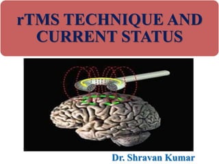
- 1. rTMS TECHNIQUE AND CURRENT STATUS Dr. Shravan Kumar
- 2. INTRODUCTION • Following the discovery of the unified and interchangeable nature of electric and magnetic forces, it became evident that electric currents generate magnetic fields while changing magnetic fields generate electric currents. • Galvani’s and Volta’s classic experiments had established that electric currents were capable of stimulating neuronal tissues. • The combination of these two concepts – the unity of electric and magnetic forces, and the responsiveness of neurons to electrical stimulation – is now being harnessed to study and manipulate the CNS from neuropsychiatric perspectives.
- 3. • TMS uses an externally generated changing magnetic field to induce electric current intracranially unlike ECT where an electric current that is generated externally and transmitted to the brain through the skull. • When electricity is forced to pass through the skull, the current used must be relatively large as the skull is a powerful insulator with an electrical resistance 8 to 15 times greater than that of soft tissues. • Externally generated electric currents cannot be focally directed as the skull dissipates the electricity globally leading to massive depolarization of cortical and subcortical structures.
- 4. • Such difficulties are minimized upon exposure of the skull to TMS, where the changing external magnetic field undergoes minimal attenuation in the skull tissues while inducing smaller, focally directed electric currents within the brain. • Barker et. al. first described in 1985 the use of a pulsed (i.e. changing) magnetic field focused over specific regions of the cerebral cortex to induce muscle action potentials. • The use of pulsed magnetic fields to induce electrical activity in peripheral nerves had been described much earlier, in the 1960’s.
- 5. • The higher frequency technique has been found to be particularly effective in neuropsychiatry and has been termed Repititive Transcranial Magnetic Stimulation (rTMS). • rTMS is believed to be unique in that rapid pulsation can induce electrical currents within neurons while they are in the refractory period, although how this relates to an altered clinical manifestation is unclear. • The magnetic field, passing largely unimpeded through the skull, induces a current within the brain tissue. • Depending on the region of the brain over which the coil is physically placed, specific circumscribed areas can be stimulated.
- 6. • TMS has been used for multiple purposes in the field of neurology including establishing hemispheric language dominance, localization of epileptogenic foci, and the study of motor pathways originating at the cerebral cortex and relevant motor pathway physiology. • It has also been utilized in neuropsychiatry in the mapping of attention, memory, movement, speech, and vision.
- 7. • Clinically, TMS has been combined with electromyographic studies (EMG) to determine impairment in CNS conduction pathways by measuring differences in the rates of muscle activation upon electromagnetic stimulation at the cerebral cortex in comparison with similar stimulation at spinal nerve roots. • This assessment is useful in the diagnosis and prognostication of demyelinating diseases such as multiple sclerosis. • More recent efforts have focused on the use of TMS in the symptomatic improvement of Parkinson’s disease.
- 8. • Being a relatively new technique, optimization of parameters such as frequency of pulsing of the magnetic field, size of the coil utilized, strength of the magnetic field generated, and duration of induction of electrical current has yet to be established. • It is likely that such parameters will vary substantially depending on the specific neurological or psychiatric applications.
- 9. PRINCIPLE OF TMS • TMS works on the Faraday’s law which states that “The induced EMF in any closed circuit is equal to the rate of change of the magnetic flux through the circuit.” TMS Magnetic field Induced neuronal current
- 10. TMS is a non-invasive method of brain stimulation in which magnetic fields are used to induce electric currents in the cerebral cortex, thereby depolarizing neurons. Uses a rapidly changing magnetic field to induce weak electric currents, through pulses. Induced by electromagnetic induction Generally reach no more than 5cm into the brain
- 11. • The stimulators and coils currently in production develop about 1.5–2 T at the face of the coil and are thought to be able to activate cortical neurons at a depth of 1.5–2 cm beneath the scalp (Epstein et al., 1990). • Even though TMS with conventional equipment appears to penetrate no deeper than the cortex, it may affect cells trans- synaptically at some distance from the site of stimulation, as evidenced by its effect on distant cortical and subcortical sites detected with PET (Kimbrell et al., 1997; Paus et al., 1997; Wassermann et al., 1997).
- 12. • Single-pulse magnetic stimulators repeat pulses no faster than once every few seconds. • Faster cycling is prevented by the charging time of the capacitors, which store the electrical charge that generates the current in the coil. • In the last 10 years, stimulators using multiple capacitors and capable of generating pulses at up to 60 Hz have been introduced. • High-frequency stimulation can also be produced by multiple stimulators that discharge through a single coil and are triggered in sequence by a microprocessor.
- 13. • The number of pulses that these systems can deliver in a single train may be limited by the number of stimulators, but very high pulse frequencies are possible. • Heating of the coil is a problem that potentially endangers subjects as well as equipment and limits the duration of stimulation at high intensities and frequencies.
- 14. • The underlying rationale for the use of TMS exploits the fact that neurons are electrochemical cells. • This means that neuronal activity can be affected either chemically, via the use of drugs, or electrically, via interventions like TMS.
- 15. Neuron TMS Directly Depolarizes Cortical Neurons Pulsed magnetic fields from TMS: • induce a local electric current in the cortex which depolarizes neurons • eliciting action potentials • causing the release of chemical neurotransmitters Neurons are “electrochemical cells” and respond to either electrical or chemical stimulation
- 16. TMS Releases Neurotransmitters in the Brain Depolarization of neurons in the DLPFC causes local neurotransmitter release Depolarization of pyramidal neurons in the DLPFC also causes neurotransmitter release in deeper brain neurons Activation of deeper brain neurons then exerts secondary effects on remaining portions of targeted mood circuits Dorsolateral prefrontal cortex Anterior cingulate cortex Kito (2008) J Neuropsychiatry Clin Neurosci These effects are associated with improvements in depressive symptoms
- 17. PHYSIOLOGICAL EFFECTS OF TMS OVER BRAIN – Neuronal excitation activity (facilitation) – Neuronal inhibition activity (silent period) – Stimulation of one site but get inhibition at another site (interhemispheric inhibition) – long-term post-stimulation effect (long-term penetration)
- 18. FACILITATION
- 21. NEUROBIOLOGICAL EFFETS OF TMS • TMS targets modulation in neuroplasticity to influence behavior consequences. NEUROPLASTICITY: • The capacity of the brain to change with learning is known as plasticity. • This is the brain’s ability to reorganize itself by forming new neural connections throughout life. • It allows the neurons in the brain to compensate for the injury and disease, and to adjust their activities in response to new situations or to changes in their environment.
- 22. • Brain reorganization takes place by mechanisms such as “axonal sprouting” in which undamaged axons grow new nerve endings to reconnect neurons whose links were injured or severed. • Undamaged axons can also sprout nerve endings and connect with other undamaged nerve cells, forming new neural pathways to accomplish a needed function. • In order to reconnect, the neurons need to be stimulated through activity.
- 23. • Neuroplasticity allows brain to compensate for irreparably damaged or dysfunctional neural pathways by strengthening or rerouting of remaining ones. COMPENSATORY MASQUERADE •Allows already constructed pathways that neighbor a damaged area to respond to changes in the body’s demands caused by lost function in some other area.
- 24. HOMOLOGOUS REGION ADOPTION •allows one entire brain area to take over functions from distant brain areas that has been damaged. CROSS MODEL REASSIGNMENT •allows the brain of a blind individual in learning to read Braille, to rewire the sense of touch so it replaces the responsibilities of vision in the brain areas linked with reading.
- 25. TMS DEVICE
- 27. TYPES OF COILS
- 28. Electric field intensity induced below the coils of different types
- 29. GENERAL RULES FOR SELECTING THE APPROPRIATE COIL FOR CERTAIN APPLICATION – Small coils have stronger field on the surface but field decreases dramatically with distance. Therefore small coils are suitable for peripheral stimulation in which magnetic field does not need to pass through skull bones.
- 30. – Big coils have lower decrease of field with distance. Therefore they are used for transcranial stimulation. – Ring coils are less focused. Therefore they are used for stimulation of wide areas (for diagnostics). – Figure-of-eight coils are more focused, therefore they are widely used for treatment.
- 31. STIMULATION POINT AND COIL ORIENTATION IN DEPRESSION TREATMENT
- 32. TYPES OF TMS • Single-Pulse TMS – Delivers one stimulus at a time • Paired-Pulse TMS – Pairs of stimuli separated by a variable interval • Repetitive TMS (rTMS) – Delivered in trains (can be of low or high frequency)
- 33. LOW FREQUENCY VS. HIGH FREQUENCY TMS Concept of Depolarization Function • Low Frequency Stimulation--inhibitory, more focal effect • High Frequency Stimulation--facilitatory, multiple spread out, global “dendritic/axonal effect”. When higher frequency rTMS is applied, a longer lasting effect can be induced which is thought to result from a long term potentiation (LTP), or depression (LTD) at the neuronal level. 33
- 34. APPLICATIONS OF TMS • Diagnostic • Therapeutic
- 35. DIAGNOSTIC • Used clinically to: – Measure activity of certain brain circuits – Survey the damage done to particular muscles following stroke, multiple sclerosis, motor neuron disease, and other injuries or disorders – Locate tumors and other lesions to generate preoperative motor maps
- 36. • Main Techniques: – Amplitude of motor evoked potential (MEP) – Amplitude ratio (motor evoked potential/M-wave) – Motor evoked potential facilitation – Central motor conduction time – Triple stimulation test
- 38. Diagnostically, the only FDA approval is the Navigated Brain Stimulation System for pre-surgical planning in patients undergoing brain surgery. Proven equally successful as traditional imaging methods in producing preoperative motor maps. When compared to direct cortical stimulation (DCS), it was found that TMS was able to recognize every motor site mapped by DCS
- 39. THERAPEUTIC TMS USE • Amblyopia • Amyotrophic lateral sclerosis • Auditory hallucinations associated with schizoaffective disorders • Chronic pain • Dysphasia • Dystonia • Epilepsy • Fibromyalgia
- 40. • Hemispatial neglect • Major depression • Migraine • Obsessive-compulsive disorder • Parkinson’s disease • Phantom limb • Stroke • Nonfluent aphasia • Tinnitus
- 41. • FDA has currently only approved rTMS as a treatment for Major Depressive Disorder (MDD).
- 42. RECENT TMS LITERATURE REVIEW • Roughly 30 controlled clinical research studies to date • Most recent meta-analysis (Slotema, et al, 2010): – Included analysis of 34 studies involving 1,383 patients – Estimated standardized effect size = 0.55 (P < 0.001) Conclusion: “…rTMS deserves a place in the standard toolbox of psychiatric treatment methods, as it is effective for depression…and has a mild side effect profile….” 1.Slotema, CW, Blom, JD, Hoek, HW, Sommer, IEC. (2010) Should we expand the toolbox of psychiatric treatment methods to include repetitive transcranial magnetic stimulation (rTMS)J Clin Psych 71(7):873-84. 1.Schutter, DJLG. (2009) Antidepressant Efficacy of High-Frequency Transcranial Magnetic Stimulation Over the Left Dorsolateral Prefrontal Cortex in Double-Blind Sham-Controlled Designs: A Meta-Analysis. Psychol Medicine, 39:65-75.
- 43. • Independent, Peer-reviewed • 15 TMS clinical trials involving nearly 500 patients – Average HAM-D decrease in depressive symptoms >5 points vs. Sham control • Meets clinical significance threshold of 3 points on the HAM-D scale – Response rate with active TMS was >3x higher than sham treatment – Remission rate with active TMS was >6x higher than sham treatment • “High strength of evidence” for efficacy from well-controlled RCTs Independent U.S. Agency for Healthcare Research And Quality (AHRQ) confirms evidence for efficacy of TMS Agency for Healthcare Research and Quality: Comparative Effectiveness Report on Non-Pharmacologic Treatments for Depression , October 2011
- 44. • Ebmeier and colleagues - conducted a 5-day study with 15 patients that called for them to be treated twice per day. At the end of the study, the participants saw a reduction of 44% on Hamilton-D scales. • Another trial testing 100 patients, however, saw most to be treatment-resistant. • Current trials include using TMS with: autism, MDD, improving speech aphasia
- 45. • In an open-label trial (most like real world clinical practice), – 1 in 2 had a significant improvement in symptoms – 1 in 3 had complete symptom resolution • Patients also experienced significant improvement in anxiety, appetite changes, aches and pains, and lack of energy associated with depression • Over 10,000 procedures performed in clinical trials • No systemic side effects such as weight gain, sexual dysfunction, nausea, dry mouth, and sedation
- 46. PROTOCOLS FOR DEPRESSION TREATMENT •The majority of trials apply Neuro-MS to treat depression using these protocols: Over the left frontal dorsolateral cortex (5 cm anterior (parallel to the sagittal line) to the area where the motor threshold is obtained) 5 Hz, 25 trains of 10 seconds each with 20-second pause between the trains. Applied at 120% of the motor threshold (protocol used by Dr. Marcolin). OR
- 47. •10 Hz, 30 trains of 4 seconds each with 20-second pause between the trains. •Applied at 100% of the motor threshold (or 120%, when possible limited by the power of the device) (protocol used by Dr. Moacyr). •The quantity of stimuli for effective treatment is considered as more than 1000 pulses •Number of sessions: 15-30 (3-6 weeks)
- 48. LONG TERM FOLLOW UPAFTER ACUTE TREATMENT Janicak, et al. Brain Stimulation, 2010. • Safety confirmed during long term, open-label 6 month follow up period • During open-label follow up on antidepressant medication monotherapy, – ~37% of patients required TMS reintroduction – ~85% of patients who received TMS reintroduction benefited • Net incidence of illness relapse under these open-label follow up conditions: 11% – Six-month relapse with antidepressant treatment alone in STAR*D study was 35- 50% (Level 2 and 3 range)
- 49. TMS OPEN-LABEL DURABILITY OF EFFECT STUDY Outcome TMS (in remission) (N=56) Outcome ECT - Combination Pharmacotherapy 1 (N=95) ECT - Continuation ECT 1 (N=89) % Early Discontinuation 16.1% % Early Discontinuation 22.1% 16.8% % Disease Recurrence 10.7% % Disease Recurrence 31.6% 37.1% % In Remission by Study Completion 73.2% % In Remission by Study Completion 46.3% 46.1% 1 Kellner, CH, Knapp, RG, Petrides, G, et al. Continuation Electroconvulsive Therapy vs Pharmacotherapy for Relapse Prevention in Major Depression: A Multisite Study From the Consortium for Research in Electroconvulsive Therapy (CORE). Arch Gen Psychiatry 2006, 63:1337-1344. Janicak, et al., Brain Stimulation (2010)
- 50. • In research settings, two large, multisite, randomized controlled trials demonstrated clinically significant antidepressant effect of TMS. • Prospective, naturalistic study confirms these results in real- world practice settings. • Overall, 1 in 2 patients respond and 1 in 3 patients achieve remission. SUMMARY OF CLINICAL OUTCOMES 50
- 51. • Meta-analyses from multiple studies shows TMS effect size of >0.5 • High level of treatment adherence , >80% of patients completed acute treatment in both research setting and in clinical practice. • Appears to be at least as effective as ECT for treatment and relapse prevention
- 52. NEW APA GUIDELINES • Transcranial magnetic stimulation is now listed in the American Psychiatric Association’s 2010 “Practice Guideline for the Treatment of Patients with Major Depressive Disorder” • It is listed as an acute phase treatment option for patients who do not respond adequately to pharmacotherapy. .
- 53. • This recent guideline states; “Acute phase treatment may include pharmacotherapy, depression-focused psychotherapy, the combination of medications and psychotherapy, or other somatic therapies such as electroconvulsive therapy (ECT), transcranial magnetic stimulation (TMS), or light therapy.” (APA Guidelines 2010; Pg 46).
- 54. BEST PRACTICES TREATMENT GUIDELINE FOR DEPRESSION Based on 2010 APA guidelines and NeuroStar TMS Therapy® indication for use. Adapted from: Practice Guideline for the Treatment of Patients with Major Depressive Disorder, 3rd Edition, APA (2010) Unmet Medical Needs
- 55. TMS THERAPY: SAFETY OVERVIEW • No systemic side effects • No adverse effect on cognition • Most common adverse event associated with treatment was scalp pain or discomfort – < 5% of patients discontinued due to adverse events • No seizures reported during clinical studies (over 10,000 treatments) • Rare risk of seizure with NeuroStar TMS in post-market use (0.003% per treatment, <0.1% per acute treatment course) (>150,000 treatments in post-marketing experience to date) • Long term safety demonstrated in 6 months follow-up
- 56. OTHER DISORDERS UNDER EVALUATION FOR TMS THERAPY • I. PSYCHIATRY: Depression (FDA approval,Oct 2008), treatment refractory cases, co-morbid, Panic Disorder, OCD, PTSD, pathologic gambling, substances use like cocaine, opiates, nicotine, schizoaffective disorder. • II. Chronic neuropathic pains, phantom pain,fibromyalgia,Migraine,headaches, Tourette's, tinnitus, painful dystonia. • III. NEUROLOGY: Rehabilitation- after stroke , recovery- Aphasia, Neglect, Brain Injury, Seizures
- 57. CURRENT RESEARCH
- 58. rTMS - CURRENT FINDINGS • rTMS upregulates BDNF in experimentally damaged area of brain in mouse model – Makowiecki K et al. J Neurosci. 2014 Aug6;34(32):10780-92 • 10Hz rTMS - improvement of freezing of gait in Parkinsonism with stimulation over M1-LL and DLPFC in single session. – Lee SY et al. Restor Neurol Neurosci. 2014 Jul 30. • Use in treatment-resistant schizophrenia – Miyajmoto S. et al. J Psychiatr Res. 2014 Jul 8.
- 59. TMS IN THE TREATMENT OF ADDITION Gorelick DA et al. Ann NY Acad Sci. 2014 Jul 28 • 19 human studies reviewed, 316 adults – Tobacco (9 studies), alcohol (6), cocaine (3), and methamphetamine (1) – Only FIVE studies were controlled tirals. • 2 out of 45 nicotine trials = decreased smoking • Cocaine trial = decreased use. • Actions – “may involved increased dopamine and glutamate function in corticomesolimbic brain circuits and modulation of neural activity in brain circuits that mediate cognitive processes relevant to addiction.” • Considered experimental at present
- 60. A DOUBLE-BLIND, RANDOMIZED TRIAL OF DEEP rTMS FOR AUTISM SPECTRUM DISORDER. Enticott PG et al. Brain Stimul. 2014 Mar-Apr;7(2):206-11 • 28 adults with high-functioning Autism or Asperger’s – Double-blind, randomized, placebo controlled design – 2 weeks of daily weekday treatment – rTMS to bilateral dorsomedial PFC • “Deep rTMS to bilateral dorsomedial PFC yielded a reduction in social relating impairment and socially-related anxiety.”
- 61. IMPROVEMENT IN ALZHEIMER’S Cotelli M et al. J Neurol Neurosurg Psychiatry. 2011 Jul;82(7):794-7 • 10 AD patients randomized – 4 weeks of rTMS vs. – 2 weeks of placebo followed by 2 weeks of real rTMS • Protocol – 25 minute rTMS, DLPFC, weekdays. • Significant difference was found between groups in terms of % of correct responses of auditory sentence comprehension. • rTMS may represent a novel approach to the treatment of language dysfunction in AD patients.”
- 62. Ahmed MA et al. J Neurol. 2012 Jan;259(1):83-92. • Study of high vs. low frequency rTMS applied bilaterally over DLPFC on cognitive function and cortical excitability of AD patients. • 45 patients studied. 3 groups: – Sham – High frequency – Low frequency • “…five sessions of high frequency rTMS over the left and then the right DLPFC improves cognitive function in patients with mild to moderate degree of AD. • This improvement was maintained for three months.”
- 63. POSITIVE EFFECTS OF rTMS ON ATTENTION IN ADHD Bloch Y et al. World J Biol Psychiatry. 2010 Aug;11(5)755-8 • Known that rTMS affects dopaminergic secretion in PFC. • Double blind crossover randomized, sham controlled study of 13 patients (7 male, 6 female) – who fulfilled DSM-IV criteria. • “There was a specific beneficial effect on attention 10 minutes after a real rTMS course.”
- 64. TMS AS A RESEARCH TOOL • A research tool to study aspects of the human brain physiology including motor function,vision,language and the pathophysiology of brain disorders. • TMS can excite or inhibit the brain allowing functional mapping of cortical regions and creation of transient functional lesions. • For eg. rTMS over the occipital lobe impaired detection of visual stimuli rTMS delivered to discrete areas in the language-dominant hemisphere can disrupt speech.
- 65. THERAPEUTIC APPLICATION IN NEUROLOGICAL DISORDERS
- 66. MOVEMENT DISORDERS • Therapeutic applications of TMS in movement disorders are preliminary. • Fast rTMS of the motor cortex has been reported to improve performance on several motor measures in Parkinson disease. • A recent meta-analysis included 12 studies and concludes that the overall literature does show a positive effect of rTMS on Parkinson motor function. • Slow rTMS has been reported to improve dystonia.
- 67. NEURO-REHABILITATION • TMS to evaluate the functional properties of the motor cortex after lesions like stroke is of special interest in the field of neuro rehabilitation. • Brain stimulation have been proposed to enhance motor function when combined with conventional neuro rehabilitative interventions after stroke
- 68. CHRONIC PAIN • rTMS of the cortex induces analgesic effects in focal chronic pain syndromes (causalgia)
- 69. ADVERSE EFFECTS OF rTMS • Risk of inducing seizures (in patients with a past or family history of seizures).current safety protocols adjust the amount of stimulation in relation to the motor threshold of the individual. • Muscle tension headache . • Short term changes in hearing threshold related to the noise generated. • Cognitive changes only during stimulation
- 70. ECT vs. TMS ECT TMS Anesthesia, LOC Yes No Induction of seizure Yes No Systemic effects Anesthetic drugs, increase HR none Treatment schedule 3X/ week (8 -15 tx) Daily, M-F, six weeks (30 tx) Rapidity of onset 2 – 3 treatments 2 – 3 weeks Mechanism of action SEIZURE. Massive NT release; rise in sz threshold Reactivation of neural circuits. Precise, LOCAL release of NT’s. Side effects Memory loss, confusion Essentially none (mild HA 1st week) Psychosocial impact can’t work Drive to and from tx’s, work improved After-effects Mild (usually transient) memory loss None. Pro-cognitive Insurance coverage Almost always Rare. Improving
- 71. CONTRAINDICATIONS TO TMS • Metallic (Ferro or Paramagnetic) hardware near the coil can be moved or heated by TMS. Therefore, the presence of metal anywhere in the head, excluding the mouth, is generally a contraindication to TMS. • Individuals with cardiac pacemakers and implanted medication pumps should not participate in rTMS studies without a clear potential benefit (e.g., treatment of severe and refractory depression).
- 72. • Children should not be used as subjects for rTMS without compelling clinical reasons, such as the treatment of refractory epilepsy or depression. • Women of childbearing age must be questioned about the possibility of pregnancy before participating in rTMS studies, and excluded if there is a chance that they may be pregnant. • Tricyclic anti-depressants, neuroleptic agents, and other drugs that lower the seizure threshold are contraindications to rTMS, except in extraordinary circumstances where the potential benefit outweighs the increased risk of a seizure.
- 73. CONCLUSION • TMS is a technique used to induce electrical current in neurons by strong magnetic field. • It is different from ECT where electrical current is applied directly over the scalp and brain is stimulated. • The induced current brings multiple immediate physiological changes in neurons’ electrical activities and in long term changes overall functioning of brain by various neurobiological modulations in neuronal circuits.
- 74. • There are many diagnostic and therapeutic application of TMS under trial in Neuropsychiatry. • Currently this is approved only as second line treatment of MDD and for resistant depression by FDA. • Numerous meta analyses are present to support use of rTMS in MDD therapy. • In some studies TMS came out with comparable efficacy as fMRI in tumor localisation and exploring focal deficits. • We can expect many other applications to get approved in near future.
- 75. REFERENCES • Agnew, W.F. and McCreery, D.B. Considerations for safety in the use of extracranial stimulation for motor evoked potentials. Neurosurgery, 1987, 20: 143–147. • Ajmone-Marsan, C. Focal electrical stimulation. In: D.P. Purpura, J.K. Penry, D.B. Tower, D.M. Woodbury and R.D. Walter (Eds.), Experimental Models of Epilepsy. Raven Press, New York, 1972, pp. 147– 172. • Amassian, V.E., Maccabee, P.J., Cracco, R.Q. and Cracco, J.B. Basic mechanisms of magnetic coil excitation of the nervous system in humans and monkeys: application of focal stimulation of different cortical areas in humans. In: S. Chokroverty (Ed.), Magnetic Stimulation in • Clinical Neurophysiology. Butterworth, Stoneham, MA, 1989, pp. 73– 111. Amassian, V.E., Maccabee, P.J., Cracco, R.Q., Cracco, J.B., Rudell, A.P. and Eberle, L. Measurement of information processing delays in human visual cortex with repetitive magnetic coil stimulation. Brain Res., 1993, 605: 317–321.
- 76. • Amassian, V.E., Henry, K., Durkin, H., Chice, S., Cracco, J.B., Somasundaram, M., Hassan, N., Cracco, R.Q., Maccabee, P.J. and Eberle, L. • Human immune functions are differentially affected by left-sided versus right-sided magnetic stimulation of temporo-parieto-occipital cortex. (abstract) Neurology, 1994, 44(Suppl. 2): A133. • Artola, A., Brocher, S. and Singer, W. Different voltage dependent thresholds for inducing long-term depression and long-term potentiation in slices of rat visual cortex. Nature, 1990, 347: 69–72. • Blaxton, T.A., Wassermann, E.M., Hoffman, E.A., Oletsky, H.S., Hallett, M. and Theodore, W.H. Functional mapping of implicit and explicit memory using repetitive transcranial magnetic stimulation (rTMS). (abstract) Soc. Neurosci. Abstr., 1996, 22: 719. • Cain, D.P. and Corcoran, M.E. Kindling with low-frequency stimulation: generality, transfer, and recruiting effects. Exp. Neurol., 1981, 73: 219– 232. • Chen, R., Classen, J., Gerloff, C., Celnik, P., Wassermann, E.M., Hallett, • M. and Cohen, L.G. Depression of motor cortex excitability by low frequency transcranial magnetic stimulation. Neurology, 1997, 48: 1398–1403. • Christie, B.R., Kerr, D.S. and Abraham, W.C. Flip side of synaptic plasticity: long-term depression mechanisms in the hippocampus. Hippocampus,1994, 4: 127–135. • Classen, J., Witte, O.W., Schlaug, G., Seitz, R.J., Holthausen, H. And Benecke, R. Epileptic seizures triggered directly by focal transcranial magnetic stimulation. Electroenceph. clin. Neurophysiol., 1995, 94: 19– 25
- 77. • Counter, S.A., Borg, E., Lofqvist, L. and Brismar, T. Hearing loss from the acoustic artifact of the coil used in extracranial magnetic stimulation. Neurology, 1990, 40: 1159–1162. • Devinsky, O. and Duchowny, M.S. Seizures after convulsive therapy: a retrospective case survey. Neurology, 1983, 33: 921–925. • Dhabhar, F.S., Miller, A.H., Stein, M., McEwen, B.S. and Spencer, R.L. Diurnal and acute stress-induced changes in distribution of peripheral blood leukocyte subpopulations. Brain Behav. Immun., 1994, 8: 66– 79. • Dhabhar, F.S., Miller, A.H., McEwen, B.S. and Spencer, R.L. Effects of stress on immune cell distribution. Dynamics and hormonal mechanisms. J. Immunol., 1995, 154: 5511–5527. • Dhuna, A.K., Gates, J.R. and Pascual-Leone, A. Transcranial magnetic stimulation in patients with epilepsy. Neurology, 1991, 41: 1067– 1071. • Edgley, S.A., Eyre, J.A., Lemon, R.N. and Miller, S. Excitation of the corticospinal tract by electromagnetic and electrical stimulation of the scalp in the macaque monkey. J. Physiol. (Lond.), 1990, 425: 301–320. • Engel, J. Seizures and Epilepsy, F.A. Davis, Philadelphia, 1989. • Epstein, C.M., Schwartzenberg, D.G., Davey, K.R. and Sudderth, D.B. Localizing the site of magnetic brain stimulation in humans. Neurology, 1990, 40: 666–670. • Epstein, C.M., Lah, J.K., Meador, K., Weissman, J.D., Gaitan, L.E. and Dihenia, B. Optimum stimulus parameters for lateralized suppression of speech with magnetic brain stimulation. Neurology, 1996, 47: 1590– 1593..
- 78. THANK YOU