
Recomendados
Más contenido relacionado
Similar a Medical Mycology.pptx
Similar a Medical Mycology.pptx (20)
Más de elphaswalela
Más de elphaswalela (20)
Último
Último (20)
Medical Mycology.pptx
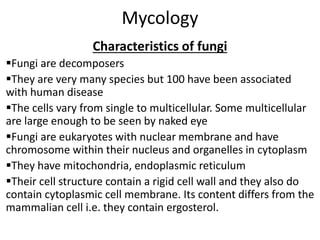
- 1. Mycology Characteristics of fungi Fungi are decomposers They are very many species but 100 have been associated with human disease The cells vary from single to multicellular. Some multicellular are large enough to be seen by naked eye Fungi are eukaryotes with nuclear membrane and have chromosome within their nucleus and organelles in cytoplasm They have mitochondria, endoplasmic reticulum Their cell structure contain a rigid cell wall and they also do contain cytoplasmic cell membrane. Its content differs from the mammalian cell i.e. they contain ergosterol.
- 2. Cont… The differs from that of mammal in that they contain chitin, mannan and glucan. Chitins are also within the cell wall and are inert compound that uncoats the rigidity of the fungal cell wall. The fungal cells grow only in organic compound, they only use carbcell wall of fungi on as the source of energy and mostly found in decaying organic matter
- 3. • The classification of fungi is based on the – Mode of reproduction – Micromorphology • Mode of reproduction – Perfect fungi: these reproduce both sexually and asexually. They reproduce sexually by means of characteristic sexual spores, which are used to classify fungi into four broad classes, thus: Ascomycetes, Basidiomycetes, Zygomycetes, Oomycetes – Imperfect fungi: also called Deuteromycetes – these reproduce only asexually • Micromorphology – Yeasts – Molds – Dimorphic Classification of fungi
- 4. Most of the human fungi fall in the Deuteromycete group. • Fungi can also be classified according to the tissue that they infect. This is done using fungal diseases that they cause. They are: – Superficial mycocete: they cause lession on the skin and appendages. And they never invade the living tissues. The organisms of these organism are dermatophytes. They have preference for keratin because they produce an enzyme that break down keratin. The enzyme is keratinase. There are three (3) genera: i. microspora, ii. Epidemophyton, iii. Trichophyton. They have septic hyphae and microconidia. – Subcutaneous fungi: they cause diseases in the inner surface to the skin. They can spread into the lymphatics. They are introduced through the skin. Examples are: Sporotherix schenkeii which causes sperotrichosis, Madunella mycetoma. Classification of fungi
- 5. • Systemic fungi: they affect the internal organs i.e. lungs, kidney e.t.c. they cause some of the most severe diseases in human. Some of them are sub clinical in that there are no clinical manifestation as such but those that cause clinical characteristics are usually severe. Most are dimorphic in nature and changes into yeast form in the body. Examples are: Cryptococcus neoformans (pure yeast), Histoplasma capsulatum (dimorphic), Blastomyces dermatitidis (dimorphic), Coccidioides and Paracoccidioides (dimorphic) • Opportunistic fungi: are occasional pathogens that attack a patient depending on the prevailing conditions of the patient. Example is Candida. They are termed as normal flora, they do not cause disease unless they are allowed to overgrow causing thrush. They form pseudohyphae in order to penetrate the skin. Another one is the Aspergillus, it is purely a mold and it does not change into other form. It causes the respiratory tract infection (chest problem) and they are toxin producers which affect the lung.
- 6. Classification of fungi • Divided into 4 main classes: • Phycomycetes: addition subdivide into the first six classes. The six classes lack regular septa in their hyphal filaments (coenocytic hyphae), resulting in the presence of many nuclei in each cell filament. Members of these six classes include organisms that are infrequent causes of human disease even though several genera in the class Zymomycetes do cause occasional serious infection in debilitated or immunologically compromised person.
- 7. • Ascomycetes: or sac fungi, form one or more sexual spores within a saclike cell called ascus. The asexual spores produced by the Ascomycetes are frequently single-celled microconidia. Microconidia may be produced in long chains extending from an aerial hypha called conidiophore. Produce regular septa which divide the mycelium into large number of individual cells. Each septum, however, has ‘a hole’ which permits the free flow of cytoplasmic and nuclear material between the cells. They grow only as single – celled yeast, and by far the best known yeast in this class are those of the genus Saccharomyces upon which both the baking and the alcoholic beverage industries are totally dependent.
- 8. • Basidiomycetes: form their sexual basidospores externally on club shaped cells called basidia. Asexual reproduction may also occur by budding, microconidia, or fragmentation of the hyphal filament. The hyphae are usually aseptate. Causes few human disease but many plant diseases. In this group is musshroom • Deuteromycetes: (fungi imperfecti) comprise of a large group of fungi for which no sexual states has been demonstrated. Some members produce sexual spores which mixed with the correct mating type and, as a result , have acquired 2 names; one for sexual classification and the other for older asexual name. many of human pathogens belong to this group. • Disease caused by deuteromycetes include superficial infection, cutaneous infection and the subcutaneous infection and deep seated systemic infection.
- 9. Mycoses • Establishment of a mycotic infection depend on the inoculums of the fungi and the resistance of the host. • Severity of infection depend on immunological factors of the host • Some of the reasons why mycoses/fungal diseases are prevalent are: – Immune states of the patient because of HIV: immunosuppressive drug – The transplantation therapy – Invasive procedures that are used today of tissue i.e. operations
- 10. Diagnosis • Wet mount: where this refers to wet preparations i.e. a drop a liquid on a slide and cover it with a cover slip. It is a common procedure for diagnosing dermatophytes. i.e. Skin scrapping, mashilingi, hair cuttings. • Prepare the wet mount and observe spores and conidia morphology. In this, common light microscope is used. Staining using electrophenol blue stain or use potassium hydroxide in skin scrapping. • Serological methods (Serology): here systemic infections are detected. Some of the test are latex agglutination and immunodiffusion. • Direct flourescent microscopy: here you stain the specimen with flourescent dyes before observing under the fluorescent microscope. Stain biopsy of specimens or section of body tissues
- 11. • The most definitive method is the culture the organism in a cultural media. The most commonly used media for fungal culture cells is the sabaroud dextrose agar (SDA). The incubation temperature depends on the organism form whether it is a yeast form or a mold form. For the yeast form, culture at 37 degrees centigrade, while for mold you culture at a lower temperature at 25 degrees centigrade. • After fungal isolation, look at the colonial morphology in terms of color, texture, size and shape. After observation, you may want to view it on the microscope without staining or stain usual electrophenol blue. NEVER USE GRAM STAINING. •
- 12. Zarqa Private UniversityBiology 4223 – The Fungi Fungal Diseases • Mycosis- fungal infection – < 100 cause human disease – Not highly contagious – Humans acquire from nature • Groups based on degree on tissue involvement and mode of entry
- 13. Zarqa Private UniversityBiology 4223 – The Fungi Introduction Fungal Infections Superficial infections: involve outermost layers of skin and its appendages [ nails or hair] ( Dermatophytosis) • Cutaneous infections: involve deeper layers of skin causing allergic or inflammatory response • Subcutaneous infections: fungi with low virulence, localized infection, or spread by mycelial growth Systemic infections: caused by true pathogenic fungi or opportunistic saprobes Opportunistic infections
- 14. Zarqa Private UniversityBiology 4223 – The Fungi Mycoses: diseases cause by fungi • Superficial Cutaneous • Subcutaneous Systemic • Opportunistic
- 15. Zarqa Private UniversityBiology 4223 – The Fungi FUNGAL DISEASES (Continued) I. Cutaneous mycoses: Fungal infections of the skin, hair, and nails. Secrete keratinase, an enzyme that degrades keratin. Infection is transmitted by direct contact or contact with infected hair (hair salon) or cells (nail files, shower floors). Examples: – Ringworm (Tinea capitis and T. corporis) – Athlete’s foot (Tinea pedis) – Jock itch (Tinea cruris)
- 16. Zarqa Private UniversityBiology 4223 – The Fungi Cutaneous Infections Dermatophytic hyphomycetes • 40 species • Epidermophyton (2 species) • Microsporum (17 species) • Trichophyton (24 species) • 50% of dermatophytes human specific
- 17. Zarqa Private UniversityBiology 4223 – The Fungi Cutaneous Mycosis Ringworm skin infection: Tinea corporis
- 18. Zarqa Private UniversityBiology 4223 – The Fungi Cutaneous Mycosis Candida albicans infection of the nails.
- 19. Zarqa Private UniversityBiology 4223 – The Fungi Cutaneous Infections – Cause common tinea (ringworm) – Grow only on humans – Reservoir not in soil or animals – Reservoir in carpets and upholstery for up to two years
- 20. Zarqa Private UniversityBiology 4223 – The Fungi Cutaneous Infections • Trichophyton rubrum • Chronic infections of the toe nails Tinea corporis
- 21. Zarqa Private UniversityBiology 4223 – The Fungi Cutaneous Infections • Microsporum canis – Reservoir in cat – May move to humans or dogs – Dies out after one or two person-person transfers
- 22. Zarqa Private UniversityBiology 4223 – The Fungi Cutaneous Infections • Disease process – Fungus stimulates epithelial cells of skin to divide more frequently – Makes more keratin available to fungus – Some species race specific in humans – Some species body location specific
- 23. Zarqa Private UniversityBiology 4223 – The Fungi FUNGAL DISEASES (Continued) II. Subcutaneous mycoses: Fungal infections beneath the skin. Caused by saprophytic fungi that live in soil or on vegetation. Infection occurs by implantation of spores or mycelial fragments into a skin wound. Can spread to lymph vessels. Superficial mycoses: Infections of hair shafts and superficial epidermal cells. Prevalent in tropical climates.
- 24. Zarqa Private UniversityBiology 4223 – The Fungi Subcutaneous mycoses Subcutaneous infections - over 35 species produce chronic inflammatory disease of subcutaneous tissues and lymphatics. e.g. sporotrichosis - ulcerated lesions at site of inoculation followed by multiple nodules - caused by a dimorphic fungus: Sporotrix schenckii.
- 25. Sporotrichosis • Agent is Sporotherix schenkii • It is a chronic infection of both the cuteneous and subcutenous tissue. They tend to form ulcers that kind of drain their fluids. Occasional mycetoma my be caused. • It is transmitted through pricks i.e. thorns • The most common parts of the body infected are the feet (primary source of infection). From the primary source of infection, the disease may pass on to the lymphatic system. • Diagnosis: clinical specimens are pass, biopsy material or sputum can be used incase pulmonary tract is infected. Staining of biopsy material may be done in addition to culture. No serological. NB// Fungi are not serologically Dx because they are pure antigens
- 26. Zarqa Private UniversityBiology 4223 – The Fungi Subcutaneous Infections Chromoblastosis • Common among barefoot peoples of the tropics • Soil hyphomycete species • Enters human by thorns or wood slivers – Fungus grows host cells respond by rapid cell division wart-like growths on feet or legs
- 27. Zarqa Private UniversityBiology 4223 – The Fungi Subcutaneous Infections Mycotic Mycetoma • Disease of barefoot tropical people • Entry: wound on foot • Attacks various tissues – Stimulates formation of tumor – Compact fungal colonies form within tumor
- 28. Zarqa Private UniversityBiology 4223 – The Fungi Subcutaneous Infections – Skin ruptures and some colonies extrude
- 29. Zarqa Private UniversityBiology 4223 – The Fungi III. Systemic Mycoses Introduction • Caused by . . . – Specialized pathogens • Dimorphic – One form outside the host – Another form inside the host – Opportunistic saprobes
- 30. Zarqa Private UniversityBiology 4223 – The Fungi Systemic Mycoses Dimorphic Pathogen Mycoses • Histoplasmosis – Histoplasma capsulatum • Grows on bird droppings, chicken manure, bat guano – Conidia inhaled primary lung infection almost always fatal until recently
- 31. Histoplasmosis The causative agent is Histoplasma capsulatum. It is a primary systemic disease that affect the liver, spleen, bone marrow and lung • Its primary sign is spleenomegally. Spleenomegally appears in children while in adult the primary sign is the pulmonary tract having lesion. • The porthole of entry for histoplasma is the lungs • In places where histoplasma is a common organism, most of the infections are subclinical • Diagnosis: Specimen to be used for lab diagnosis depends on the part of the body infected. For the dimorphic fungi, one has to culture them two temperatures: 25 and 37 degrees centigrade. In serological test, the following can be used: ELISA, Complement fixation test, Immunodiffusion.
- 32. Zarqa Private UniversityBiology 4223 – The Fungi Systemic mycoses: Fungal infections deep within the body. Can affect a number if tissues and organs. Usually caused by fungi that live in the soil and are inhaled. Not contagious. Examples: – Histoplasmosis (Histoplasma capsulatum): Initial infection in lungs. Later spreads through blood to most organs. – Coccidiomycosis (Coccidioides immites): Resembles tuberculosis.
- 33. Zarqa Private UniversityBiology 4223 – The Fungi Systemic Mycosis: Histoplasmosis Disseminated Histoplasma capsulatum, lung infection.
- 34. Systemic Mycoses Coccidiomycosis • Coccidioides immitis • Also a pulmonary disease and most of the patients in which it occurs, it will resolve on their own (60%). It is mainly found in USA and also S. America, N. America • The organisms are mostly found in desert soils and rodent barrows. • Diagnosis: the clinical specimens are the sputum, and pass from the skin since it affects the skin also. Serological test can be done similar to histoplasma.
- 35. Zarqa Private UniversityBiology 4223 – The Fungi Systemic Mycoses • Infection, disease process, and clinical symptoms similar to histoplasmosis • Can be effectively treated with fluconazole
- 36. Paracoccidiomycosis • Agent is Paracoccidioides brasiliensis. • It affects the mucus membrane and the skin. • Diagnosis: can be done by KOH microscopy on sputum specimen or on skin scrapping and also on pass. The organism can also be cultured at 25 degrees centigrade using SDA as the medium culture. Serological test used is the immodiffusion.
- 37. Zarqa Private UniversityBiology 4223 – The Fungi FUNGAL DISEASES (Continued) IV. Opportunistic mycoses: Caused by organisms that are generally harmless unless individual has weakened defenses: – AIDS and cancer patients – Individuals treated with broad spectrum antibiotics – Very old or very young individuals (newborns). Examples: – Aspergillosis: Inhalation of Aspergillus spores. – Yeast Infections or Candidiasis: Caused mainly by Candida albicans. Part of normal mouth, esophagus, and vaginal flora.
- 38. Zarqa Private UniversityBiology 4223 – The Fungi Opportunistic Pathogens/Disease General • Pathogens all grow well at 37C • None cause disease in well individuals • Require breakdown in resistance system • Complication of diabetes, AIDS, advanced cancer, sequel to steroid or antibiotic treatments
- 39. Zarqa Private UniversityBiology 4223 – The Fungi Opportunistic Pathogens/Disease Zygomycosis • Species of Zygomycota – Rhizopus, Mucor, Rhizomucor • Rhinocerebral mycosis – Spores enter through sinuses – Grows rapidly outward to the eyes and inward towards the brain
- 40. Zarqa Private UniversityBiology 4223 – The Fungi Opportunistic Pathogens/Disease Aspergillosis • Aspergillus sp. • Bronchiopulmonary aspergillosis – Mucus within the bronchi severe allergic reaction • Aspergilloma – Forms a mycelia ball in lung cavity formed from earlier TB
- 41. Aspergillosis Aspergilus cause a variety of diseases. One of them being allergic reaction due to the hypersensitivity of aspergillus spores • The organism can invade the lungs the disease my invade other organs like the heart causing endocardiasis. They may also invade the bones • There is also fungal ball: this is when the apergillus organisms invade the carvities that are left by tuberculosis (TB). Hepatitis: toxins produced by Aspergillus accumulate in the liver. • There are many species of Aspergillus but only 3 are associated with human diseases namely: A. flavus, A. Niger and A. fumigatus • Aspergillus are worldwide distributed found in soil e.t.c. They also cause opportunistic diseases rather than frank infections/true. • To culture Aspergilllus, depends on the part of tissue from which the specimen is taken. • The incubation period is 3 weeks because some are slow growers and others fast growers
- 42. Zarqa Private UniversityBiology 4223 – The Fungi Opportunistic Pathogens/Disease – Surgical intervention often required • Invasive aspergillosis – Severely debilitated – Immunosuppressed (AIDS) • Almost always fatal until recently
- 43. Zarqa Private UniversityBiology 4223 – The Fungi AIDS and Mycoses • Aspergillosis • Candidiases (Candidiasis seen in 2/3 of AIDS patients • Cryptococcosis • Zygomycosis • Esophogeal candidiasis and cryptococcosis are strong indicators of AIDS
- 44. Zarqa Private UniversityBiology 4223 – The Fungi PRIMARY ANTI-FUNGAL AGENTS 1. Polyene derivatives – Amphotericin B – Nystatin 2. Azoles – Ketoconazole – Fluconazole – Itraconazole – Voriconazole
- 45. Zarqa Private UniversityBiology 4223 – The Fungi
- 46. Zarqa Private UniversityBiology 4223 – The Fungi Azoles There are a few rare serious side effects from Itraconazole and Fluconazole
- 47. Zarqa Private UniversityBiology 4223 – The Fungi 5-fluorocytosine (5-FC) Interferes With RNA Synthesis
- 48. Zarqa Private UniversityBiology 4223 – The Fungi MECHANISMS OF ACTION • Polyenes • Azoles • Griseofulvin • 5 - FC • Ergosterol in cell membrane • Interfere with ergosterol synthesis • Forms a barrier to fungal growth • Inhibits RNA synthesis