1. Have you seen?
Prion permissive pathways: extracellular
matrix genes control susceptibility to
prion infection
Thibaut Imberdis & David A Harris
There are wide variations in the susceptibil-ity
of humans, animals, and cultured cell
lines to infection by prions. In this issue of
The EMBO Journal, Marbiah et al (2014)
identified a gene regulatory network that
regulates the susceptibility of cultured cells
to prion infection. Surprisingly, a number of
these genes impact the structure of the
extracellular matrix. These results have
important implications for understanding
mechanisms of prion infection and also
suggest new therapeutic targets.
See also: MM Marbiah et al (July 2014)
Prion diseases are transmissible neuro-degenerative
disorders of humans and
animals characterized by dementia,
motor dysfunction, and the accumulation of
an abnormal isoform of the prion protein
(PrPSc) in the central nervous system. PrPSc
is an infectious protein that propagates itself
via its ability to promote conversion of PrPC
(the normal, cellular form of the prion
protein) into additional PrPSc molecules via
a sequence-specific, templating mechanism
(Prusiner, 1998). Examples of prion disor-ders
include Creutzfeldt-Jakob disease and
kuru in humans, scrapie in sheep, and
bovine spongiform encephalopathy in cattle.
A number of factors control susceptibility
to prion diseases, most notably the endoge-nous
gene that encodes PrPC. Mice that lack
PrPC are completely resistant to prion infec-tion
(Bu¨ eler et al, 1993), and coding poly-morphisms
in the PrPC gene affect disease
susceptibility and incubation times in
animals and humans (Westaway et al, 1987;
Collinge et al, 1996). However, it is clear
from genetic studies in mice and humans
that additional, non-PrP loci affect incuba-tion
times and susceptibility to infection
(Lloyd et al, 2013). Exactly how the corre-sponding
gene products function in the PrPSc
propagation pathway remains unknown.
Prions can be propagated in cultured cell
lines, as well as in laboratory animals. This
is generally done by exposing cells to prion-infected
brain homogenate, passaging the
cells, and then assessing the presence of
PrPSc via Western or cell blotting. Interest-ingly,
only certain cell lines are susceptible
to infection, while others are not. For exam-ple,
N2a mouse neuroblastoma cells are
easily infectible and are a commonly used
model in the prion field, while CHO or HEK
cells are resistant to infection (Butler et al,
1988). Amazingly, from a single cell line, it
is possible to isolate some subclones that are
highly infectible, as well as other subclones
that are almost totally resistant to infection
(Klohn et al, 2003). Importantly, these
differences are not correlated with PrP
expression levels and are presumably due to
genetic or and/or transcriptional differences
that are inherited within each subclone.
Until now, there was very little insight into
the molecular factors that control these vari-ations
in susceptibility. Identifying these
factors is of great importance, both for
understanding basic pathogenic mechanisms
and for developing effective therapies. Genes
and proteins that influence prion susceptibil-ity
represent potential new targets for treat-ment
of these invariably fatal diseases.
In this paper, Marbiah et al (2014)
employed a clever strategy to elucidate a
gene regulatory network that controls prion
infectibility in cultured cells. The authors
used different subclones of N2a neuroblas-toma
cells that are either susceptible or
resistant to infection by a particular prion
strain. Using transcriptional profiling, they
compared three subclones that are suscepti-ble
to prion infection with three other ones
that are resistant (called revertants because
they were derived from susceptible N2a
cells). Employing this approach, they identi-fied
a set of 95 genes that are differentially
expressed in the two groups. Based on their
observations that this set was enriched in
genes involved in cellular differentiation and
development and that the susceptible cells
over-expressed genes that promoted a differ-entiated
phenotype, the authors tested the
effect of the pro-differentiation agent, reti-noic
acid, on prion infectibility. Treatment
of resistant subclones with retinoic acid
increased prion propagation up to 40-fold,
rendering the cells highly susceptible to
infection.
The authors then used this phenomenon
as the basis for an additional filter to identify
relevant genes. They first compared the tran-scriptional
signatures of the resistant cells
treated or not with retinoic acid and identi-fied
97 genes that were over-expressed in the
treated group. They then compared this list
of genes with the list of 95 genes identified
from their original analysis of susceptible vs.
resistant subclones, yielding a small set of 18
overlapping genes that were found on both
lists. They proceeded to validate this set of
genes, first by quantitative, real-time PCR,
and then functionally using shRNA-mediated
knockdown. Strikingly, knockdown of any
one of 9 genes in prion-resistant cells
Department of Biochemistry, Boston University School of Medicine, Boston, MA, USA. E-mail: daharris@bu.edu
DOI 10.15252/embj.201489071 | Published online 21 June 2014
1506 The EMBO Journal Vol 33 | No 14 | 2014 ª 2014 The Authors
2. Thibaut Imberdis & David A Harris Prion permissive pathways The EMBO Journal
caused the cells to become several-fold more
susceptible to infection. These genes
included fibronectin 1 (Fn1), integrin a8
(Itga8), chromogranin A (Chga), IQ motif
containing GTPase-activating protein 2
(Iqgap2), interleukin 11 receptor alpha chain 1
(Il11ra1), Micalc C-terminal like (Micalcl),
regulator of G-protein signaling 4 (Rgs4), 30-
phosphoadenosine 50-phosphosulfate synthase
2 (Papss2), and galactosyltransferase (Galt).
These genes thus defined a regulatory
network whose upregulation suppresses prion
infection.
Next, the authors carried out a series of
experiments to explore the cellular roles of
the corresponding gene products. Using
immunostaining, they found that several of
the nine proteins were associated with the
extracellular matrix (ECM), including Fn1,
Chga, Il11ra1, Itga8, and Micalcl. Using an
improved method for visualizing extracellu-lar
PrPSc, they demonstrated that expression
of some of the proteins, notably Fn1 and
Chga, was negatively correlated with deposi-tion
of PrPSc in the ECM. To further docu-ment
the connection between the ECM and
prion resistance, the authors showed that
treatment of resistant cells with the RGD
peptide, known to block Fn1–integrin inter-action,
caused the cells to become more
susceptible to prion infection. This was
accompanied by reduced secretion of the
MMP2/9 metalloproteinase. To assess the
role of Papss2, an enzyme critical for glycos-aminoglycan
(GAG) sulfation, they used
Papss2 siRNA or sodium chlorate, a chemi-cal
inhibitor of glycosaminoglycan sulfation.
Both agents inhibited sulfation of heparin
sulfate proteoglycans and increased the
prion susceptibility of resistant cells. More-over,
silencing of Fn1 or Papss2 expression
caused increased deposition of PrPC at the
ECM level, an alteration that can explain
why these cells are more susceptible to prion
infection, because more substrate is avail-able
to be converted into PrPSc.
Overall, the results of Marbiah et al
indicate that the ECM plays a critical role
in controlling the susceptibility of cultured
cells to prion infection. This conclusion is
consistent with a number of lines of
evidence linking PrPSc and PrPC to sulfated
GAGs, prominent components of the ECM.
For example, exogenously administered,
sulfated GAGs are potent inhibitors of prion
propagation in cultured cells and animal
models (Caughey & Raymond, 1993). In
addition, GAGs are known to bind to the
N-terminal half of PrPC, thereby enhancing
its endocytosis from the cell surface (Shyng
et al, 1995). GAGs also co-localize with
PrPSc deposits in brain.
This work raises a host of interesting
questions for future study. Perhaps the most
pressing is exactly how upregulation of
certain ECM components inhibits prion
infection. One possibility is that endogenous
GAGs in ECM normally bind to the N-termi-nal
part of PrPC, thereby inhibiting conver-sion
to PrPSc. ECM GAGs may also bind
PrPSc in the prion inoculum, impeding its
access to PrPC on the cell surface and its
ability to initiate infection. In either case,
downregulating GAG sulfation, or otherwise
remodeling the ECM, may reverse these
inhibitory processes. These mechanisms
would be consistent with the effect of Papss2
gene knockdown, which reduces GAG sulf-ation.
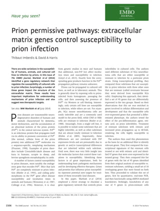
Another hypothesis, suggested by the
authors (Fig 1), is that PrPC deposited in the
ECM serves as substrate for the initiation of
infection. If this were the case, remodeling
of the ECM may allow more PrPC to be
deposited there, thereby enhancing PrPC
A Prion susceptible B Prion resistant
Integrin α8
Itga8
Fibronectin 1
Fn1
Other ECM components
(collagen, proteoglycans, etc.)
PrPC PrPSc
Integrin
β chain
Figure 1. Model for how two ECM components, the a8 chain of integrin (Itga8) and fibronectin 1 (Fn1), regulate susceptibility to prion infection.
In prion-susceptible cells (A), Fn1 and Itga8 are expressed at low levels, leading to a poorly developed ECM structure, and more deposition of PrPC in the matrix that can be
converted into PrPSc, thereby facilitating infection. The RGD domains of Fn1 (turquoise) are shown binding to an integrin dimer, consisting of an Itga8 a chain and a b chain
(not analyzed in this paper). In prion-resistant cells (B), Fn1 and Itga8 are highly expressed, the ECM is denser, and less PrPC is deposited, impeding generation of PrPSc.
Consistent with this model, disruption of Fn1-integrin interaction by incubation of resistant cells with soluble RGD peptide rendered the cells more prion-susceptible.
ª 2014 The Authors The EMBO Journal Vol 33 | No 14 | 2014
1507
3. The EMBO Journal Prion permissive pathways Thibaut Imberdis & David A Harris
formation. This scenario is consistent with
the observed changes in PrPC localization
observed upon knockdown of Fn1 and
Papss2. It is known that PrPC attached to the
plasma membrane via its glycolipid anchor
is rapidly converted to PrPSc upon contact
with exogenous prions (Goold et al, 2011),
but how PrPC might be released into the
ECM and what role this form may play in
prion propagation are open questions. How
the other gene products identified in the
study affect prion infection is unclear. Some
of them, such as Chga, Iqgap2, Il11ra1,
Micalcl, and Rgs4, are membrane or cyto-plasmic
proteins that are not known to be
directly involved in ECM biology. These
proteins could have indirect effects on the
ECM, or alternatively, they may act via a
completely different mechanism. Finally, it
will be important to determine to what
extent the current results can be extrapo-lated
from cultured cells to tissues and
organs. It seems likely that the factors that
control cellular accessibility to prions in an
in vivo setting differ from those operative in
a culture dish.
The study of Marbiah et al (2014) has
potentially important therapeutic implica-tions.
There are currently no effective treat-ments
for prion diseases, although the utility
of PrP knockdown approaches has been
demonstrated in experimental animals
(White et al, 2008). The proteins identified
in this paper represent novels targets for
anti-prion drugs. In this regard, compounds
that cause enhanced deposition or stabiliza-tion
of the ECM might be predicted to reduce
or prevent prion infection. In their study, the
authors demonstrated that knockdown of
ECM-related genes rendered resistant cells
more susceptible to infection, but they did
not determine whether over-expression of
the same genes made susceptible cells resis-tant.
This would clearly be an important first
step in developing the novel therapeutic
approach suggested here. Aside from their
potential clinical relevance, the results
presented here may also be helpful to prion
biologists, by providing a way to enhance
the prion susceptibility of cell types, such as
primary neurons, that have been tradition-ally
difficult to infect in culture.
Acknowledgements
Prion work in the Harris laboratory is supported by
N.I.H. grants R01 NS065244 and R01 NS040975.
Conflict of interest
The authors declare that they have no conflict of
interest.
References
Büeler H, Aguzzi A, Sailer A, Greiner RA, Autenried
P, Aguet M, Weissmann C (1993) Mice devoid of
PrP are resistant to scrapie. Cell 73: 1339 – 1347
Butler DA, Scott MRD, Bockman JM, Borchelt DR,
Taraboulos A, Hsiao KK, Kingsbury DT, Prusiner
SB (1988) Scrapie-infected murine
neuroblastoma cells produce protease-resistant
prion proteins. J Virol 62: 1558 – 1564
Caughey B, Raymond GJ (1993) Sulfated polyanion
inhibition of scrapie-associated PrP
accumulation in cultured cells. J Virol 67:
643 – 650
Collinge J, Beck J, Campbell T, Estibeiro K, Will RG
(1996) Prion protein gene analysis in new
variant cases of Creutzfeldt-Jakob disease.
Lancet 348: 56
Goold R, Rabbanian S, Sutton L, Andre R, Arora P,
Moonga J, Clarke AR, Schiavo G, Jat P, Collinge
J, Tabrizi SJ (2011) Rapid cell-surface prion
protein conversion revealed using a novel cell
system. Nat Commun 2: 281
Klohn PC, Stoltze L, Flechsig E, Enari M,
Weissmann C (2003) A quantitative, highly
sensitive cell-based infectivity assay for mouse
scrapie prions. Proc Natl Acad Sci USA 100:
11666 – 11671
Lloyd SE, Mead S, Collinge J (2013) Genetics of
prion diseases. Curr Opin Genet Dev 23:
345 – 351
Marbiah MM, Harvey A, West BT, Louzolo A,
Banerjee P, Alden J, Grigoriadis A, Hummerich
H, Kan HM, Cai Y, Bloom GS, Jat P, Collinge J,
Klöhn PC (2014) Identification of a gene
regulatory network associated with prion
replication. EMBO J 33: 1527 – 1547
Prusiner SB (1998) Prions. Proc Natl Acad Sci USA
95: 13363 – 13383
Shyng S-L, Lehmann S, Moulder KL, Harris DA
(1995) Sulfated glycans stimulate endocytosis
of the cellular isoform of the prion protein,
PrPC, in cultured cells. J Biol Chem 270:
30221 – 30229
Westaway D, Goodman PA, Mirenda CA, McKinley
MP, Carlson GA, Prusiner SB (1987) Distinct
prion proteins in short and long scrapie
incubation period mice. Cell 51: 651 – 662
White MD, Farmer M, Mirabile I, Brandner S,
Collinge J, Mallucci GR (2008) Single treatment
with RNAi against prion protein rescues early
neuronal dysfunction and prolongs survival in
mice with prion disease. Proc Natl Acad Sci USA
105: 10238 – 10243
The EMBO Journal Vol 33 | No 14 | 2014 ª 2014 The Authors
1508