11. MACROSCOPIC TERMS
Macule: Circumscribed lesion of <5 mm in diameter characterized by flatness
and usually discolored (often red)
Patch: Circumscribed lesion of >5 mm in diameter characterized by flatness
and usually discolored (often red)
Papule: Elevated dome-shaped or flat-topped lesion <5 mm across.
Nodule: Elevated lesion with spherical contour >5 mm across.
Plaque: Elevated flat-topped lesion, usually >5 mm across (may be caused by
coalescent papules).
Vesicle: Fluid-filled raised lesion <5 mm across.
Bulla: Fluid-filled raised lesion >5 mm across.
Blister: Common term used for vesicle or bulla.
Pustule: Discrete, pus-filled, raised lesion.
Wheal: Itchy, transient, elevated lesion with variable blanching and erythema
formed as the result of dermal edema.
Scale: Dry, horny, plate-like excrescence; usually the result of imperfect
cornification (i.e., keratinization).
Lichenification: Thickened and rough skin characterized by prominent skin
markings; usually the result of repeated rubbing in susceptible persons.
Excoriation: Traumatic lesion characterized by breakage of the epidermis,
causing a raw linear area (i.e., a deep scratch)
Onycholysis: Separation of nail plate from nail bed.
13. MICROSCOPIC TERMS
Hyperkeratosis: Thickening of the stratum corneum, often associated with a qualitative
abnormality of the keratin.
Parakeratosis: Modes of keratinization characterized by the retention of the nuclei in the
stratum corneum. On mucous membranes, parakeratosis is normal.
Hypergranulosis: Hyperplasia of the stratum granulosum, often due to intense rubbing.
Acanthosis: Diffuse epidermal hyperplasia.
Papillomatosis: Surface elevation caused by hyperplasia and enlargement of
contiguous dermal papillae.
Dyskeratosis: Abnormal keratinization occurring prematurely within individual cells or
groups of cells below the stratum granulosum. Generally the same as DYSPLASIA.
Acantholysis: Loss of intercellular connections resulting in loss of cohesion between
keratinocytes.
Spongiosis: Intercellular edema of the epidermis.
Hydropic swelling (ballooning): Intracellular edema of keratinocytes.
Exocytosis: Infiltration of the epidermis by inflammatory or circulating blood cells.
Erosion: Discontinuity of the skin exhibiting incomplete loss of the epidermis.
Ulceration: Discontinuity of the skin exhibiting complete loss of the epidermis and often
of portions of the dermis and even subcutaneous fat.
Vacuolization: Formation of vacuoles within or adjacent to cells; often refers to basal
cell-basement membrane zone area.
Lentiginous: Referring to a linear pattern of melanocyte proliferation within the
epidermal basal cell layer. Lentiginous melanocytic hyperplasia can occur as a reactive
change or as part of a neoplasm of melanocytes.
22. NEVI
• Many, many adjectives and classifications.
• The MAIN things to differentiate from
melanomas
• Junctional (more pigmented,
more closely associated with
melanoma)
• Intradermal
• Compound (both)
23.
24.
25.
26.
27. MALIGNANT MELANOMA
• Incidence rising, VERY much
• Related to SUN like ALL skin cancers are
• The only primary skin cancer that can kill
you (except for the RARE Merkel cell tumor)
• QUICKLY METASTASIZES
• Has both VERTICAL and HORIZONTAL
growth phase but prognosis is 100% related
to the VERTICAL, (BRESLOW staging, TNM
too)
• DIFFICULT to differentiate from NEVUS
clinically and often microscopically
45. PREMALIGNANT/MALIGNANT
• ACTINIC (Solar) KERATOSIS, i.e. precursor
to SCC
• SQUAMOUS CELL CARCINOMA,
squamous “pearls”, intercellular bridges
• BASAL CELL CARCINOMA, by far, MOST
COMMON, BLUE palisading nests
• MERKEL CELL CARCINOMA (TUMOR),
VERY MALIGNANT AND LETHAL, LOOK
LIKE SMALL CELL CA. OF LUNG
46. GENERAL COMMENTS
• BOTH SCC and BCC related to SUN (i.e.,
radiation) exposure. (as is MM also)
• SCC also related to As, carcinogens, chaw,
betel nut, HPV, familial, etc.
• BOTH SCC and BCC can do local damage
but very rarely metastasize or kill.
• MERKEL CELL tumors metastasize early
and extensively, like melanomas.
68. URTICARIA
• DERMAL EDEMA
• DILATATION of VASCULAR
SPACES
• EARLY PERIVASCULAR
CUFFING OF
INFLAMMATORY CELLS
69.
70. ECZEMA
(aka, acute eczematous dermatitis)
• A myriad of ACUTE
inflammatory disorders, with
allergic, drug related, sun
related etiologies
• The common histologic feature
is
SPONGIOSIS
71.
72.
73.
74.
75.
76. PSORIASIS
• 1-2% of USA
• Elbows, Knees
• Parakeratosis, generalized
epidermal hyperplasia,
elongation of the rete pegs,
extensive chronic inflammatory
cell infiltrates, “MUNRO”
intraepidermal microabscesses
88. ACNE VULGARIS
• Bread and Butter of dermatology
practice
• Sebaceous duct blockage with
secondary inflammation is main
feature
• bacterial lipases of
Propionibacterium acnes break
down sebaceous oils, and the
resulting fatty acids acts as
irritants
Basic microscopic and macroscopic descriptive terms, the essentials of describing skin lesions properly
Classifications and major examples of the classifications.
Classifications and major examples of the classifications.
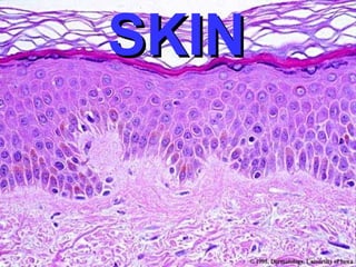
Histology review (no labels)
Histology review (labels)
Voila!
Appendages: hair follicles, sebaceous glands, sweat glands (eccrine, apocrine). Is this thick skin or thin skin?
These are just descriptive terms, NOT diagnoses.
These are just descriptive terms, NOT diagnoses.
Vitiligo
Freckles (ephelis)
Melasma, also called “mask of pregnancy”
Lentigo, (plural: lentigenes), is generally considered a brown pigmented spot on the skin. It is a harmless (benign) hyperplasia of melanocytes which is linear in its spread
Lentigo. Would lentigo be palpable? Ans: NO
Intradermal nevus
Intradermal nevus.
Note the lack of “junctional” activity.
Junctional nevus
Junctional nevus. Why is this called “Junctional”?
What would a “compound” nevus be? Ans: BOTH junctional and intradermal.
Malignant melanomas are malignant proliferations of melanocytes.
What is the ABCDEEFG principle?
Asymmetry
Borders (irregular)
Color (variegated), and
Diameter (greater than 6 mm (0.24 in.), about the size of a pencil eraser)
Evolving over time
These classifications do not however apply to the most dangerous form of melanoma, nodular melanoma, which has its own classifications:
Elevated above the skin surface
Firm to the touch
Growing
Why do only idiots learn the ABCDEEFG acronym? Ans: Because all these features are already basic in understanding malignancy.
What kind of malignant cell would have pigment IN it? What could this pigment be?
Clark vs. Breslow (better) classification
Seborrheic keratosis
Seborrheic keratosis, a bit more pigmented than the previous one, pigmentation is very common in ALL types of benign keratoses.
Squamous “horn cysts” in seborrheic keratosis
Acanthosis nigricans, often associated with diabetes mellitus
Acanthosis nigricans, often associated with diabetes mellitus
Fibroepithelial polyp, or “skin tag”
Fibroepithelial polyp, or “skin tag”.
Would you call this a papilloma? Why, or why not?
Epidermal inclusion cyst, the overlying skin looks normal.
Epidermal inclusion cyst
Keratoacanthoma, the MAIN lesion to differentiate from squamous cell carcinoma
Keratoacanthoma, the MAIN lesion to differentiate from squamous cell carcinoma
Keratoacanthoma, the MAIN lesion to differentiate from squamous cell carcinoma. What is a collarette?
Is a collarette the classical feature which differentiates KAs from SCCs? Ans: YES
Actinic keratosis
Actinic keratosis vs. squamous cell carcinoma
Squamous cell carcinoma, infiltrating. What is squamous cell carcinoma-in-situ usually called? Ans: Bowen’s disease.
Is there a collarette here? Ans: NO
Squamous cell carcinoma, infiltrating. Note the “pearls”. Does the presence of pearls make this well differentiated? Ans: Yes.
Squamous dysplasia, perhaps actinic keratosis, or something leading into squamous cell carcinoma.
By far, the commonest malignancy of skin, BCC, i.e., Basal Cell Carcinoma, typical appearance.
By far, the commonest malignancy of skin, BCC, i.e., Basal Cell Carcinoma, typical appearance. Note the PERIPHERAL PALISADING!!!
PALISADING!!!
PALISADING!!!
PALISADING!!!
Merkel cell tumor, very highly malignant RARE and usually fatal, looks EXACTLY like a small cell carcinoma of the lung? Ans: yes.
Note the LACK of the term “Fibroma”. Why” Ans: Because tumors of pure “fibroblasts” of the dermis are, somehow, rare.
Benign fibrous histiocytoma, or dermatofibroma
Benign fibrous histiocytoma, or dermatofibroma
Large fibrous histiocytoma, perhaps a dermatofibrosarcoma protuberans?
Malignant fibrous histiocytoma.
Xanthomas filled with cholesterol and lipids, to give the “foamy” appearance.
Xanthoma filled with cholesterol and lipids, to give the “foamy” appearance. Would you suspect these are associated, often, with hypercholesterolemia? Ans: YES
Hemangioma, often a congenital “birth mark”, which can regress significantly with aging. A red lesion which “blanches” when you put pressure on it, is always suspected to be a vascular tumor.
Kaposi’s sarcoma
Ichthyosis, usually genetic. Do you see the lamellae? Would you guess the term “lamellar” ichthiosis is often used? Ans: Yes
What is the Greek word for fish?
Inflammatory skin diseases are called dermatoses. Emphasis will NOT be on etiology, chiefly because most are unknown and are classically described as being related to a host of many things, like drugs, sun, allergies, etc.
While the neoplastic skin diseases can often be easily diagnosed by slide only, the dermatoses can rarely be diagnosed by slide only.
The diagnosis of inflammatory skin diseases requires SEEING THE PATIENT!!!
Acute and chronic, often refer to clinical features, because acute and chronic inflammatory cells, i.e., polys and monos respectively, do not often apply here.
Urticaria = hives = wheals = Type I Hypersensitivity = triggered off by perivascular mast cells releasing histamine
Is urticaria the classic skin response to type 1 hypersensitivity? Ans: YES
Often the word or classification “atopic” precedes the word “eczema”, implying an allergic etiology
(Atopic) Eczema
Eczema with spongiosis. Spongiosis: = Intercellular edema of the epidermis.
Pustules can be thought of as extreme “spongiosis”
Pustules, ulcerated.
Pustules, like vesicles and bullae, have an “evolution” of clinical and histologic appearances, generally following the acute__>chronic inflammatory evolution.
Erythema multiforme is a skin condition of unknown cause, possibly mediated by deposition of immune complex (mostly IgM) in the superficial microvasculature of the skin and oral mucous membrane that usually follows an infection or drug exposure. It is a common disorder, with peak incidence in the second and third decades of life. This severe form may be related to Stevens-Johnson syndrome.
Does this look like extreme urticaria?
Name the FOUR histopathologic findings in most of the forms of psoriasis: 1,2,3,4
1) Parakeratosis, 4) hyperplasia, 3) rete peg elongation, 4) MUNRO abscesses.
Please note that the classical diseases which cause “pigmentary incontinence” are the lichenoid disorders.
Note the pigmented cells are MELANOPHAGES, not MELANOCYTES.
Possibly the commonest skin disease you will see every day, so I’m giving you 5 classic views.
Do you think stasis dermatitis is commonest in the areas of tissues often most compromised by atherosclerosis?
Know the various types of “bullous” diseases. What is a bulla? What is acantholysis? Bulla: Fluid-filled raised lesion greater than 5 mm across.
Acantholysis: Loss of intercellular connections resulting in loss of cohesion between keratinocytes.
Pemphigus, fresh bullae
Pemphigus, ruptured, scabbed bullae
Acantholysis in the bullous family of diseases. Notice that the “seperation” can be within the acanthocytes, i.e., the stratum spinosum, or at the dermal-epidermal junction. So would you imagine many of the bullous disorders are diseases of basement membrane and tonofibrils (i.e., desmosomes), and may be autoimmune?
So is acne chiefly a folliculitis? Ans: Yes!
In about 30-50% of cases, the cause of E.N. is unknown. EN may be associated with a wide variety of diseases, including infections (e.g., TB, Strep, Mycoplasma, Yersinia, and EBV), coccidiomycosis, sarcoid, IBD, autoimmune disorders , pregnancy, medications (sulfas, oral contraceptives, bromides), and cancer. Do you see a lot of granulomatous diseases here? Ans: YES.
E.I. is OBSCURE, RARE, and mainly in WOMEN, also felt to be related to TB.
A panniculitis is a primary inflammation of the subdermal connective tissues, i.e., the hypodermis, or subcutis.
How many viruses besides HPV can you think of which cause squamous proliferations in ANY part of the body? Ans: Not many.
Papillomatous epidermal hyperplasia is the most consistent feature of verrucae (warts), Also note the “hypergranulosis”.
“hypergranulosis” “hypergranulosis” “hypergranulosis”
Molluscum contagiosum, a pox virus
Molluscum contagiosum.
Some things in pathology can only best be described by pictures, not words.
Impetigo, caused by staph and strep, usually in small kids.
Ringworm of scalp, Tinea capitis
Tinea barbae
Ringworm of the body, Tinea corporis
Tinea cruris, or jock itch
Athlete’s foot, or tinea pedis.
Is this interspace the most common place for tinea pedis?
Why? If your patient has a gangrenous toe, which one is most likely?
Onychomycosis (Note the LACK of the word tinea)
PAS stain of hyphae, probably scrapings.
PAS stain of hyphae, probably a histologic slice, NOT scrapings.
Scabies in it’s most common location
Body lice (pediculosis)
Pubic louse (phthirus pubis)
Demodex follicularis, a mite larva, notice how it likes to share a hair follicle with a hair shaft.
Why is this an arachnid, and not an insect? Ans: 8 legs.