Case Review #13: 13 year old female softball player with Adolescent Idiopathic Scoliosis
•
1 recomendación•2,325 vistas

A 13 year old female softball player presented with Adolescent Idiopathic Scoliosis. The degree of her scoliosis curve progressed to 48 degrees and she required a spinal fusion.

Recomendados
Recomendados
Más contenido relacionado
La actualidad más candente
La actualidad más candente (20)
Destacado
Destacado (10)
Similar a Case Review #13: 13 year old female softball player with Adolescent Idiopathic Scoliosis
Similar a Case Review #13: 13 year old female softball player with Adolescent Idiopathic Scoliosis (20)
Más de Robert Pashman
Más de Robert Pashman (15)
Último
Último (20)
Case Review #13: 13 year old female softball player with Adolescent Idiopathic Scoliosis
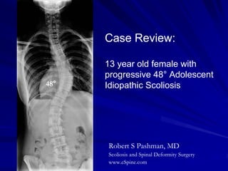
- 1. Case Review: 13 year old female with progressive 48° Adolescent 48° Idiopathic Scoliosis Robert S Pashman, MD Scoliosis and Spinal Deformity Surgery www.eSpine.com
- 2. Patient History 13-year-old post menarchal female No other congenital non- idiopathic or familial history of scoliosis in the family The patient is a competitive softball pitcher and athlete and although she reports bedtime soreness of her pitching arm, she denies symptoms referable to her upper or lower back and she has no allured neurologic sequela. On physical examination, the patient is well-developed female who is balanced in the frontal and sagittal planes. She has a few cm right rib hump, minimal lumbar fullness. She has no evidence of non-idiopathic etiologies for scoliosis such as connective tissue disorders or dermatological issues. Neurologically, she is intact without upper motor neuron signs.
- 3. Pre-op X-rays X-rays reveal a 48° type 1CN right thoracic adolescent idiopathic curve with significant rotation. The patient has level shoulders which might indicate structurality the proximal 48° thoracic curve making it a type 2 curve. This can only be seen on bending films. The patient has a lumbosacral transitional vertebra, so the count was carefully modulated.
- 4. Bending X-rays This is a fully compensatory lumbar curve, as evidenced on right and left side bending I felt that distal instrumentation should not encroach on the Cobb angle of the distal lumbar curve and therefore, L1 was chosen as the logical distal invertebrate. Proximally, the patient has a depression of the left shoulder and therefore, there is no structurally to the proximal thoracic curve, and the upper level was picked as T4.
- 5. Indications for Surgery 1. Type 1 progressive Adolescent Idiopathic Scoliosis, right thoracic, 50°. 2. Failed conservative therapy. 3. Thoracic low back pain secondary to the primary diagnosis.
- 6. Surgical Strategy Segmental spinal instrumentation, thoracic T4 to lumbar L1 using 1/4-inch stainless steel pedicle screw-rod construct. Posterior spinal fusion, T4 to L1, using locally harvested autogenous bone and allograft croutons. Spinal osteotomy for ankylosed facet joints, thoracic T4 to T10, and mobilization of curve. Interlaminar decompression, T12-L1, for visualization of L1 lumbar pedicle. Intraoperative somatosensory evoked potential, motor evoked potential analysis. Intraoperative fluoroscopy.
- 7. Post-Op Films The patient did very well after sugery. The AP and lateral x-rays look great. Posterior spinal fusion is balanced in the frontal sagittal plane without issue.