Surgicaldrainsand their types
•Descargar como PPTX, PDF•
92 recomendaciones•19,596 vistas

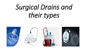
Surgical drains have several purposes and types. They help evacuate fluids from surgical sites to prevent infection and allow wounds to heal. Common types include closed suction drains like Jackson-Pratt drains and open drains like Penrose drains. Placement, securing, and care of drains is important to avoid complications like infection, displacement, or blockage. Drains are removed once drainage decreases significantly or they are no longer needed.

Recomendados
Más contenido relacionado
La actualidad más candente
La actualidad más candente (20)
Similar a Surgicaldrainsand their types
Similar a Surgicaldrainsand their types (20)
Más de Faryal Tebani
Más de Faryal Tebani (11)
Último
Último (20)
Surgicaldrainsand their types
- 1. Surgical Drains and their types
- 2. Definition of Drain A channel by which surplus liquid is drained or gradually carried out. An appliance or piece of material that acts as a channel for the escape (exit) of gases, fluids and other material from a cavity, wound, infected area or focus of suppuration. Drains inserted after surgery help the wound to heal faster and assist in preventing infection.
- 3. Qualities of a good drain (Ideal drain ) Soft to firm-Minimal damage to surrounding tissues Smooth -Efficiently evacuate effluent and easy removal Sterile- not potentiate infection or allow introduction of infection from external environment Stable- Inert, non allergenic, not degraded by body Simple to manage by both patient and staff
- 4. How drains work??? • According to Poiseuille’s law the laminar flow rate of an incompressible fluid along a tube is proportional to the fourth power of radius of tube and suction pressure. Flow is inversely proportional to viscosity of the fluid and length of the tube. • It means that wider and small length tube have more flow rate.
- 5. •Gravity •Capillary action •Tissue pressure •Negative pressure Factors governing effluent movement
- 6. INDICATIONS OF DRAINS THERE ARE DIFFERENT INDICATIONS.IT INCLUDE 1. THERAPEUTIC 2. DIAGNOSTIC 3. PROPHYLACTIC 4. MONITORING 5. PALLIATIVE
- 7. THERAPEUTIC • TENSION PNEUMOTHORAX • PLEURAL FLUID • ABSCESS CAVITY • SEROMA • ACUTE URINARY RETENTION
- 8. DIAGNOSTIC • T-TUBE CHOLANGIOGRAM FOR RETAINED GALL STONES IN COMMON BILE DUCT • BILIARY FISTULA.
- 9. PROPHYLACTIC • POST THYROIDECTOMY • THORACOTOMY • SPLENECTOMY • PANCREATECTOMY • ESOPHAGEAL RESECTION • CARDIOTHORACIC PROCEDURES
- 10. MONITORING AND PALLIATIVE FOR MONITORING IT IS USE FOR. GASTROINTESTINAL BLEEDING. URETHRAL CATHERIZATION. FOR PALLIATIVE. ADVANCED CA ESOPHAGUS. HYDROCEPHALUS.
- 11. CLASSIFICATION OF DRAINS SURGICAL DRAINS CAN BE A. OPEN OR CLOSED B. ACTIVE OR PASSIVE C. SILASTIC OR RUBBER DRAIN D.EXTERNAL OR INTERNAL DRAIN. E.WITH SUCTION OR WITHOUT SUCTION F.WITH CLOSED SUCTION OR WITH SUMP SUCTION.
- 12. Open drains • Include corrugated rubber or plastic sheets • Drain fluid collects into gauze pad or stoma bag • They increase the risk of infection. • E.g. penrose drain Closed drain• Consist of tubes draining into a bag or bottle. • They include chest and abdominal drains. • The risk of infection is reduced. • e.g. Jackson pratt drain
- 13. PASSIVE VS ACTIVE DRAINS PASSIVE DRAINS ACTIVE DRAINS
- 14. Passive drains (without suction) Passive drains have no suction. Drains by means of pressure differential ,overflow and gravity between body cavities and exterior . Closed: NGT , Foleys catheter, T-tube Open: penrose drain, corrugated drain.
- 15. Active drains (with suction) Active drains are maintained under suction. They can be under low or high pressure. Closed: Jackson pratt drain, hemovac drain. Open: sump drain.
- 16. 6.According to material • Irritant drains • composed of materials irritant to tissues • excite fibrous tissue response leading to fibrosis and tract formation • E.g. latex, plastic and rubber drains • Inert drains • Non irritant drains • Provoke minimal tissue fibrosis • E.g. polyvinyl chloride(PVC),polyurethane(PU) silicon elastomer (silastic)
- 17. According to type of suction:- • a. Sump suction :-in this double lumen tubes are there. Second tube act as a vent to allow air flow down to the tip of a drain. This prevent negative pressure at the tip and causes less tissue erosion and less blockage. • b. closed suction
- 18. According to fluid diversion • Internal drains • Divert retain fluids form a body cavity to another • Useful in neurosurgery,G.I surgery • E.g. Celestine tube, V-P shunt. • External drains • Channel discharge from cavity to external environment CELESTINE TUBE
- 19. Packs • Abscess cavity • Infected wound • must be replaced frequently. wicks
- 20. Cigarette drain • Penrose drain that has gauze within it is called cigarette drain. • in cigeratte drain, the ooze exist along the gauze. Penrose drain is a soft and flexible. It empites into a absorptive dressing material. A sterile large pin is often attached to the outer portion Penrose drain (obsolete)
- 21. Corrugated rubber drain • Passive, open drain. • Passive, open drain • Parallel tubes . • Side and end holes. • Capillary action Yeates drain
- 22. a) Corrugated; b) Penrose; c) Yeates
- 23. Robinson drain(simple drain or tube drain) • Passive and closed drain. • Made of latex Indication Anticipated fluid collection in a closed space after major abdominal surgery, to prevent seroma formation e.g. Pelvic surgery ,laparotomy for perforated viscus
- 24. Jackson-pratt drain A Jackson pratt drain is an active,close drain. is used to remove fluids that build up in an area of the body after surgery . Common uses in • abdominal surgery • Mastectomy • Thoracic surgery
- 25. Redivac drain • Closed suction drainage system. • Drainage of wound to prevent hematoma e.g.after thyroid surgery, repair of incisional harnia .
- 26. Pigtail drain Used in • Nephrostomy • Pleural fluid drainage • Intraperitoneal fluid drainage
- 27. Foleys cathater Types • Latex Foley: Short term drainage, yellow • Silicon-treated Foley: Minimize inflammatory reaction for long term placement, transparent • 3-way Foley: To be used in hematuria patient, water can be used to flush when necessary • Curved or coude catheter have curved tip used in older male with enlarged prostate which partialy obstruct the urethra
- 28. • Drainage of urine • Foley catheter can be used to drain suprapubic bladder • Chest drain in emergency • Stop nose bleeding by inserting into nasopharynx • Stop anorectal bleeding Indications of foleys catheter • Urethral traumaContraindication
- 29. sizes • 16-20 fr (adults) • 5-8 fr in children
- 30. Nasogastric tube • Ryles tube • Levin tube(gastroduodenal tube) Commonly used NG TUBE • Indications: • To drain gastric content to decompress stomach and prevent aspiration e.g IO or intraop use • Feeding (Not for long-term uses due to microaspiration pneumonia and discomfort
- 31. Nasogastric tube Placement First check for the patency of nostrils. Measure the distance from naris to ear lobule and to the xiphi sternum. Lubricate the tube . Pass tube through nostrils. Ask the patient to swallow water . Insertion of tube should point towards the xiphoid process. Asses the placement of distal end by air instillation and aspiration
- 32. Salem sump drain • Active and OPEN system • 1st lumen for suction ppf gastric content • 2nd lumen is air vent
- 33. Sangstaken blakemore 3 channels • Esophageal balloon • Gastric aspiration (Monitor bleeding) • Cardiac (Gastric) balloon 2 balloons • Cardiac balloon As a tamponade to stop bleeding Inflated by 200-300 cc contrast + water to ease assessment of tube position by X-ray Water + methylene blue to visualize leakage if the balloon burst • Esophageal balloon
- 34. T-TUBE
- 35. CARE OF SURGICAL DRAIN IT INCLUDES • INTRA OPERATIVE CARE • SECURING A SURGICAL DRAIN • POST OPERATIVE CARE
- 36. Intraoperative care Should not exit cavity through same surgical incision. Reach skin by safest shortest route Appropriate size and length A gravity drain must be placed in the safest and most dependent recess in cavity. Must be inserted away from delicate structures Firmly secured at exit wound Appropriate care-dressing,emptying. Must be removed when no longer useful-at once or by progressive shortening
- 37. B. SECURING A SURGICAL DRAIN DRAINS HAVE BEEN SECURED USING VARIOUS TECHNIQUES AND MATERIALS. • ROMAN GARTER TECHNIQUE WHICH USES SILK TO SECURE THE DRAIN. • USES OF NYLON SUTURES. • SAFETY PIN. • DRAIN CLIP. • ADHESIVE.
- 38. C. POST OPERATIVE CARE • SKIN AROUND THE WOUND MUST BE KEPT CLEAN,AND DRY TO PREVENT INFECTION AND SKIN IRRITATION. • METICULOUS SKIN CARE AND ASEPTIC TECHNIQUES MUST BE OBSERED DURING APPLICATION AND CHANGE OF DRESSING OVER DRAIN. • GAUZE DRESSING ARE USED AROUND AND OVER DRAINAGE TUBES.. • AN ACCURATE MEASUREMENT AND RECORD KEEPING OF DRAINAGE OUTPUT. • DRAIN CONTAINER SHOULD BE EMPTIED AT LEAST ONCE A DAY.
- 39. WHEN TO DISCONTINUOUS A SURGICAL DRAIN. • ONCE THE DRAINAGE HAS STOPED. • ITS OUT PUT HAS BECOME <25ml/DAY. • THE DRAIN HAS STOPPED SERVING THE DESIRED FUNCTION.
- 40. COMPLICATIONS. • IMMEDIATE. • PAIN • IRRITATION • BLEEDING • PERFORATTION OR INJURY TO ADJACENT STRUCTURES. • EARLY. • OCCLUSION • LEAKING AROUND DRAIN • DISPLACEMENT • INFECTION • LOSS OF FLUID,ELECTROLYTES AND PROTEIN
- 41. COMPLICATIONS…. • LATE. • PRESSURE/SUCTION NECROSIS OF BOWL OR VESSEL. • FISTULA. • SCAR. • HERNIA. • COMPLICATIONS DURING REMOVAL. • PAIN • INFECTION(CELLULITIS/ABSCESS) • INJURY TO ADJACENT STRUCTURES. • RETAINED OR FRAGMENTATION OF TUBE.
- 42. THANK YOU