Pneumonia
•
14 recomendaciones•4,786 vistas

Subject: Pediatric Nursing Topic: Pneumonia

Recomendados
Más contenido relacionado
La actualidad más candente
La actualidad más candente (20)
Destacado
Destacado (20)
Similar a Pneumonia
Similar a Pneumonia (20)
Último
Último (20)
Pneumonia
- 2. Pneumonia, an inflammation of the lungs caused by infections agent in which the air sacs are filled with pus or exudates so that air is excluded and lungs become solid. Or Pneumonia is an inflammation of the lung parenchyma
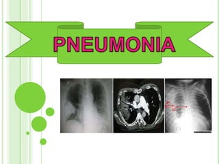
- 3. NORMAL CHEST X-RAY Courtesy of Up To Date
- 4. REVIEW OF LUNG ANATOMY RUL RML RLL LUL LLL Lingula http://www.meddean.luc.edulumenMedEdGrossAnatomythor ax0thor_lecthorax1.jpg
- 5. Bacterial : H.influenza streptococcal klebsella staphylococcal tuberculous Viral : Influenza virus, Adenoviruses, Rhinovirus
- 6. Anatomical classification. A – lobar pneumonia . The consolidation involves all or part of lobe B – Bronchopneumonia the consolidation involves scattered lobules C - Interstitial pneumonia . As in viral pneumonia where inflammatory . Infiltrate involve mainly interstitial tissue between alveoli. 2 : Etiological classfication. the cause of pneumonia in patient is often difficult to determine because direct culture of lung tissue invasive and rarely performed. - culture obtained from upper respiratory tract or sputum generally not accurately.
- 8. BRONCHOPNEUMONIA (Bronchitis and Pneumonia occur together)
- 10. CHEST X-RAY FOR LOBAR PNEUMONIA Lobarpneumonia Consolidation confined to one or more lobes (or segments of lobes) of lungs. 12/12/2011 Pneumonia 10
- 11. High fever, Shaking Chills Shortness of breath (Dyspnoea) Increased breathing rate Chest pain when you breathe deeply or cough Dusky or purplish skin colour (cyanosis) from poorly oxygenated blood
- 12. Fatigue and muscle aches Nausea, vomiting or diarrhoea chattering teeth cough that produces rust-colored or greenish mucus sweating rapid breathing rapid pulse rate
- 13. Cigarette smoking , -Abuse alcohol. Recent viral respiratory infection—a cold, laryngitis, influenza etc. Difficulty swallowing (due to stroke, dementia, or other neurological conditions) Chronic lung disease such as COPD, emphysema, asthma Other serious illnesses, such as heart disease, liver cirrhosis, or diabetes
- 14. Impaired consciousness (loss of brain function due to dementia, stroke, or other neurologic conditions) Are younger than 1 year of age or older than 65. Have a weakened or impaired immune system. Are malnourished. -Have been exposed to certain chemicals or pollutants.
- 15. Mode of transmission: Droplet infection - From the mouth and nose of an infected person via nasopharynx, through intimate contact with carriers. Indirect contact- By contaminated object is possible; systemic infection inhalation of caustic or toxic chemicals, aspiration of food, fluid or vomitus.
- 16. Chest X-ray Blood test Bronchoscopy Sputum analysis, smear, culture
- 17. If sever problem immediate hospitalization Give oxygen inhalation if: 1. central cyanosis 2. not able to drink Give antibiotic according to doctors order If age < 2 months a. benzyle penicilline. b. gentamicin If age 2 months to 5 years a. chloramphenicol Ttreat fever (if present) : paracetamol 500 mg 6 hourly a. 2 months – 3years ->¼tab b. 3 years – 5 years ->½tab Treatment of wheezing or stridor (if present):
- 18. a. If in respiratory distress- rapid acting bronchodilators nebulized salbutamol (5mg/ml) b. If not respiratory distress – oral salbutamol tds for 5 days 2 months -3 years ->¼tab (1 tab= 2mg) 12 months – 5 years -> 1 tab c. Supportive care d. Reassess daily
- 19. Maintain a patient airway and adequate oxygenation. Teach the patient how to cough and perform deep breathing exercises to clear secretions. Obtain sputum specimen as needed. Maintain adequate nutrition to offset high caloric utilization.
- 20. Control the spread of infection by disposing secretions properly. Control temperature by doing cooling measures. Monitor vital signs closely. Advice mother to give – a. Feed the child during illness b. Increase feeding after illness c. Clear nose if it interferes with feeding
- 21. Vaccinations a. Pneumococcal Vaccine b. Pneumococcal polysaccharide vaccine - PPV c. Hib vaccine (haemophilus influenzae vaccine type b) in children Healthy lifestyle Diet and nutrition Regular exercise
- 22. Enough sleep Quit smoke Prevent common colds, influenza, and other upper respiratory infection Environmental factors such as exposure to cold, pollution and physical conditions of fatigue or alcoholism.
- 23. Acute respiratory distress syndrom (ARDS) Pleural effusion Lung abscess Respiratory failure (which requires a breathing machine or ventilator) Sepsis (which may lead to organ failure Otitis media in children