SlideShare una empresa de Scribd logo
1 de 23
Urologic Complications

In early reports of renal transplantation, the prevalence of urologic complications varied from 10% to
25%, with a mortality rate ranging from 20% to 30% . In these patients, ureteroureterostomy or
pyeloureterostomy was used to restore urinary tract continuity. Patients who undergo
ureteroneocystostomy, the reconstructive technique used in most active kidney transplantation
programs today, have a lower incidence of urine leak or obstruction, compared with patients who
underwent the older procedures. Approximately two-thirds of the early urologic complications (urine
leak or obstruction) are apparent in the first month after transplantation and are treated by the
transplantation team. Currently, urologic complication rates are 4%–8% with very low patient mortality
.

Urine Leak and Urinomas
Urine leaks and urinomas are relatively rare complications of transplantation and usually constitute an
early postoperative problem. Extravasation of urine may occur from the renal pelvis, ureter, or
ureteroneocystostomy site due to ureteral necrosis caused by vascular insufficiency or increased
urinary pressures caused by obstruction. Caliceal leakage is an uncommon cause and occurs secondary
to segmental infarction in patients with accessory renal arteries or due to ligation of a polar artery.
Urinomas vary in size and are usually found in the first 2 postoperative weeks between the transplanted
kidney and the bladder. They may occur, however, in unexpected locations such as the scrotum or
thigh.


A decrease in urine output suggests that a urine leak may have occurred, and clinical signs may include
fullness and tenderness around the graft, ipsilateral leg swelling, or scrotal or labial edema. Early
detection and repair have been instrumental in reducing patient mortality.


At ultrasonography (US), a urine leak or urinoma appears as a well-defined, anechoic fluid collection
with no septations that increases in size rapidly. Drainage may be performed with US guidance, and the
higher creatinine level of the fluid compared with its serum concentration differentiates a urine leak
from a seroma or lymphocele. Large urinomas can rupture to produce urinary ascites. They can also
become infected and eventually form abscesses. Radionuclide studies show extravasation of radiotracer
into an area that was initially ―cold.― Delayed scintigrams should be obtained, since accumulation of
radiotracer may be slow. Antegrade pyelography is necessary to provide detailed information about the
site of origin of the urinoma and in planning appropriate intervention (6).


Figure 1a. Urinoma from ruptured ureterovesical junction stenosis. (a) Gray-scale sonogram reveals a
renal transplant that is obstructed because of a ruptured ureterovesical anastomosis and an
urinoma. (b) Postvoid sonogram demonstrates a fluid collection that represents extravasated urine
(arrow) and a small bladder BL). (c) Cystogram demonstrates extravasation (arrow). (Reprinted, with
permission, from reference 5.)
Figure 1b. Urinoma from ruptured ureterovesical junction stenosis. (a) Gray-scale sonogram reveals a
renal transplant that is obstructed because of a ruptured ureterovesical anastomosis and an
urinoma. (b) Postvoid sonogram demonstrates a fluid collection that represents extravasated urine
(arrow) and a small bladder (BL). (c) Cystogram demonstrates extravasation (arrow). (Reprinted, with
permission, from reference 5.)


Figure 1c. Urinoma from ruptured ureterovesical junction stenosis. (a) Gray-scale sonogram reveals a
renal transplant that is obstructed because of a ruptured ureterovesical anastomosis and an
urinoma. (b) Postvoid sonogram demonstrates a fluid collection that represents extravasated urine
(arrow) and a small bladder (BL). (c) Cystogram demonstrates extravasation (arrow). (Reprinted, with
permission, from reference 5.)


Small urine leaks may be treated with percutaneous nephrostomy and stent placement. Caliceal leakage
caused by infarction is treated with percutaneous nephrostomy alone. Ureteral stents must be kept in
place for 6–8 weeks after cessation of leakage to allow complete healing of the ureter and to preserve
long-term patency. If healing is unsuccessful, reimplantation of the ureter may be required; if distal
necrosis is present, pyelocystostomy or pyeloureterostomy with the native ureter is performed (6).

Urinary Obstruction
Urinary obstruction occurs in approximately 2% of transplantations and almost always within the first
6 months after the procedure. Obstruction of the transplanted kidney may occur at any location but is
most frequent at the site of implantation of the ureter into the bladder. More than 90% of ureteral
stenoses occur within the distal third of the ureter. Narrowing at the ureterovesical junction may be
caused by scarring secondary to ischemia or rejection, by technical error during the
ureteroneocystostomy, or by kinking. These events account for more than 50% of obstructions that
cause ureteral stricture. Less common causes include pelvic fibrosis, calculi, papillary necrosis, fungus
ball, clots, and compression from an extrinsic mass such as adjacent peritransplant collections.
Occasionally, obstruction that develops years after transplantation—especially in patients who have
undergone multiple procedures—may be related to adhesions, vascular insufficiency, or fibrosis.
Figure 2a. Hydronephrosis secondary to ureteral stricture. (a) Gray-scale sonogram demonstrates mild
hydronephrosis secondary to ureteral stricture. (b)Retrograde pyelogram shows the area of narrowing
at the site of ureteral implantation into the bladder (arrow). (Fig 2a and 2b reprinted, with permission,
from reference5.)


Figure 2b. Hydronephrosis secondary to ureteral stricture. (a) Gray-scale sonogram demonstrates mild
hydronephrosis secondary to ureteral stricture. (b)Retrograde pyelogram shows the area of narrowing
at the site of ureteral implantation into the bladder (arrow). (Fig 2a and 2b reprinted, with permission,
from reference).
The transplanted kidney is denervated; thus, the patient will not complain of typical renal colic when
obstruction occurs. Urinary obstruction manifests by a rising level of serum creatinine. Obstruction
may be difficult to differentiate from chronic rejection, since both cause rising creatinine levels. In
addition, mild dilatation of the collecting system may occasionally be seen in cases of chronic
rejection.


US can be used to confirm the diagnosis of hydronephrosis; however, intrarenal edema and fibrosis
associated with rejection may prevent the normal hydronephrotic response. Some transplant recipients
may have substantial obstruction but little or no renal dilatation. Mild to moderate dilatation of the
transplanted pyelocaliceal system and ureter may occur secondary to a full bladder. In all cases, when
the patient’s bladder is full, the bladder should be emptied and the transplant imaged again. In addition,
a denervated, transplanted collecting system has no tone; thus, any transient episode of hydronephrosis
may cause a persistent appearance of mild dilatation. The single renal graft is responsible for excreting
the entire urine output from the transplant recipient’s body, and, over time, increasing output may
exacerbate pelvocaliectasis. Studies have attempted to differentiate obstructive from
nonobstructivepyelocaliectasis by using the resistive index. Extrapolation of these results from native
to transplanted kidneys has not, however, been entirely successful. US also demonstrateslymphoceles,
hematomas, abscesses, and urinomas that may cause ureteral obstruction. Any echogenicity within a
dilated collecting system is usually clinically significant.

Scintigraphy may demonstrate urinary obstruction. In a patient with early partial obstruction, good
perfusion and prompt uptake of the radiotracer may be seen; however, in a patient with functionally
significant hydronephrosis, radioactivity is retained in the collecting system. Delayed images (obtained
2–4 hours after injection) are useful for differentiating an obstructed ureter from a dilated but
unobstructed ureter, since an unobstructed system shows clearance into the bladder. Diuretic
renography has proved useful for determining the functional significance of dilated collecting systems.
Conventional clearance times (furosemide clearance t1/2>20 minutes ) can be used in the assessment of
urinary tract patency.

Excretory urography is only infrequently successful for locating a urinary obstruction, and the site is
usually best determined with an antegrade contrast material examination. The superficial anterior
position of the allograft allows easy access for antegrade pyelography performed with real-time US
guidance. The Whitaker test is helpful in equivocal cases. The combination of normal results from the
Whitaker test and antegrade pyelography virtually excludes the presence of obstruction.

Percutaneous nephrostomy is used to relieve obstruction and allow the deployment of other radiologic
interventions such as ureteral stent placement and balloon ureteroplasty. Balloon dilation of
posttransplantation ureteral strictures produces an overall success rate of ureteral dilatation in 90% of
cases, with the best results obtained in fresh surgical strictures and the poorest in chronic ischemic
strictures or areas of periureteral fibrosis. Strictures of the middle and upper ureters result most
commonly from ischemic or periureteral fibrosis and may require surgical intervention. High balloon
pressures of 10–12 atm or greater are usually needed for successful dilation, and the balloon is left
inflated for 20 minutes or until balloon ―waisting‖ has disappeared. After dilation, a 10-F stent is left in
place for only 10 days. In the immunocompromised transplant recipient, the risk of infection precludes
long-term stent placement unless a totally internal, double pigtail stent is used. Recurrences most
commonly develop within the first year after transplantation.


Figure 3a. Hydronephrosis relieved by stent placement.(a) Gray-scale sonogram reveals severe
hydronephrosis resulting from narrowing at the ureterovesical anastomosis. Obstruction was relieved
by stent placement. (b)Sonogram obtained after placement of a retrograde stent (arrow) demonstrates
complete resolution of the hydronephrosis.


Figure 3b. Hydronephrosis relieved by stent placement.(a) Gray-scale sonogram reveals severe
hydronephrosis resulting from narrowing at the ureterovesical anastomosis. Obstruction was relieved
by stent placement. (b)Sonogram obtained after placement of a retrograde stent (arrow) demonstrates
complete resolution of the hydronephrosis.
Figure 4a. Dilation of a ureteral stenosis. Sequence of fluoroscopic images (a–c)demonstrates
placement of a JJ stent across a midureteral stricture.


Figure 4b. Dilation of a ureteral stenosis. Sequence of fluoroscopic images (a–c)demonstrates
placement of a JJ stent across a midureteral stricture.


Figure 4c. Dilation of a ureteral stenosis. Sequence of fluoroscopic images (a–c)demonstrates
placement of a JJ stent across a midureteral stricture.


Peritransplant Fluid Collections

Peritransplant fluid collections have been reported in up to 50% of renal transplantations and include
urinomas, hematomas, lymphoceles, and abscesses. The clinical significance of these collections is
largely determined by their size, location, and possible growth. In the immediate postoperative period,
small hematomas or seromas manifesting as crescenticperitransplant collections are almost expected.
Their size should be documented at baseline examination, since any increase in size may warrant
intervention. Growing collections may be indicative of urine leak, abscess, or vascular injury. Different
types of peritransplant fluid collections can be partially differentiated based on the time interval after
transplantation. Urinomas and hematomas are most likely to develop immediately after transplantation,
whereas lymphoceles generally occur 4–8 weeks after the surgical procedure. The sonographic
characteristics of peritransplant fluid collections, however, are entirely nonspecific, and ultimately,
diagnosis may be made only with percutaneous aspiration.

Hematomas
Hematomas are common in the immediate postoperative period, but they may also develop
spontaneously or as a consequence of trauma or biopsy. They are usually small and resolve
spontaneously. Large hematomas can displace the transplanted kidney and produce hydronephrosis.


At US, hematomas demonstrate a complex appearance. Acute hematomas are echogenic and become
less echogenic with time. Older hematomas even appear anechoic, more closely resembling fluid, and
septations may develop. Similarly, at computed tomography (CT), the appearance of a hematoma also
is time dependent. An acute hematoma has high-attenuation components, and older hematomas contain
liquefied and serous portions of intermediate attenuation . At magnetic resonance (MR) imaging, an
acute hematoma can show high signal intensity with both T1-weighted and T2-weighted pulse
sequences. At scintigraphy, hematomas demonstrate a ―cold defect.‖


Figure 5a. Subcapsular hematoma in a patient who sustained blunt trauma to the abdomen while
playing a sport. (a) US image demonstrates an isoechoicsubcapsular fluid collection (arrows). (b) CT
scan shows a heterogeneous crescenticsubcapsular collection in the transplanted kidney secondary to
hematoma (arrows).


Figure 5b. Subcapsular hematoma in a patient who sustained blunt trauma to the abdomen while
playing a sport. (a) US image demonstrates an isoechoicsubcapsular fluid collection (arrows). (b) CT
scan shows a heterogeneous crescenticsubcapsular collection in the transplanted kidney secondary to
hematoma (arrows).


Diagnostic aspiration can be performed to rule out abscess formation. However, percutaneous drainage
of the entire fluid collection is often neither efficacious (due to its multiloculated nature) nor advisable
because of the self-limited nature of the complication and risk of infection.


Lymphoceles
Lymphoceles are the most common peritransplant fluid collections, with a prevalence of 0.5%–20%.
They may develop at any time, from weeks to years after transplantation. However, they are usually an
early complication, occurring within 1–2 months after transplantation. Lymphoceles are caused by
leakage of lymph from surgically disrupted lymphatic channels along the iliac vessels or from the
lymphatics of the transplanted kidney. Risk factors include inadequate ligation of the lymphatic
channels across the iliac vessels, administration of heparin, and possibly increased lymphatic flow
secondary to edema of the lower extremities. These fluid collections usually occur medial to the
transplant, between the graft and the bladder .

Lymphoceles are the most common fluid collection that causes transplant hydronephrosis. Patients with
a failing allograft may develop ipsilateral lower extremity edema caused by compression of the femoral
vein. In rare cases, lymphoceles may develop in the scrotum and lymphatic drainage may occur
through the wound.


At US, lymphoceles are anechoic) and may have septations. Similar to other peritransplant fluid
collections, they can become infected and can develop a more complex appearance . At CT,
lymphoceles have variable characteristics and are usually sharply circumscribed. Their CT attenuation
values are typical of those of water and usually lower than those of recent hematomas and abscesses.
Radionuclide and MR imaging studies are helpful for excluding the presence of urine and blood,
respectively.


Figure 6. Lymphocele. US image demonstrates mild hydronephrosis of the transplanted kidney. The
anechoic area represents a lymphocele (L) adjacent to the kidney.


Figure 7. Lymphocele. Sonogram of another patient shows a multilocular fluid collection (arrows)
that surrounds the renal transplant. The collection proved to be a lymphocele.
Figure 8. Infected lymphoceles. US image demonstrates a complex echogenic fluid collection
(arrows) adjacent to the transplanted kidney.


Small lymphoceles are monitored sonographically, and large ones, if they grow or cause
hydronephrosis, should be drained. Aspirated fluid should have a creatinine content equal to that of
serum, and the cell count and differential may show relatively few white blood cells with a
predominance of lymphocytes. Results of Gram stain and culture of the fluid should be negative.


Lymphoceles commonly recur after simple percutaneous or surgical drainage. Permanent resolution of
lymphoceles may require prolonged catheter drainage and transcatheter instillation of sclerosing agents
such as povidine-iodine, absolute alcohol, or doxycycline (17). Repeated aspirations run the risk of
infecting an otherwise sterile fluid collection. However, success rates of up to 97% have been achieved
safely and effectively with percutaneous transcathetersclerotherapy (17). If a lymphocele becomes
infected, external drainage and antibiotic treatment are needed. Surgical treatment of a sterile fluid
collection involves removal of a generous window of peritoneum for marsupialization. The
transplanted kidney effectively becomes an intraperitoneal structure. Future leakage is therefore
resorbed in the peritoneal cavity. Retrograde pyelography helps identify the course of the transplanted
ureter before marsupialization, since the ureter could be displaced anteromedially and injured when the
peritoneal window is constructed.
Abscesses and Infection
More than 80% of renal transplant recipients suffer at least one case of infection during the first year
after transplantation. Early diagnosis of and intervention for infectious diseases can help prevent loss of
graft function and improve patient outcome.


Infections that occur in the first weeks after transplantation, such as pneumonia, surgical wound
infections, and urinary tract infections, are similar to those that typically develop in
nonimmunocompromised patients who have undergone surgery. Infections with opportunistic
pathogens and cytomegalovirus often develop 1–6 months after surgery, and infections common in the
general population are seen after 6 months. Patients frequently have multiple infections, and
immunosuppressive medications, indwelling catheters, and frequent glycosuria are all risk factors.
Peritransplant abscesses are an uncommon complication and usually develop within the first few weeks
after transplantation. These abscesses may be caused by pyelonephritis or bacterial seeding of a
lymphocele, hematoma, or urinoma. Acute bacterial nephritis, renal abscess, or perinephric abscess
may occur.

Patients may have few signs or symptoms of infection because of their immunosuppressed state. They
may present with fever of unknown origin, pain, or symptoms related to the pressure of the abscess on
the transplanted system. In a febrile transplant recipient, any peritransplant fluid collection must be
presumed to be infected. Acute pyelonephritis can mimic acute graft rejection, and their imaging
appearances may be the same.


The sonographic appearances of infections and abscesses are quite variable. Focal pyelonephritis may
appear as focal areas of increased or decreased echogenicity. These findings are nonspecific and can
represent infarction or rejection. Intrarenal masses are infrequently visualized and are also
sonographically nonspecific. They may represent primary renal cell carcinoma or
posttransplantationlymphoproliferative disorder. Abscesses have a complex, cystic, nonspecific
appearance at US; however, at CT, they manifest with gas, which serves to differentiate them from
other collections. In emphysematous pyelonephritis, gas in the parenchyma of the renal graft produces
an echogenic line with distal reverberation artifacts. This finding can be confirmed with abdominal
radiography or CT. Any echogenicity within a dilated collecting system is usually clinically significant.
Highly echogenic, weakly shadowing masses within a transplanted collecting system are fairly specific
for fungus balls). The presence of low-level echoes in a dilated pyelocaliceal system in a febrile patient
suggests pyonephrosis. Papillary necrosis results in sonographic changes in the calices, and intravenous
or retrograde pyelography is the definitive study for diagnosing this condition. Retrograde pyelography
may also demonstrate distortion of the calices, a feature seen in tuberculosis of the renal transplant.
Gallium-67 citrate and indium-111–tagged leukocyte studies are helpful for diagnosing infections that
develop 1 month or more after graft healing is complete .


Figure 9. Perinephric abscess. US image demonstrates a loculatedperinephric fluid collection with
complex sonographic features (arrow). The collection proved to be an infected urinoma.


Figure 10a. Fungus ball of the transplanted kidney. (a)US image demonstrates a soft-tissue echogenic
mass(m) in the renal pelvis of a transplanted kidney. (b)Retrograde pyelogram shows filling defects
(arrows) within the renal pelvis. These findings proved to be a fungus ball. (Fig 10a and 10b reprinted,
with permission, from reference 5.)
Figure 10b. Fungus ball of the transplanted kidney. (a)US image demonstrates a soft-tissue echogenic
mass(m) in the renal pelvis of a transplanted kidney. (b)Retrograde pyelogram shows filling defects
(arrows) within the renal pelvis. These findings proved to be a fungus ball. (Fig 10a and 10b reprinted,
with permission, from reference.)


Figure 11. Hydronephrosis secondary to a fungus ball. Color Doppler US image demonstrates an
echogenic mass (m) in the transplanted kidney that is causing hydronephrosis. The mass proved to be a
fungus ball.


Figure 12. Renal transplant tuberculosis. Retrograde pyelogram demonstrates a renal transplant with
irregular margins from renal tuberculosis. (Reprinted, with permission, from reference).


Abscesses may be treated with either US- or CT-guided percutaneous drainage. Both intrarenal and
extrarenal abscesses usually respond to external drainage and systemic antibiotics.
Vascular Complications

Renal Artery Stenosis
Renal artery stenosis occurs usually in the first year after transplantation. The stenosis may be located
before the anastomosis (because of atherosclerotic disease in the donor vessel), at the anastomosis
(secondary to vessel perfusion injury, faulty suture technique, or reaction to suture material), or after
the anastomosis (due to rejection, turbulent flow from kidney malposition, or arterial twisting, kinking,
or compression). Approximately half of renal artery stenoses occur at the anastomosis, and end-to-end
anastomoses have a threefold greater risk of stenosis than end-to-side anastomoses.

About 80% of patients with end-stage renal disease are hypertensive, and after renal transplantation
two-thirds of this group experience a reduction in hypertension. In patients with persistent
hypertension, renal artery stenosis may not be considered as a cause owing to concomitant graft failure.
Several clinical scenarios should prompt a search for stenosis: (a) severe hypertension refractory to
medical therapy, (b) hypertension and the presence of an audible bruit over the graft,
and (c)hypertension associated with unexplained graft dysfunction.

Color Doppler US demonstrates stenotic segments as focal areas of color aliasing due to increased flow
velocity. These areas can be selectively evaluated with duplex Doppler techniques to characterize and
grade the flow disturbance. Doppler criteria for significant stenoses include (a) focal frequency shifts
greater than 7.5 KHz (when a 3-MHz transducer is used) or velocities greater than 2 m/sec, (b) a
velocity gradient between stenotic and prestenotic segments of more than 2:1, and (c)marked distal
disturbance (spectral broadening). In the renal parenchyma, tardus-parvus waveform abnormalities can
be observed.

MR angiography can be used to diagnose renal artery stenosis in renal transplants. MR angiography
has the advantage of requiring either no contrast material or a gadolinium chelate that is not
nephrotoxic. Renal scintigraphy and time activity curves show reduced perfusion in transplants with
complete vascular obstruction and renal artery stenosis. This finding is nonspecific, however, as it may
be seen with other causes of parenchymal failure, including graft rejection and urinary obstruction.


Figure 13. Renal artery stenosis. Maximum intensity projection (MIP) image of a 35-year-old patient
with recurrent severe hypertension who underwent a second renal transplantation shows severe
proximal stenosis (arrow) in the renal artery of the first transplant. Notice the severe decrease in the
perfusion of the first transplant.


Surgical correction of graft renal artery stenosis is usually successful, but it is associated with
substantial morbidity. Primary treatment of graft renal artery stenosis by means of percutaneous
transluminal angioplasty with or without stent placement results in good intermediate-term patency and
is associated with significant early improvement in blood pressure and creatinine level. Because of its
low level of morbidity, relatively modest cost, and effectiveness, percutaneous transluminal
angioplasty is accepted as the initial treatment of choice. Clinical success rates resulting in substantial
initial improvement or cure have been reported in 73% of patients. Decrease in hypertension may be
seen in 1 day, and the prevalence of periprocedural complications is substantially lower than that
associated with surgical repair. Up to 20% of stenoses may require repeat dilation for maximum
success.
Figure 14a. Percutaneous transluminal angioplasty of a renal artery stenosis. (a)Angiogram shows
narrowing of the proximal renal artery (arrow). (b) Another angiogram shows a catheter placed across
the area of stenosis. (c) Angiogram obtained after angioplasty was performed shows restoration of a
near normal arterial lumen.


Figure 14b. Percutaneous transluminal angioplasty of a renal artery stenosis. (a)Angiogram shows
narrowing of the proximal renal artery (arrow). (b) Another angiogram shows a catheter placed across
the area of stenosis. (c) Angiogram obtained after angioplasty was performed shows restoration of a
near normal arterial lumen.


Figure 14c. Percutaneous transluminal angioplasty of a renal artery stenosis. (a)Angiogram shows
narrowing of the proximal renal artery (arrow). (b) Another angiogram shows a catheter placed across
the area of stenosis. (c) Angiogram obtained after angioplasty was performed shows restoration of a
near normal arterial lumen.


Infarction
Renal artery thrombosis may result from hyperacute rejection, anastomotic occlusion, arterial kinking,
or intimal flap. Segmental infarcts in the renal transplant may be focal or diffuse and may occur as part
of rejection or as a result of an unassociated vascular thrombosis. Vasculitis may induce small
segmental infarcts.


Patients with renal transplant infarction present with absence of urinary output and often with swelling
and tenderness over the graft and anuria. Although the graft itself is denervated, the inflammation
within the transplanted kidney may incite an inflammatory response in the adjacent visceral
peritoneum, with local pain in this location.
At US, a segmental infarct appears as a poorly marginated, hypoechoic mass or a hypoechoic mass
with a well-defined echogenic wall. If the infarction is global, the kidney will appear hypoechoic and
be diffusely enlarged. At color or power Doppler imaging, segmental infarcts appear as wedge-shaped
areas without color flow in , although these findings may also be seen in severe pyelonephritis or
transplant rupture. In total vascular obstruction, there is no perfusion to the transplanted kidney, and no
arterial or venous blood flow is seen in the graft at US. At technetium-99m dynamic radionuclide
studies, a photopenic region may be seen. However, these findings are not specific, since hyperacute or
accelerated acute rejection can have similar clinical, Doppler US, and scintigraphic features.


Figure 15. Infarct of a renal graft. Power Doppler US image demonstrates segmental loss of perfusion
in the transplanted kidney (arrows), a finding compatible with infarct.


CT can be used to detect perfusion deficits in the renal graft parenchyma, but it is generally not
employed in patients with elevated creatinine values because of the risk of compromising renal
function. If any doubt exists as to the nature of the US findings, angiography or MR angiography may
be performed. MR angiography is increasingly used to screen for vascular abnormalities in renal
transplants. Phased-array surface coils provide excellent signal-to-noise information that permits rapid
acquisition of high-quality images without the use of potentially nephrotoxic agents. Dynamic
enhanced MR imaging can be useful for diagnosing both segmental and global infarctions as well as
renal artery thrombosis. In addition, MR imaging has been helpful for demonstrating small infarcts
caused by iatrogenic drug-induced vasculitis.


Figure 16. Infarct of a renal graft. CT scan demonstrates segmental hypoattenuation (arrow) in the
transplanted kidney, a finding that proved to represent an infarct.
Figure 17a. Occlusion of a graft renal artery with lower pole infarction in a 38-year-old
patient. (a) Nontargeted MIP image reveals one graft renal artery that is normal. However, a total of
three graft renal arteries were anastomosed to the external iliac artery, and nonvisualization of two of
the three arteries indicates perioperative thrombosis. This vascular anatomy could not be ascertained
with Doppler US. (b) Another MIP image demonstrates the venous anatomy.(c) Contrast material–
enhanced three-dimensional MIP image demonstrates perfusion to the upper renal pole with lack of
perfusion to the lower pole, a finding that indicates lower pole infarction from arterial thrombosis.


Figure 17b. Occlusion of a graft renal artery with lower pole infarction in a 38-year-old
patient. (a) Nontargeted MIP image reveals one graft renal artery that is normal. However, a total of
three graft renal arteries were anastomosed to the external iliac artery, and nonvisualization of two of
the three arteries indicates perioperative thrombosis. This vascular anatomy could not be ascertained
with Doppler US. (b) Another MIP image demonstrates the venous anatomy.(c) Contrast material–
enhanced three-dimensional MIP image demonstrates perfusion to the upper renal pole with lack of
perfusion to the lower pole, a finding that indicates lower pole infarction from arterial thrombosis.


Figure 17c. Occlusion of a graft renal artery with lower pole infarction in a 38-year-old
patient. (a) Nontargeted MIP image reveals one graft renal artery that is normal. However, a total of
three graft renal arteries were anastomosed to the external iliac artery, and nonvisualization of two of
the three arteries indicates perioperative thrombosis. This vascular anatomy could not be ascertained
with Doppler US. (b) Another MIP image demonstrates the venous anatomy.(c) Contrast material–
enhanced three-dimensional MIP image demonstrates perfusion to the upper renal pole with lack of
perfusion to the lower pole, a finding that indicates lower pole infarction from arterial thrombosis.
Figure 18a. Complete occlusion of a graft renal artery with allograft infarction in a 47-year-old patient
with elevated serum creatinine levels. (a) Axial T1-weighted breath-hold gradient-echo image reveals
peripheral high signal intensity involving the renal cortex (arrow), a finding compatible with
hemorrhage. The patient had cortical necrosis proved at surgery. (b) Axial T2-weighted breath-hold
short inversion time inversion recovery image demonstrates peripheral low signal intensity
(arrow). (c) MIP image, obtained during the arterial phase of three-dimensional contrast-enhanced MR
angiography, demonstrates complete occlusion of the graft renal artery beyond its origin
(arrow). (d) Contrast-enhanced three-dimensional MIP image demonstrates lack of a cortical
nephrogram with a peripheral rim sign (arrow), an appearance that indicates total infarction of the
kidney.


Figure 18b. Complete occlusion of a graft renal artery with allograft infarction in a 47-year-old patient
with elevated serum creatinine levels. (a) Axial T1-weighted breath-hold gradient-echo image reveals
peripheral high signal intensity involving the renal cortex (arrow), a finding compatible with
hemorrhage. The patient had cortical necrosis proved at surgery. (b) Axial T2-weighted breath-hold
short inversion time inversion recovery image demonstrates peripheral low signal intensity
(arrow). (c) MIP image, obtained during the arterial phase of three-dimensional contrast-enhanced MR
angiography, demonstrates complete occlusion of the graft renal artery beyond its origin
(arrow). (d) Contrast-enhanced three-dimensional MIP image demonstrates lack of a cortical
nephrogram with a peripheral rim sign (arrow), an appearance that indicates total infarction of the
kidney.


Figure 18c. Complete occlusion of a graft renal artery with allograft infarction in a 47-year-old patient
with elevated serum creatinine levels. (a) Axial T1-weighted breath-hold gradient-echo image reveals
peripheral high signal intensity involving the renal cortex (arrow), a finding compatible with
hemorrhage. The patient had cortical necrosis proved at surgery.(b) Axial T2-weighted breath-hold
short inversion time inversion recovery image demonstrates peripheral low signal intensity
(arrow). (c) MIP image, obtained during the arterial phase of three-dimensional contrast-enhanced MR
angiography, demonstrates complete occlusion of the graft renal artery beyond its origin
(arrow). (d) Contrast-enhanced three-dimensional MIP image demonstrates lack of a cortical
nephrogram with a peripheral rim sign (arrow), an appearance that indicates total infarction of the
kidney.


Figure 18d. Complete occlusion of a graft renal artery with allograft infarction in a 47-year-old patient
with elevated serum creatinine levels. (a) Axial T1-weighted breath-hold gradient-echo image reveals
peripheral high signal intensity involving the renal cortex (arrow), a finding compatible with
hemorrhage. The patient had cortical necrosis proved at surgery.(b) Axial T2-weighted breath-hold
short inversion time inversion recovery image demonstrates peripheral low signal intensity
(arrow). (c) MIP image, obtained during the arterial phase of three-dimensional contrast-enhanced MR
angiography, demonstrates complete occlusion of the graft renal artery beyond its origin
(arrow). (d) Contrast-enhanced three-dimensional MIP image demonstrates lack of a cortical
nephrogram with a peripheral rim sign (arrow), an appearance that indicates total infarction of the
kidney.


Figure 19. Multiple cortical infarctions from drug-induced vasculitis in a 42-year-old patient. Early
contrast-enhanced three-dimensional MIP image reveals a large upper pole infarction with multiple
smaller focal cortical areas of signal loss, findings compatible with small infarctions.


Percutaneous angiographic interventional techniques may be valuable in treating infarctions and renal
artery thrombosis. Early diagnosis and treatment are vital for allograft salvage. Depending on the cause
and duration of the occlusion, thrombolytic therapy may prolong survival.
Arteriovenous Fistulas and Pseudoaneurysms
Percutaneous biopsy is commonly performed in transplant recipients when rejection is suspected.
Arteriovenous fistulas and pseudoaneurysms are occasionally seen after graft biopsies. Gross hematuria
is seen after 5%–7% of biopsies and is usually self limiting; however, massive or persistent hematuria
may occur.

Color and duplex Doppler US easily demonstrate arteriovenous fistulas. Arteriovenous fistulas and
pseudoaneurysms appear as localized areas of disorganized color that extend outside the confines of the
normal vessel. This appearance is caused by perifistula vibration and is detected with the more
sensitive color Doppler units. Arteriovenous fistulas also appear as abnormal high-velocity turbulent
flow isolated to a single segmental or interlobar artery and paired vein that produces aliasing on color
Doppler images. The feeding artery shows a high-velocity low-resistance waveform, and the draining
vein demonstrates arterialization.


Figure 20a. Arteriovenous fistula. (a) Color Doppler US image demonstrates a highly vascular
lesion. (b) Color duplex Doppler image shows the classic waveform of an arteriovenous fistula, with
high velocities and low impedance.


Figure 20b. Arteriovenous fistula. (a) Color Doppler US image demonstrates a highly vascular
lesion. (b) Color duplex Doppler image shows the classic waveform of an arteriovenous fistula, with
high velocities and low impedance.


Figure 21a. Arteriovenous fistula. (a) Duplex Doppler US image of the lower pole segmental artery
shows increased velocity and decreased resistive index.(b) Duplex Doppler US image of the adjacent
vein shows arterialization of flow, a finding consistent with arteriovenous fistula.


Figure 21b. Arteriovenous fistula. (a) Duplex Doppler US image of the lower pole segmental artery
shows increased velocity and decreased resistive index.(b) Duplex Doppler US image of the adjacent
vein shows arterialization of flow, a finding consistent with arteriovenous fistula.
On gray-scale US images, pseudoaneurysms appear as simple or complex renal cysts, but with color
imaging, highly vascular intracystic flow is seen. Pseudoaneurysms with a narrow neck and no venous
communication demonstrate a classic machinelike to-and-fro Doppler spectrum at their necks. Those
associated with arteriovenous fistulas exhibit a high-velocity low-resistance spectrum at their necks,
with minimally pulsatile high-velocity flow in the draining vein. When the degree of perifistula
vibration limits US assessment of the vascular anatomy, MR imaging enables evaluation.


Figure 22. Arteriovenous fistula following transplant biopsy. A color Doppler study in a 41-year-old
hypertensive patient with a bruit audited over the renal allograft revealed tissue vibration. Because of
marked tissue vibration, it was difficult to interrogate the transplant vasculature. Non-targeted MIP
images during the arterial phase of contrast-enhanced three-dimensional MR angiography reveal a
focal severe stenosis of the graft renal artery beyond its origin (arrow). The transplanted renal venous
system is enhancing during the early arterial phase along with the iliac vasculature and inferior vena
cava. The cortical nephrogram is very faint due to the marked arteriovenous shunting.


Figure 23. Arteriovenous fistula. Contrast-enhanced three-dimensional MR angiogram demonstrates
the renal artery and veins simultaneously, an appearance suggestive of fistula formation. The diagnosis
was confirmed at angiography (not shown).


Most complications after biopsy are treated conservatively, and most pseudoaneurysms resolve
spontaneously. If, however, a pseudoaneurysm demonstrates progressive enlargement or unusual size
(more than 2 cm in diameter), intervention is warranted. Selective transvascular catheterization may be
used. The size and location of the arteriovenous fistula or pseudoaneurysm determine which embolic
agent to use. For example, a large arteriovenous malformation is more easily treated with steel coils,
since particulate matter such as absorbable gelatin sponge (Gelfoam) may pass through the fistulous
communication and embolize the systemic circulation. Peripheral lesions may be difficult to catheterize
selectively, but a flow-directed detachable balloon can be delivered to the site. Microcatheter systems
may also be helpful for superselective embolization of small, peripheral feeding vessels. In treatment of
transplant-related arteriovenous fistulas (unlike that of peripheral arteriovenous fistulas), occlusion of
the proximal bleeding vessel is sufficient.

Renal Vein Thrombosis
Renal vein thrombosis is an unusual complication of transplantation; it occurs in less than 5% of
patients and usually in the first postoperative week. Renal vein thrombosis is heralded by an abrupt
cessation of urinary function and swelling and tenderness over the graft. Hypovolemia, venous
compression from a peritransplant fluid collection, dysfunctional anastomosis, and slow flow
secondary to rejection or other allograft disease can also precipitate renal vein thrombosis. An
increased prevalence of renal vein thrombosis in left lower quadrant allografts has also been attributed
to compression of the left common iliac vein between the sacrum and the left common iliac artery
(silent iliac artery compression syndrome.

At gray-scale US, renal vein thrombosis may manifest with an enlarged kidney. Venous flow is
reduced or absent, and there is increased resistance on the arterial side, often resulting in reversed
diastolic flow on Doppler images. MR venography helps confirm this complication in transplants.


Figure 24. Renal vein thrombosis. Duplex Doppler image demonstrates reversal of flow in diastole
(arrows) in the transplanted kidney due to renal vein thrombosis secondary to deep venous thrombosis.


Figure 25a. Iliac venous compression from a lymphocele in a 39-year-old patient with right lower
extremity edema. (a) MIP image produced from subtracting the arterial phase data from the equilibrium
phase data demonstrates a normal renal vein and poor visualization of the external iliac vein due to
compression. (b) Axial source image (from a two-dimensional time-of-flight MR venographic study)
helps confirm that the external iliac vein (solid arrow) is compressed by the underlying lymphocele
(open arrow).


Figure 25b. Iliac venous compression from a lymphocele in a 39-year-old patient with right lower
extremity edema. (a) MIP image produced from subtracting the arterial phase data from the equilibrium
phase data demonstrates a normal renal vein and poor visualization of the external iliac vein due to
compression. (b) Axial source image (from a two-dimensional time-of-flight MR venographic study)
helps confirm that the external iliac vein (solid arrow) is compressed by the underlying lymphocele
(open arrow).
Early recognition of renal vein thrombosis is crucial because the allograft may sometimes be salvaged
by prompt thrombectomy. Even with prompt diagnosis, however, graft infarction may develop, and a
transplant nephrectomy is usually performed to prevent secondary infection.
Calculous Disease

Kidney transplant recipients, compared with the general population, are at increased risk for developing
urinary calculi and approximately 1%–2% develop a clinically significant stone. Persistent secondary
hyperparathyroidism is seen in 38%–77% of end-stage renal disease patients after transplantation. In
the first year after transplantation, 15% of patients may be hypercalcemic, which increases the risk of
renal stones

The diagnosis of urinary calculi is suspected when renal function suddenly deteriorates. The patient
does not experience typical renal colic because the transplanted kidney is denervated.


US, radionuclide imaging, and contrast material studies are all useful diagnostic methods. Percutaneous
nephrostomy tube placement is valuable because it provides low-pressure drainage and allows renal
function to stabilize. Antegrade pyelography can then be used to give exact definition of the collecting
system and position of the stone .


Figure 26a. Renal transplant calculus in a 34-year-old patient with hematuria. (a) US image
demonstrates hydronephrosis with a shadowing echogenic focus seen in the upper middle renal pole
(arrow). (b) US image of the distal ureter shows an echogenic focus with shadowing (arrow) a finding
consistent with an obstructing calculus.


Figure 26b. Renal transplant calculus in a 34-year-old patient with hematuria. (a) US image
demonstrates hydronephrosis with a shadowing echogenic focus seen in the upper middle renal pole
(arrow). (b) US image of the distal ureter shows an echogenic focus with shadowing (arrow) a finding
consistent with an obstructing calculus.


Most stones can be removed with endourologic techniques. Upper tract stones are best treated by
means of percutaneous nephrostolithotomy. Cystoscopy with electrohydraulic lithotripsy is appropriate
for treating bladder stones.

Neoplasms
Prolonged immunosuppression following renal transplantation places the transplant recipient at about
100 times the normal risk for developing cancer. The average reported prevalence of malignancy in
these patients is 6% (38), with cases consisting predominantly of skin cancers and lymphomas. The
degree of immunosuppression and its duration are both important factors in the development of
malignancy. The frequency of urologic tumors such as prostatic adenocarcinoma and germ cell tumors
of the testis does not appear to be increased in this population. However, the prevalence of renal
adenocarcinoma may be increased, with 90% of the tumors occurring in the native kidney and 10%
occurring in the transplanted kidney (39). One reason for the increased risk of renal adenocarcinoma is
that approximately half of the patients who undergo hemodialysis because of chronic renal failure
develop acquired renal cystic disease and 9% develop tumors (40). Patients with longer use of
hemodialysis are at greater risk for developing acquired cystic disease. Fortunately, less than 1% of
these patients develop malignant tumors that metastasize. In addition, patients with a significant past
exposure to cyclophosphamide have an increased risk for developing of urothelial tumors (41);
although cyclophosphamide is less commonly used now because of the availability of cyclosporin A,
there are many renal transplant patients with a history of cyclophosphamide exposure.

A complete urologic evaluation for gross hematuria is recommended in all renal transplant recipients.
Intravenous pyelography may be useful if there is good transplant function, although it will not provide
information about the native kidney. US or CT of the native and transplanted kidneys provides more
complete information, and a solid heterogeneous enhancing mass that may be cystic is often identified.
Cystoscopy with retrograde pyelography of all present kidneys and ureters is advisable.


Figure 27. Renal transplant adenocarcinoma. CT scan shows a cystic mass (arrow) arising from the
renal transplant that proved to be renal cell carcinoma.


Transplant glomerulopathy and recurrence of preexisting glomerulonephritis frequently cause
microhematuria often associated with proteinuria. However, since these patients have an increased risk
of developing neoplasms, a complete urologic work-up is advisable.


Development of renal adenocarcinoma in the transplanted kidney is uncommon, with the majority of
malignancies related to skin cancers and lymphomas. Malignancy in the transplanted kidney is treated
by total removal of the renal graft and cessation of immunosuppression. Urothelial malignancies can
likewise be treated in a standard fashion. In the presence of immunosuppression, intravesical
instillation of Calmette-Guérin bacillus is not recommended.

Gastrointestinal and Herniation Complications

Gastrointestinal complications may occur in transplantation patients. The most common of these
complications is gastrointestinal hemorrhage due to peptic ulcer (42). Prevalence of the complications
may vary with use of an intraperitoneal or extraperitoneal approach in placement of the allograft (43).
Postoperative adhesions may lead to intestinal obstruction. Herniation of bowel through a transplant
peritoneal defect may lead to compromise of the intestine or of the transplant itself (Fig 28). Urinary
obstruction from obturator herniation of the ureter may also occur (44). Pseudomembranous colitis
secondary to overgrowth of Clostridium difficle in patients receiving antibiotic therapy is another
complication that can be seen in renal transplant recipients. A typical ―accordion sign‖ secondary to
massive edema of the intestinal wall is highly suggestive of the diagnosis on CT images of the
abdomen.
Figure 28. Renal transplant herniation. CT scan demonstrates multiple distended small bowel loops
around the transplanted kidney, findings compatible with obstruction. Small bowel had herniated
through the peritoneal defect related to the renal graft, a diagnosis that was surgically proved.


PosttransplantationLymphoproliferative Disorder

Impaired immune surveillance and the oncogenic effects of immunosuppressive therapy itself may be
causative factors of the posttransplantationlymphoproliferative disorder (PLTD). An association with
Epstein Barr virus is usually demonstrated. PLTD complicates 8% of transplantations and is diagnosed
at a median of 80 months after transplantation. The liver, brain, and lung are more common sites of
involvement than the gastrointestinal tract. PLTD lesions are polymorphic collections of B cells but
may evolve into forms indistinguishable from non-Hodgkin lymphoma. If there is involvement of the
small intestines, patients often have generalized disease.


Figure 29a. Posttransplantationlymphoproliferative disease in a 25-year-old renal allograft recipient
who presented with abdominal pain. (a) Contrast-enhanced CT scan demonstrates circumferential
thickening of the jejunum (arrows). (b)Contrast-enhanced CT scan obtained at a lower level shows
encasement of the superior mesenteric artery by lymphadenopathy (arrowheads), in addition to the
jejunal thickening (arrow).


Figure 29b. Posttransplantationlymphoproliferative disease in a 25-year-old renal allograft recipient
who presented with abdominal pain. (a) Contrast-enhanced CT scan demonstrates circumferential
thickening of the jejunum (arrows). (b)Contrast-enhanced CT scan obtained at a lower level shows
encasement of the superior mesenteric artery by lymphadenopathy (arrowheads), in addition to the
jejunal thickening (arrow).
Figure 30. Posttransplantationlymphoproliferative disease in a kidney transplant recipient who
presented with fever. Contrast-enhanced CT scan demonstrates a necrotic mass in the pelvis posterior
to the transplanted kidney (arrows).


Conclusions

Transplantation is currently one of the accepted treatments of irreversible kidney disease.
Improvements in surgical techniques and more sophisticated, potent immunosuppressive drugs have
resulted in remarkable advances in survival of patients and renal grafts. Nevertheless, substantial
complications occur in both the immediate postoperative period and later. Imaging has a critical role in
the evaluation of these complications, and interventional radiologic techniques are often successfully
applied to their treatment.

Más contenido relacionado

La actualidad más candente

Urethral & bladder injury
Urethral & bladder injuryUrethral & bladder injury
Urethral & bladder injuryQiba Hospital
 
Flexible Uretero-renoscopy or RIRS
Flexible Uretero-renoscopy or RIRSFlexible Uretero-renoscopy or RIRS
Flexible Uretero-renoscopy or RIRSGAURAV NAHAR
 
Principles of Endourology
Principles of EndourologyPrinciples of Endourology
Principles of EndourologyEko indra
 
Post obstructive diuresis
Post obstructive diuresisPost obstructive diuresis
Post obstructive diuresisEko indra
 
Genitourinary trauma
Genitourinary traumaGenitourinary trauma
Genitourinary traumaAmit Rauniyar
 
Urinary Diversion after cystectomy [Dr.Edmond Wong]
Urinary Diversion after cystectomy  [Dr.Edmond Wong]Urinary Diversion after cystectomy  [Dr.Edmond Wong]
Urinary Diversion after cystectomy [Dr.Edmond Wong]Edmond Wong
 
Ectopic ureter and ureterocele - Embryology and Presentation
Ectopic ureter and ureterocele - Embryology and PresentationEctopic ureter and ureterocele - Embryology and Presentation
Ectopic ureter and ureterocele - Embryology and PresentationAbhishekPandey1012
 
Post operative complications of renal transplant
Post operative complications of renal transplantPost operative complications of renal transplant
Post operative complications of renal transplantHabrol Afzam
 
Management of stricture urethra
Management of stricture urethra Management of stricture urethra
Management of stricture urethra SomendraBansal
 
Principles of urethral reconstructive surgery
Principles of urethral reconstructive surgeryPrinciples of urethral reconstructive surgery
Principles of urethral reconstructive surgeryAhmed Eliwa
 
Etiopathogenesis, Evaluation & Management of Posterior Urethral Valve
Etiopathogenesis, Evaluation & Management of Posterior Urethral ValveEtiopathogenesis, Evaluation & Management of Posterior Urethral Valve
Etiopathogenesis, Evaluation & Management of Posterior Urethral ValveShubham Lavania
 
Urinary Diversion
Urinary DiversionUrinary Diversion
Urinary Diversionbbthapa
 
Urethra stricture etiopathogenesis & evaluation
Urethra stricture  etiopathogenesis & evaluationUrethra stricture  etiopathogenesis & evaluation
Urethra stricture etiopathogenesis & evaluationGovtRoyapettahHospit
 

La actualidad más candente (20)

Urethral & bladder injury
Urethral & bladder injuryUrethral & bladder injury
Urethral & bladder injury
 
Flexible Uretero-renoscopy or RIRS
Flexible Uretero-renoscopy or RIRSFlexible Uretero-renoscopy or RIRS
Flexible Uretero-renoscopy or RIRS
 
Principles of Endourology
Principles of EndourologyPrinciples of Endourology
Principles of Endourology
 
Post obstructive diuresis
Post obstructive diuresisPost obstructive diuresis
Post obstructive diuresis
 
Genitourinary trauma
Genitourinary traumaGenitourinary trauma
Genitourinary trauma
 
Puj obstruction
Puj obstructionPuj obstruction
Puj obstruction
 
Urinary Diversion after cystectomy [Dr.Edmond Wong]
Urinary Diversion after cystectomy  [Dr.Edmond Wong]Urinary Diversion after cystectomy  [Dr.Edmond Wong]
Urinary Diversion after cystectomy [Dr.Edmond Wong]
 
Ectopic ureter and ureterocele - Embryology and Presentation
Ectopic ureter and ureterocele - Embryology and PresentationEctopic ureter and ureterocele - Embryology and Presentation
Ectopic ureter and ureterocele - Embryology and Presentation
 
First stage of johanson procedures
First stage of johanson proceduresFirst stage of johanson procedures
First stage of johanson procedures
 
Post operative complications of renal transplant
Post operative complications of renal transplantPost operative complications of renal transplant
Post operative complications of renal transplant
 
Upj obstruction
Upj obstructionUpj obstruction
Upj obstruction
 
Management of stricture urethra
Management of stricture urethra Management of stricture urethra
Management of stricture urethra
 
Principles of urethral reconstructive surgery
Principles of urethral reconstructive surgeryPrinciples of urethral reconstructive surgery
Principles of urethral reconstructive surgery
 
Etiopathogenesis, Evaluation & Management of Posterior Urethral Valve
Etiopathogenesis, Evaluation & Management of Posterior Urethral ValveEtiopathogenesis, Evaluation & Management of Posterior Urethral Valve
Etiopathogenesis, Evaluation & Management of Posterior Urethral Valve
 
Pfudd
PfuddPfudd
Pfudd
 
Ureteric injury (1)
Ureteric injury (1)Ureteric injury (1)
Ureteric injury (1)
 
Urinary Diversion
Urinary DiversionUrinary Diversion
Urinary Diversion
 
COMPLICATIONS OF PCNL
COMPLICATIONS OF PCNLCOMPLICATIONS OF PCNL
COMPLICATIONS OF PCNL
 
Urethra stricture etiopathogenesis & evaluation
Urethra stricture  etiopathogenesis & evaluationUrethra stricture  etiopathogenesis & evaluation
Urethra stricture etiopathogenesis & evaluation
 
Bladder injuries
Bladder injuriesBladder injuries
Bladder injuries
 

Destacado

Renal Transplantation Dr. Jorge L. Posada
Renal Transplantation   Dr. Jorge L. PosadaRenal Transplantation   Dr. Jorge L. Posada
Renal Transplantation Dr. Jorge L. PosadaMedicineAndHealthCancer
 
Kidney transplantation
Kidney transplantationKidney transplantation
Kidney transplantationJijo G John
 
KIDNEY TRANSPLANTATION SEMINAR PRESENTATION
KIDNEY TRANSPLANTATION SEMINAR PRESENTATIONKIDNEY TRANSPLANTATION SEMINAR PRESENTATION
KIDNEY TRANSPLANTATION SEMINAR PRESENTATIONfareedresidency
 
Kidney Transplant
Kidney TransplantKidney Transplant
Kidney Transplanthelunchis
 
Kidney Transplantation
Kidney TransplantationKidney Transplantation
Kidney TransplantationMohammed Rajab
 
Organ transplant ppt
Organ transplant pptOrgan transplant ppt
Organ transplant pptRichard Frank
 
Kidney transplant process for children
Kidney transplant process for childrenKidney transplant process for children
Kidney transplant process for childrenguest4748f4
 
Post Transplant Malignancy – A Brief Review
Post Transplant Malignancy – A Brief ReviewPost Transplant Malignancy – A Brief Review
Post Transplant Malignancy – A Brief ReviewApollo Hospitals
 
Post transplant diabetes patient education class
Post transplant diabetes patient education classPost transplant diabetes patient education class
Post transplant diabetes patient education classawclarke
 
Jose Maria Morales - Spain - Tuesday 29 - HLA for Renal Allocation
Jose Maria Morales - Spain - Tuesday 29 - HLA for Renal AllocationJose Maria Morales - Spain - Tuesday 29 - HLA for Renal Allocation
Jose Maria Morales - Spain - Tuesday 29 - HLA for Renal Allocationincucai_isodp
 
Journal club solid organ transplant (New Onset Diabetes)
Journal club solid organ transplant (New Onset Diabetes)Journal club solid organ transplant (New Onset Diabetes)
Journal club solid organ transplant (New Onset Diabetes)Daniel Le
 
Doppler ultrasound in transplant renal artery stenosis
Doppler ultrasound in transplant renal artery stenosisDoppler ultrasound in transplant renal artery stenosis
Doppler ultrasound in transplant renal artery stenosisSamir Haffar
 
Principles of organ transplant and Renal transplant
Principles of organ transplant and Renal transplantPrinciples of organ transplant and Renal transplant
Principles of organ transplant and Renal transplantDr Navil Sharma
 
Artifacts in esophageal high resolution manometry
Artifacts in esophageal high resolution manometryArtifacts in esophageal high resolution manometry
Artifacts in esophageal high resolution manometrySamir Haffar
 
Mechanism of tissue graft rejection
Mechanism of tissue graft rejectionMechanism of tissue graft rejection
Mechanism of tissue graft rejectionNilesh Chandra
 

Destacado (20)

Renal Transplantation Dr. Jorge L. Posada
Renal Transplantation   Dr. Jorge L. PosadaRenal Transplantation   Dr. Jorge L. Posada
Renal Transplantation Dr. Jorge L. Posada
 
Kidney transplantation
Kidney transplantationKidney transplantation
Kidney transplantation
 
Renal transplantation
Renal transplantationRenal transplantation
Renal transplantation
 
KIDNEY TRANSPLANTATION SEMINAR PRESENTATION
KIDNEY TRANSPLANTATION SEMINAR PRESENTATIONKIDNEY TRANSPLANTATION SEMINAR PRESENTATION
KIDNEY TRANSPLANTATION SEMINAR PRESENTATION
 
Kidney Transplant
Kidney TransplantKidney Transplant
Kidney Transplant
 
Kidney Transplantation
Kidney TransplantationKidney Transplantation
Kidney Transplantation
 
Organ transplant ppt
Organ transplant pptOrgan transplant ppt
Organ transplant ppt
 
Hematuria post transplant
Hematuria post transplantHematuria post transplant
Hematuria post transplant
 
Kidney transplant process for children
Kidney transplant process for childrenKidney transplant process for children
Kidney transplant process for children
 
Post Transplant Malignancy – A Brief Review
Post Transplant Malignancy – A Brief ReviewPost Transplant Malignancy – A Brief Review
Post Transplant Malignancy – A Brief Review
 
Nodat
NodatNodat
Nodat
 
Post transplant diabetes patient education class
Post transplant diabetes patient education classPost transplant diabetes patient education class
Post transplant diabetes patient education class
 
Jose Maria Morales - Spain - Tuesday 29 - HLA for Renal Allocation
Jose Maria Morales - Spain - Tuesday 29 - HLA for Renal AllocationJose Maria Morales - Spain - Tuesday 29 - HLA for Renal Allocation
Jose Maria Morales - Spain - Tuesday 29 - HLA for Renal Allocation
 
Journal club solid organ transplant (New Onset Diabetes)
Journal club solid organ transplant (New Onset Diabetes)Journal club solid organ transplant (New Onset Diabetes)
Journal club solid organ transplant (New Onset Diabetes)
 
Doppler ultrasound in transplant renal artery stenosis
Doppler ultrasound in transplant renal artery stenosisDoppler ultrasound in transplant renal artery stenosis
Doppler ultrasound in transplant renal artery stenosis
 
Principles of organ transplant and Renal transplant
Principles of organ transplant and Renal transplantPrinciples of organ transplant and Renal transplant
Principles of organ transplant and Renal transplant
 
Renal doppler
Renal dopplerRenal doppler
Renal doppler
 
Artifacts in esophageal high resolution manometry
Artifacts in esophageal high resolution manometryArtifacts in esophageal high resolution manometry
Artifacts in esophageal high resolution manometry
 
Nephrectomy
NephrectomyNephrectomy
Nephrectomy
 
Mechanism of tissue graft rejection
Mechanism of tissue graft rejectionMechanism of tissue graft rejection
Mechanism of tissue graft rejection
 

Similar a Main renal transplants complications

Post operative complications of renal transplant
Post operative complications of renal transplantPost operative complications of renal transplant
Post operative complications of renal transplantHabrol Afzam
 
URINARY SYSTEM - Copy.pptx
URINARY SYSTEM - Copy.pptxURINARY SYSTEM - Copy.pptx
URINARY SYSTEM - Copy.pptxGokulnathMbbs
 
Direct contrast investigations
Direct contrast investigationsDirect contrast investigations
Direct contrast investigationsairwave12
 
urology trauma english.pptx
urology trauma english.pptxurology trauma english.pptx
urology trauma english.pptxVelmaPranayReddy
 
MCU- Micturating cysto-urethrogram
MCU- Micturating cysto-urethrogramMCU- Micturating cysto-urethrogram
MCU- Micturating cysto-urethrogramDr. Mohit Goel
 
Pediatric urinary tract infection..the role of imaging
Pediatric urinary tract infection..the role of imagingPediatric urinary tract infection..the role of imaging
Pediatric urinary tract infection..the role of imagingAhmed Bahnassy
 
Pediatric urinary tract infection..the role of imaging
Pediatric urinary tract infection..the role of imagingPediatric urinary tract infection..the role of imaging
Pediatric urinary tract infection..the role of imagingAlexandria Urology Hospital
 
Renal infections radiology
Renal infections radiology Renal infections radiology
Renal infections radiology docaashishgupt
 
Bile leaks after lapchole Nir Hus MD., PhD.
Bile leaks after lapchole Nir Hus MD., PhD.Bile leaks after lapchole Nir Hus MD., PhD.
Bile leaks after lapchole Nir Hus MD., PhD.Nir Hus MD, PhD, FACS
 
LECTURE NOTE ON URETHRAL STRICTURES AND STENOSIS.pdf
LECTURE NOTE ON URETHRAL STRICTURES AND STENOSIS.pdfLECTURE NOTE ON URETHRAL STRICTURES AND STENOSIS.pdf
LECTURE NOTE ON URETHRAL STRICTURES AND STENOSIS.pdfSolomon Lakew
 
Management of pelviureteric junction obstruction onyeze copy
Management of pelviureteric junction obstruction onyeze   copyManagement of pelviureteric junction obstruction onyeze   copy
Management of pelviureteric junction obstruction onyeze copyChigozie Onyeze
 
Posterior urethral valve
Posterior urethral valvePosterior urethral valve
Posterior urethral valveMakafui Yigah
 
Biliary tract
Biliary tractBiliary tract
Biliary tractairwave12
 

Similar a Main renal transplants complications (20)

Post operative complications of renal transplant
Post operative complications of renal transplantPost operative complications of renal transplant
Post operative complications of renal transplant
 
URINARY SYSTEM - Copy.pptx
URINARY SYSTEM - Copy.pptxURINARY SYSTEM - Copy.pptx
URINARY SYSTEM - Copy.pptx
 
Direct contrast investigations
Direct contrast investigationsDirect contrast investigations
Direct contrast investigations
 
urology trauma english.pptx
urology trauma english.pptxurology trauma english.pptx
urology trauma english.pptx
 
Mcu
McuMcu
Mcu
 
MCU- Micturating cysto-urethrogram
MCU- Micturating cysto-urethrogramMCU- Micturating cysto-urethrogram
MCU- Micturating cysto-urethrogram
 
Hydronephrosis
HydronephrosisHydronephrosis
Hydronephrosis
 
Pediatric urinary tract infection..the role of imaging
Pediatric urinary tract infection..the role of imagingPediatric urinary tract infection..the role of imaging
Pediatric urinary tract infection..the role of imaging
 
Pediatric urinary tract infection..the role of imaging
Pediatric urinary tract infection..the role of imagingPediatric urinary tract infection..the role of imaging
Pediatric urinary tract infection..the role of imaging
 
Renal infections radiology
Renal infections radiology Renal infections radiology
Renal infections radiology
 
Bile leaks after lapchole Nir Hus MD., PhD.
Bile leaks after lapchole Nir Hus MD., PhD.Bile leaks after lapchole Nir Hus MD., PhD.
Bile leaks after lapchole Nir Hus MD., PhD.
 
LECTURE NOTE ON URETHRAL STRICTURES AND STENOSIS.pdf
LECTURE NOTE ON URETHRAL STRICTURES AND STENOSIS.pdfLECTURE NOTE ON URETHRAL STRICTURES AND STENOSIS.pdf
LECTURE NOTE ON URETHRAL STRICTURES AND STENOSIS.pdf
 
Management of pelviureteric junction obstruction onyeze copy
Management of pelviureteric junction obstruction onyeze   copyManagement of pelviureteric junction obstruction onyeze   copy
Management of pelviureteric junction obstruction onyeze copy
 
CT Urography
CT UrographyCT Urography
CT Urography
 
Lap pyeloplasty
Lap pyeloplastyLap pyeloplasty
Lap pyeloplasty
 
Ureteral injury)
Ureteral injury)Ureteral injury)
Ureteral injury)
 
Posterior urethral valve
Posterior urethral valvePosterior urethral valve
Posterior urethral valve
 
UTI 2.pptx
UTI 2.pptxUTI 2.pptx
UTI 2.pptx
 
Biliary tract
Biliary tractBiliary tract
Biliary tract
 
Radiology 5th year, 4th lecture (Dr. Nasrin Alatrushi)
Radiology 5th year, 4th lecture (Dr. Nasrin Alatrushi)Radiology 5th year, 4th lecture (Dr. Nasrin Alatrushi)
Radiology 5th year, 4th lecture (Dr. Nasrin Alatrushi)
 

Más de Habrol Afzam

Mechanical low back pain
Mechanical low back painMechanical low back pain
Mechanical low back painHabrol Afzam
 
Ankle and foot pain
Ankle and foot painAnkle and foot pain
Ankle and foot painHabrol Afzam
 
Presentation elbow
Presentation elbowPresentation elbow
Presentation elbowHabrol Afzam
 
Bladder cancer,tbl mcq
Bladder cancer,tbl  mcqBladder cancer,tbl  mcq
Bladder cancer,tbl mcqHabrol Afzam
 
9 renal transplant
9 renal transplant9 renal transplant
9 renal transplantHabrol Afzam
 
9 hn,rf,transplant 2003
9 hn,rf,transplant 20039 hn,rf,transplant 2003
9 hn,rf,transplant 2003Habrol Afzam
 
7 prostate lecture
7 prostate lecture7 prostate lecture
7 prostate lectureHabrol Afzam
 
6 obstructive uropathy,acute urinary retention,hematuria
6 obstructive uropathy,acute urinary retention,hematuria6 obstructive uropathy,acute urinary retention,hematuria
6 obstructive uropathy,acute urinary retention,hematuriaHabrol Afzam
 
4 urinary tract infection
4 urinary tract infection4 urinary tract infection
4 urinary tract infectionHabrol Afzam
 
3 urological emergency
3 urological emergency3 urological emergency
3 urological emergencyHabrol Afzam
 
3 scrotal swellings
3 scrotal swellings3 scrotal swellings
3 scrotal swellingsHabrol Afzam
 
2 congenital anomalies undergraduate
2 congenital anomalies  undergraduate2 congenital anomalies  undergraduate
2 congenital anomalies undergraduateHabrol Afzam
 
2 congenital anomalies lower
2 congenital anomalies lower2 congenital anomalies lower
2 congenital anomalies lowerHabrol Afzam
 

Más de Habrol Afzam (20)

Neck pain 03
Neck pain 03Neck pain 03
Neck pain 03
 
Mechanical low back pain
Mechanical low back painMechanical low back pain
Mechanical low back pain
 
Magnetotherapy
MagnetotherapyMagnetotherapy
Magnetotherapy
 
Low back pain
Low back painLow back pain
Low back pain
 
Hip pain1
Hip pain1Hip pain1
Hip pain1
 
Electrotherapy
ElectrotherapyElectrotherapy
Electrotherapy
 
Ankle and foot pain
Ankle and foot painAnkle and foot pain
Ankle and foot pain
 
Presentation elbow
Presentation elbowPresentation elbow
Presentation elbow
 
Bladder cancer,tbl mcq
Bladder cancer,tbl  mcqBladder cancer,tbl  mcq
Bladder cancer,tbl mcq
 
9 renal transplant
9 renal transplant9 renal transplant
9 renal transplant
 
9 hn,rf,transplant 2003
9 hn,rf,transplant 20039 hn,rf,transplant 2003
9 hn,rf,transplant 2003
 
8 bladder tumor
8 bladder tumor8 bladder tumor
8 bladder tumor
 
7 prostate lecture
7 prostate lecture7 prostate lecture
7 prostate lecture
 
6 obstructive uropathy,acute urinary retention,hematuria
6 obstructive uropathy,acute urinary retention,hematuria6 obstructive uropathy,acute urinary retention,hematuria
6 obstructive uropathy,acute urinary retention,hematuria
 
5 renal tumor
5 renal tumor 5 renal tumor
5 renal tumor
 
4 urinary tract infection
4 urinary tract infection4 urinary tract infection
4 urinary tract infection
 
3 urological emergency
3 urological emergency3 urological emergency
3 urological emergency
 
3 scrotal swellings
3 scrotal swellings3 scrotal swellings
3 scrotal swellings
 
2 congenital anomalies undergraduate
2 congenital anomalies  undergraduate2 congenital anomalies  undergraduate
2 congenital anomalies undergraduate
 
2 congenital anomalies lower
2 congenital anomalies lower2 congenital anomalies lower
2 congenital anomalies lower
 

Main renal transplants complications

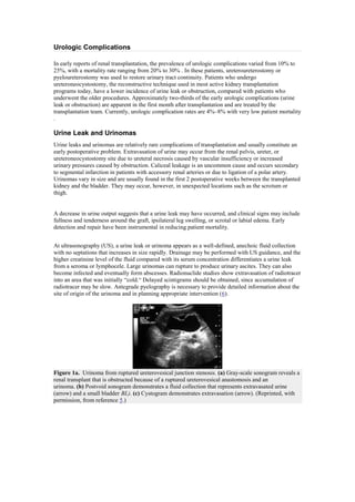
  • 1. Urologic Complications In early reports of renal transplantation, the prevalence of urologic complications varied from 10% to 25%, with a mortality rate ranging from 20% to 30% . In these patients, ureteroureterostomy or pyeloureterostomy was used to restore urinary tract continuity. Patients who undergo ureteroneocystostomy, the reconstructive technique used in most active kidney transplantation programs today, have a lower incidence of urine leak or obstruction, compared with patients who underwent the older procedures. Approximately two-thirds of the early urologic complications (urine leak or obstruction) are apparent in the first month after transplantation and are treated by the transplantation team. Currently, urologic complication rates are 4%–8% with very low patient mortality . Urine Leak and Urinomas Urine leaks and urinomas are relatively rare complications of transplantation and usually constitute an early postoperative problem. Extravasation of urine may occur from the renal pelvis, ureter, or ureteroneocystostomy site due to ureteral necrosis caused by vascular insufficiency or increased urinary pressures caused by obstruction. Caliceal leakage is an uncommon cause and occurs secondary to segmental infarction in patients with accessory renal arteries or due to ligation of a polar artery. Urinomas vary in size and are usually found in the first 2 postoperative weeks between the transplanted kidney and the bladder. They may occur, however, in unexpected locations such as the scrotum or thigh. A decrease in urine output suggests that a urine leak may have occurred, and clinical signs may include fullness and tenderness around the graft, ipsilateral leg swelling, or scrotal or labial edema. Early detection and repair have been instrumental in reducing patient mortality. At ultrasonography (US), a urine leak or urinoma appears as a well-defined, anechoic fluid collection with no septations that increases in size rapidly. Drainage may be performed with US guidance, and the higher creatinine level of the fluid compared with its serum concentration differentiates a urine leak from a seroma or lymphocele. Large urinomas can rupture to produce urinary ascites. They can also become infected and eventually form abscesses. Radionuclide studies show extravasation of radiotracer into an area that was initially ―cold.― Delayed scintigrams should be obtained, since accumulation of radiotracer may be slow. Antegrade pyelography is necessary to provide detailed information about the site of origin of the urinoma and in planning appropriate intervention (6). Figure 1a. Urinoma from ruptured ureterovesical junction stenosis. (a) Gray-scale sonogram reveals a renal transplant that is obstructed because of a ruptured ureterovesical anastomosis and an urinoma. (b) Postvoid sonogram demonstrates a fluid collection that represents extravasated urine (arrow) and a small bladder BL). (c) Cystogram demonstrates extravasation (arrow). (Reprinted, with permission, from reference 5.)
  • 2. Figure 1b. Urinoma from ruptured ureterovesical junction stenosis. (a) Gray-scale sonogram reveals a renal transplant that is obstructed because of a ruptured ureterovesical anastomosis and an urinoma. (b) Postvoid sonogram demonstrates a fluid collection that represents extravasated urine (arrow) and a small bladder (BL). (c) Cystogram demonstrates extravasation (arrow). (Reprinted, with permission, from reference 5.) Figure 1c. Urinoma from ruptured ureterovesical junction stenosis. (a) Gray-scale sonogram reveals a renal transplant that is obstructed because of a ruptured ureterovesical anastomosis and an urinoma. (b) Postvoid sonogram demonstrates a fluid collection that represents extravasated urine (arrow) and a small bladder (BL). (c) Cystogram demonstrates extravasation (arrow). (Reprinted, with permission, from reference 5.) Small urine leaks may be treated with percutaneous nephrostomy and stent placement. Caliceal leakage caused by infarction is treated with percutaneous nephrostomy alone. Ureteral stents must be kept in place for 6–8 weeks after cessation of leakage to allow complete healing of the ureter and to preserve long-term patency. If healing is unsuccessful, reimplantation of the ureter may be required; if distal necrosis is present, pyelocystostomy or pyeloureterostomy with the native ureter is performed (6). Urinary Obstruction Urinary obstruction occurs in approximately 2% of transplantations and almost always within the first 6 months after the procedure. Obstruction of the transplanted kidney may occur at any location but is most frequent at the site of implantation of the ureter into the bladder. More than 90% of ureteral stenoses occur within the distal third of the ureter. Narrowing at the ureterovesical junction may be caused by scarring secondary to ischemia or rejection, by technical error during the ureteroneocystostomy, or by kinking. These events account for more than 50% of obstructions that cause ureteral stricture. Less common causes include pelvic fibrosis, calculi, papillary necrosis, fungus ball, clots, and compression from an extrinsic mass such as adjacent peritransplant collections. Occasionally, obstruction that develops years after transplantation—especially in patients who have undergone multiple procedures—may be related to adhesions, vascular insufficiency, or fibrosis.
  • 3. Figure 2a. Hydronephrosis secondary to ureteral stricture. (a) Gray-scale sonogram demonstrates mild hydronephrosis secondary to ureteral stricture. (b)Retrograde pyelogram shows the area of narrowing at the site of ureteral implantation into the bladder (arrow). (Fig 2a and 2b reprinted, with permission, from reference5.) Figure 2b. Hydronephrosis secondary to ureteral stricture. (a) Gray-scale sonogram demonstrates mild hydronephrosis secondary to ureteral stricture. (b)Retrograde pyelogram shows the area of narrowing at the site of ureteral implantation into the bladder (arrow). (Fig 2a and 2b reprinted, with permission, from reference). The transplanted kidney is denervated; thus, the patient will not complain of typical renal colic when obstruction occurs. Urinary obstruction manifests by a rising level of serum creatinine. Obstruction may be difficult to differentiate from chronic rejection, since both cause rising creatinine levels. In addition, mild dilatation of the collecting system may occasionally be seen in cases of chronic rejection. US can be used to confirm the diagnosis of hydronephrosis; however, intrarenal edema and fibrosis associated with rejection may prevent the normal hydronephrotic response. Some transplant recipients may have substantial obstruction but little or no renal dilatation. Mild to moderate dilatation of the transplanted pyelocaliceal system and ureter may occur secondary to a full bladder. In all cases, when the patient’s bladder is full, the bladder should be emptied and the transplant imaged again. In addition, a denervated, transplanted collecting system has no tone; thus, any transient episode of hydronephrosis may cause a persistent appearance of mild dilatation. The single renal graft is responsible for excreting the entire urine output from the transplant recipient’s body, and, over time, increasing output may exacerbate pelvocaliectasis. Studies have attempted to differentiate obstructive from nonobstructivepyelocaliectasis by using the resistive index. Extrapolation of these results from native to transplanted kidneys has not, however, been entirely successful. US also demonstrateslymphoceles, hematomas, abscesses, and urinomas that may cause ureteral obstruction. Any echogenicity within a dilated collecting system is usually clinically significant. Scintigraphy may demonstrate urinary obstruction. In a patient with early partial obstruction, good perfusion and prompt uptake of the radiotracer may be seen; however, in a patient with functionally significant hydronephrosis, radioactivity is retained in the collecting system. Delayed images (obtained 2–4 hours after injection) are useful for differentiating an obstructed ureter from a dilated but
  • 4. unobstructed ureter, since an unobstructed system shows clearance into the bladder. Diuretic renography has proved useful for determining the functional significance of dilated collecting systems. Conventional clearance times (furosemide clearance t1/2>20 minutes ) can be used in the assessment of urinary tract patency. Excretory urography is only infrequently successful for locating a urinary obstruction, and the site is usually best determined with an antegrade contrast material examination. The superficial anterior position of the allograft allows easy access for antegrade pyelography performed with real-time US guidance. The Whitaker test is helpful in equivocal cases. The combination of normal results from the Whitaker test and antegrade pyelography virtually excludes the presence of obstruction. Percutaneous nephrostomy is used to relieve obstruction and allow the deployment of other radiologic interventions such as ureteral stent placement and balloon ureteroplasty. Balloon dilation of posttransplantation ureteral strictures produces an overall success rate of ureteral dilatation in 90% of cases, with the best results obtained in fresh surgical strictures and the poorest in chronic ischemic strictures or areas of periureteral fibrosis. Strictures of the middle and upper ureters result most commonly from ischemic or periureteral fibrosis and may require surgical intervention. High balloon pressures of 10–12 atm or greater are usually needed for successful dilation, and the balloon is left inflated for 20 minutes or until balloon ―waisting‖ has disappeared. After dilation, a 10-F stent is left in place for only 10 days. In the immunocompromised transplant recipient, the risk of infection precludes long-term stent placement unless a totally internal, double pigtail stent is used. Recurrences most commonly develop within the first year after transplantation. Figure 3a. Hydronephrosis relieved by stent placement.(a) Gray-scale sonogram reveals severe hydronephrosis resulting from narrowing at the ureterovesical anastomosis. Obstruction was relieved by stent placement. (b)Sonogram obtained after placement of a retrograde stent (arrow) demonstrates complete resolution of the hydronephrosis. Figure 3b. Hydronephrosis relieved by stent placement.(a) Gray-scale sonogram reveals severe hydronephrosis resulting from narrowing at the ureterovesical anastomosis. Obstruction was relieved by stent placement. (b)Sonogram obtained after placement of a retrograde stent (arrow) demonstrates complete resolution of the hydronephrosis.
  • 5. Figure 4a. Dilation of a ureteral stenosis. Sequence of fluoroscopic images (a–c)demonstrates placement of a JJ stent across a midureteral stricture. Figure 4b. Dilation of a ureteral stenosis. Sequence of fluoroscopic images (a–c)demonstrates placement of a JJ stent across a midureteral stricture. Figure 4c. Dilation of a ureteral stenosis. Sequence of fluoroscopic images (a–c)demonstrates placement of a JJ stent across a midureteral stricture. Peritransplant Fluid Collections Peritransplant fluid collections have been reported in up to 50% of renal transplantations and include urinomas, hematomas, lymphoceles, and abscesses. The clinical significance of these collections is largely determined by their size, location, and possible growth. In the immediate postoperative period, small hematomas or seromas manifesting as crescenticperitransplant collections are almost expected. Their size should be documented at baseline examination, since any increase in size may warrant intervention. Growing collections may be indicative of urine leak, abscess, or vascular injury. Different types of peritransplant fluid collections can be partially differentiated based on the time interval after transplantation. Urinomas and hematomas are most likely to develop immediately after transplantation,
  • 6. whereas lymphoceles generally occur 4–8 weeks after the surgical procedure. The sonographic characteristics of peritransplant fluid collections, however, are entirely nonspecific, and ultimately, diagnosis may be made only with percutaneous aspiration. Hematomas Hematomas are common in the immediate postoperative period, but they may also develop spontaneously or as a consequence of trauma or biopsy. They are usually small and resolve spontaneously. Large hematomas can displace the transplanted kidney and produce hydronephrosis. At US, hematomas demonstrate a complex appearance. Acute hematomas are echogenic and become less echogenic with time. Older hematomas even appear anechoic, more closely resembling fluid, and septations may develop. Similarly, at computed tomography (CT), the appearance of a hematoma also is time dependent. An acute hematoma has high-attenuation components, and older hematomas contain liquefied and serous portions of intermediate attenuation . At magnetic resonance (MR) imaging, an acute hematoma can show high signal intensity with both T1-weighted and T2-weighted pulse sequences. At scintigraphy, hematomas demonstrate a ―cold defect.‖ Figure 5a. Subcapsular hematoma in a patient who sustained blunt trauma to the abdomen while playing a sport. (a) US image demonstrates an isoechoicsubcapsular fluid collection (arrows). (b) CT scan shows a heterogeneous crescenticsubcapsular collection in the transplanted kidney secondary to hematoma (arrows). Figure 5b. Subcapsular hematoma in a patient who sustained blunt trauma to the abdomen while playing a sport. (a) US image demonstrates an isoechoicsubcapsular fluid collection (arrows). (b) CT scan shows a heterogeneous crescenticsubcapsular collection in the transplanted kidney secondary to hematoma (arrows). Diagnostic aspiration can be performed to rule out abscess formation. However, percutaneous drainage of the entire fluid collection is often neither efficacious (due to its multiloculated nature) nor advisable because of the self-limited nature of the complication and risk of infection. Lymphoceles
  • 7. Lymphoceles are the most common peritransplant fluid collections, with a prevalence of 0.5%–20%. They may develop at any time, from weeks to years after transplantation. However, they are usually an early complication, occurring within 1–2 months after transplantation. Lymphoceles are caused by leakage of lymph from surgically disrupted lymphatic channels along the iliac vessels or from the lymphatics of the transplanted kidney. Risk factors include inadequate ligation of the lymphatic channels across the iliac vessels, administration of heparin, and possibly increased lymphatic flow secondary to edema of the lower extremities. These fluid collections usually occur medial to the transplant, between the graft and the bladder . Lymphoceles are the most common fluid collection that causes transplant hydronephrosis. Patients with a failing allograft may develop ipsilateral lower extremity edema caused by compression of the femoral vein. In rare cases, lymphoceles may develop in the scrotum and lymphatic drainage may occur through the wound. At US, lymphoceles are anechoic) and may have septations. Similar to other peritransplant fluid collections, they can become infected and can develop a more complex appearance . At CT, lymphoceles have variable characteristics and are usually sharply circumscribed. Their CT attenuation values are typical of those of water and usually lower than those of recent hematomas and abscesses. Radionuclide and MR imaging studies are helpful for excluding the presence of urine and blood, respectively. Figure 6. Lymphocele. US image demonstrates mild hydronephrosis of the transplanted kidney. The anechoic area represents a lymphocele (L) adjacent to the kidney. Figure 7. Lymphocele. Sonogram of another patient shows a multilocular fluid collection (arrows) that surrounds the renal transplant. The collection proved to be a lymphocele.
  • 8. Figure 8. Infected lymphoceles. US image demonstrates a complex echogenic fluid collection (arrows) adjacent to the transplanted kidney. Small lymphoceles are monitored sonographically, and large ones, if they grow or cause hydronephrosis, should be drained. Aspirated fluid should have a creatinine content equal to that of serum, and the cell count and differential may show relatively few white blood cells with a predominance of lymphocytes. Results of Gram stain and culture of the fluid should be negative. Lymphoceles commonly recur after simple percutaneous or surgical drainage. Permanent resolution of lymphoceles may require prolonged catheter drainage and transcatheter instillation of sclerosing agents such as povidine-iodine, absolute alcohol, or doxycycline (17). Repeated aspirations run the risk of infecting an otherwise sterile fluid collection. However, success rates of up to 97% have been achieved safely and effectively with percutaneous transcathetersclerotherapy (17). If a lymphocele becomes infected, external drainage and antibiotic treatment are needed. Surgical treatment of a sterile fluid collection involves removal of a generous window of peritoneum for marsupialization. The transplanted kidney effectively becomes an intraperitoneal structure. Future leakage is therefore resorbed in the peritoneal cavity. Retrograde pyelography helps identify the course of the transplanted ureter before marsupialization, since the ureter could be displaced anteromedially and injured when the peritoneal window is constructed. Abscesses and Infection More than 80% of renal transplant recipients suffer at least one case of infection during the first year after transplantation. Early diagnosis of and intervention for infectious diseases can help prevent loss of graft function and improve patient outcome. Infections that occur in the first weeks after transplantation, such as pneumonia, surgical wound infections, and urinary tract infections, are similar to those that typically develop in nonimmunocompromised patients who have undergone surgery. Infections with opportunistic pathogens and cytomegalovirus often develop 1–6 months after surgery, and infections common in the general population are seen after 6 months. Patients frequently have multiple infections, and immunosuppressive medications, indwelling catheters, and frequent glycosuria are all risk factors. Peritransplant abscesses are an uncommon complication and usually develop within the first few weeks after transplantation. These abscesses may be caused by pyelonephritis or bacterial seeding of a lymphocele, hematoma, or urinoma. Acute bacterial nephritis, renal abscess, or perinephric abscess may occur. Patients may have few signs or symptoms of infection because of their immunosuppressed state. They may present with fever of unknown origin, pain, or symptoms related to the pressure of the abscess on the transplanted system. In a febrile transplant recipient, any peritransplant fluid collection must be presumed to be infected. Acute pyelonephritis can mimic acute graft rejection, and their imaging appearances may be the same. The sonographic appearances of infections and abscesses are quite variable. Focal pyelonephritis may appear as focal areas of increased or decreased echogenicity. These findings are nonspecific and can represent infarction or rejection. Intrarenal masses are infrequently visualized and are also
  • 9. sonographically nonspecific. They may represent primary renal cell carcinoma or posttransplantationlymphoproliferative disorder. Abscesses have a complex, cystic, nonspecific appearance at US; however, at CT, they manifest with gas, which serves to differentiate them from other collections. In emphysematous pyelonephritis, gas in the parenchyma of the renal graft produces an echogenic line with distal reverberation artifacts. This finding can be confirmed with abdominal radiography or CT. Any echogenicity within a dilated collecting system is usually clinically significant. Highly echogenic, weakly shadowing masses within a transplanted collecting system are fairly specific for fungus balls). The presence of low-level echoes in a dilated pyelocaliceal system in a febrile patient suggests pyonephrosis. Papillary necrosis results in sonographic changes in the calices, and intravenous or retrograde pyelography is the definitive study for diagnosing this condition. Retrograde pyelography may also demonstrate distortion of the calices, a feature seen in tuberculosis of the renal transplant. Gallium-67 citrate and indium-111–tagged leukocyte studies are helpful for diagnosing infections that develop 1 month or more after graft healing is complete . Figure 9. Perinephric abscess. US image demonstrates a loculatedperinephric fluid collection with complex sonographic features (arrow). The collection proved to be an infected urinoma. Figure 10a. Fungus ball of the transplanted kidney. (a)US image demonstrates a soft-tissue echogenic mass(m) in the renal pelvis of a transplanted kidney. (b)Retrograde pyelogram shows filling defects (arrows) within the renal pelvis. These findings proved to be a fungus ball. (Fig 10a and 10b reprinted, with permission, from reference 5.)
  • 10. Figure 10b. Fungus ball of the transplanted kidney. (a)US image demonstrates a soft-tissue echogenic mass(m) in the renal pelvis of a transplanted kidney. (b)Retrograde pyelogram shows filling defects (arrows) within the renal pelvis. These findings proved to be a fungus ball. (Fig 10a and 10b reprinted, with permission, from reference.) Figure 11. Hydronephrosis secondary to a fungus ball. Color Doppler US image demonstrates an echogenic mass (m) in the transplanted kidney that is causing hydronephrosis. The mass proved to be a fungus ball. Figure 12. Renal transplant tuberculosis. Retrograde pyelogram demonstrates a renal transplant with irregular margins from renal tuberculosis. (Reprinted, with permission, from reference). Abscesses may be treated with either US- or CT-guided percutaneous drainage. Both intrarenal and extrarenal abscesses usually respond to external drainage and systemic antibiotics. Vascular Complications Renal Artery Stenosis Renal artery stenosis occurs usually in the first year after transplantation. The stenosis may be located before the anastomosis (because of atherosclerotic disease in the donor vessel), at the anastomosis
  • 11. (secondary to vessel perfusion injury, faulty suture technique, or reaction to suture material), or after the anastomosis (due to rejection, turbulent flow from kidney malposition, or arterial twisting, kinking, or compression). Approximately half of renal artery stenoses occur at the anastomosis, and end-to-end anastomoses have a threefold greater risk of stenosis than end-to-side anastomoses. About 80% of patients with end-stage renal disease are hypertensive, and after renal transplantation two-thirds of this group experience a reduction in hypertension. In patients with persistent hypertension, renal artery stenosis may not be considered as a cause owing to concomitant graft failure. Several clinical scenarios should prompt a search for stenosis: (a) severe hypertension refractory to medical therapy, (b) hypertension and the presence of an audible bruit over the graft, and (c)hypertension associated with unexplained graft dysfunction. Color Doppler US demonstrates stenotic segments as focal areas of color aliasing due to increased flow velocity. These areas can be selectively evaluated with duplex Doppler techniques to characterize and grade the flow disturbance. Doppler criteria for significant stenoses include (a) focal frequency shifts greater than 7.5 KHz (when a 3-MHz transducer is used) or velocities greater than 2 m/sec, (b) a velocity gradient between stenotic and prestenotic segments of more than 2:1, and (c)marked distal disturbance (spectral broadening). In the renal parenchyma, tardus-parvus waveform abnormalities can be observed. MR angiography can be used to diagnose renal artery stenosis in renal transplants. MR angiography has the advantage of requiring either no contrast material or a gadolinium chelate that is not nephrotoxic. Renal scintigraphy and time activity curves show reduced perfusion in transplants with complete vascular obstruction and renal artery stenosis. This finding is nonspecific, however, as it may be seen with other causes of parenchymal failure, including graft rejection and urinary obstruction. Figure 13. Renal artery stenosis. Maximum intensity projection (MIP) image of a 35-year-old patient with recurrent severe hypertension who underwent a second renal transplantation shows severe proximal stenosis (arrow) in the renal artery of the first transplant. Notice the severe decrease in the perfusion of the first transplant. Surgical correction of graft renal artery stenosis is usually successful, but it is associated with substantial morbidity. Primary treatment of graft renal artery stenosis by means of percutaneous transluminal angioplasty with or without stent placement results in good intermediate-term patency and is associated with significant early improvement in blood pressure and creatinine level. Because of its low level of morbidity, relatively modest cost, and effectiveness, percutaneous transluminal angioplasty is accepted as the initial treatment of choice. Clinical success rates resulting in substantial initial improvement or cure have been reported in 73% of patients. Decrease in hypertension may be seen in 1 day, and the prevalence of periprocedural complications is substantially lower than that associated with surgical repair. Up to 20% of stenoses may require repeat dilation for maximum success.
  • 12. Figure 14a. Percutaneous transluminal angioplasty of a renal artery stenosis. (a)Angiogram shows narrowing of the proximal renal artery (arrow). (b) Another angiogram shows a catheter placed across the area of stenosis. (c) Angiogram obtained after angioplasty was performed shows restoration of a near normal arterial lumen. Figure 14b. Percutaneous transluminal angioplasty of a renal artery stenosis. (a)Angiogram shows narrowing of the proximal renal artery (arrow). (b) Another angiogram shows a catheter placed across the area of stenosis. (c) Angiogram obtained after angioplasty was performed shows restoration of a near normal arterial lumen. Figure 14c. Percutaneous transluminal angioplasty of a renal artery stenosis. (a)Angiogram shows narrowing of the proximal renal artery (arrow). (b) Another angiogram shows a catheter placed across the area of stenosis. (c) Angiogram obtained after angioplasty was performed shows restoration of a near normal arterial lumen. Infarction Renal artery thrombosis may result from hyperacute rejection, anastomotic occlusion, arterial kinking, or intimal flap. Segmental infarcts in the renal transplant may be focal or diffuse and may occur as part of rejection or as a result of an unassociated vascular thrombosis. Vasculitis may induce small segmental infarcts. Patients with renal transplant infarction present with absence of urinary output and often with swelling and tenderness over the graft and anuria. Although the graft itself is denervated, the inflammation within the transplanted kidney may incite an inflammatory response in the adjacent visceral peritoneum, with local pain in this location.
  • 13. At US, a segmental infarct appears as a poorly marginated, hypoechoic mass or a hypoechoic mass with a well-defined echogenic wall. If the infarction is global, the kidney will appear hypoechoic and be diffusely enlarged. At color or power Doppler imaging, segmental infarcts appear as wedge-shaped areas without color flow in , although these findings may also be seen in severe pyelonephritis or transplant rupture. In total vascular obstruction, there is no perfusion to the transplanted kidney, and no arterial or venous blood flow is seen in the graft at US. At technetium-99m dynamic radionuclide studies, a photopenic region may be seen. However, these findings are not specific, since hyperacute or accelerated acute rejection can have similar clinical, Doppler US, and scintigraphic features. Figure 15. Infarct of a renal graft. Power Doppler US image demonstrates segmental loss of perfusion in the transplanted kidney (arrows), a finding compatible with infarct. CT can be used to detect perfusion deficits in the renal graft parenchyma, but it is generally not employed in patients with elevated creatinine values because of the risk of compromising renal function. If any doubt exists as to the nature of the US findings, angiography or MR angiography may be performed. MR angiography is increasingly used to screen for vascular abnormalities in renal transplants. Phased-array surface coils provide excellent signal-to-noise information that permits rapid acquisition of high-quality images without the use of potentially nephrotoxic agents. Dynamic enhanced MR imaging can be useful for diagnosing both segmental and global infarctions as well as renal artery thrombosis. In addition, MR imaging has been helpful for demonstrating small infarcts caused by iatrogenic drug-induced vasculitis. Figure 16. Infarct of a renal graft. CT scan demonstrates segmental hypoattenuation (arrow) in the transplanted kidney, a finding that proved to represent an infarct.
  • 14. Figure 17a. Occlusion of a graft renal artery with lower pole infarction in a 38-year-old patient. (a) Nontargeted MIP image reveals one graft renal artery that is normal. However, a total of three graft renal arteries were anastomosed to the external iliac artery, and nonvisualization of two of the three arteries indicates perioperative thrombosis. This vascular anatomy could not be ascertained with Doppler US. (b) Another MIP image demonstrates the venous anatomy.(c) Contrast material– enhanced three-dimensional MIP image demonstrates perfusion to the upper renal pole with lack of perfusion to the lower pole, a finding that indicates lower pole infarction from arterial thrombosis. Figure 17b. Occlusion of a graft renal artery with lower pole infarction in a 38-year-old patient. (a) Nontargeted MIP image reveals one graft renal artery that is normal. However, a total of three graft renal arteries were anastomosed to the external iliac artery, and nonvisualization of two of the three arteries indicates perioperative thrombosis. This vascular anatomy could not be ascertained with Doppler US. (b) Another MIP image demonstrates the venous anatomy.(c) Contrast material– enhanced three-dimensional MIP image demonstrates perfusion to the upper renal pole with lack of perfusion to the lower pole, a finding that indicates lower pole infarction from arterial thrombosis. Figure 17c. Occlusion of a graft renal artery with lower pole infarction in a 38-year-old patient. (a) Nontargeted MIP image reveals one graft renal artery that is normal. However, a total of three graft renal arteries were anastomosed to the external iliac artery, and nonvisualization of two of the three arteries indicates perioperative thrombosis. This vascular anatomy could not be ascertained with Doppler US. (b) Another MIP image demonstrates the venous anatomy.(c) Contrast material– enhanced three-dimensional MIP image demonstrates perfusion to the upper renal pole with lack of perfusion to the lower pole, a finding that indicates lower pole infarction from arterial thrombosis.
  • 15. Figure 18a. Complete occlusion of a graft renal artery with allograft infarction in a 47-year-old patient with elevated serum creatinine levels. (a) Axial T1-weighted breath-hold gradient-echo image reveals peripheral high signal intensity involving the renal cortex (arrow), a finding compatible with hemorrhage. The patient had cortical necrosis proved at surgery. (b) Axial T2-weighted breath-hold short inversion time inversion recovery image demonstrates peripheral low signal intensity (arrow). (c) MIP image, obtained during the arterial phase of three-dimensional contrast-enhanced MR angiography, demonstrates complete occlusion of the graft renal artery beyond its origin (arrow). (d) Contrast-enhanced three-dimensional MIP image demonstrates lack of a cortical nephrogram with a peripheral rim sign (arrow), an appearance that indicates total infarction of the kidney. Figure 18b. Complete occlusion of a graft renal artery with allograft infarction in a 47-year-old patient with elevated serum creatinine levels. (a) Axial T1-weighted breath-hold gradient-echo image reveals peripheral high signal intensity involving the renal cortex (arrow), a finding compatible with hemorrhage. The patient had cortical necrosis proved at surgery. (b) Axial T2-weighted breath-hold short inversion time inversion recovery image demonstrates peripheral low signal intensity (arrow). (c) MIP image, obtained during the arterial phase of three-dimensional contrast-enhanced MR angiography, demonstrates complete occlusion of the graft renal artery beyond its origin (arrow). (d) Contrast-enhanced three-dimensional MIP image demonstrates lack of a cortical nephrogram with a peripheral rim sign (arrow), an appearance that indicates total infarction of the kidney. Figure 18c. Complete occlusion of a graft renal artery with allograft infarction in a 47-year-old patient with elevated serum creatinine levels. (a) Axial T1-weighted breath-hold gradient-echo image reveals peripheral high signal intensity involving the renal cortex (arrow), a finding compatible with hemorrhage. The patient had cortical necrosis proved at surgery.(b) Axial T2-weighted breath-hold short inversion time inversion recovery image demonstrates peripheral low signal intensity (arrow). (c) MIP image, obtained during the arterial phase of three-dimensional contrast-enhanced MR
  • 16. angiography, demonstrates complete occlusion of the graft renal artery beyond its origin (arrow). (d) Contrast-enhanced three-dimensional MIP image demonstrates lack of a cortical nephrogram with a peripheral rim sign (arrow), an appearance that indicates total infarction of the kidney. Figure 18d. Complete occlusion of a graft renal artery with allograft infarction in a 47-year-old patient with elevated serum creatinine levels. (a) Axial T1-weighted breath-hold gradient-echo image reveals peripheral high signal intensity involving the renal cortex (arrow), a finding compatible with hemorrhage. The patient had cortical necrosis proved at surgery.(b) Axial T2-weighted breath-hold short inversion time inversion recovery image demonstrates peripheral low signal intensity (arrow). (c) MIP image, obtained during the arterial phase of three-dimensional contrast-enhanced MR angiography, demonstrates complete occlusion of the graft renal artery beyond its origin (arrow). (d) Contrast-enhanced three-dimensional MIP image demonstrates lack of a cortical nephrogram with a peripheral rim sign (arrow), an appearance that indicates total infarction of the kidney. Figure 19. Multiple cortical infarctions from drug-induced vasculitis in a 42-year-old patient. Early contrast-enhanced three-dimensional MIP image reveals a large upper pole infarction with multiple smaller focal cortical areas of signal loss, findings compatible with small infarctions. Percutaneous angiographic interventional techniques may be valuable in treating infarctions and renal artery thrombosis. Early diagnosis and treatment are vital for allograft salvage. Depending on the cause and duration of the occlusion, thrombolytic therapy may prolong survival. Arteriovenous Fistulas and Pseudoaneurysms Percutaneous biopsy is commonly performed in transplant recipients when rejection is suspected. Arteriovenous fistulas and pseudoaneurysms are occasionally seen after graft biopsies. Gross hematuria is seen after 5%–7% of biopsies and is usually self limiting; however, massive or persistent hematuria may occur. Color and duplex Doppler US easily demonstrate arteriovenous fistulas. Arteriovenous fistulas and pseudoaneurysms appear as localized areas of disorganized color that extend outside the confines of the normal vessel. This appearance is caused by perifistula vibration and is detected with the more
  • 17. sensitive color Doppler units. Arteriovenous fistulas also appear as abnormal high-velocity turbulent flow isolated to a single segmental or interlobar artery and paired vein that produces aliasing on color Doppler images. The feeding artery shows a high-velocity low-resistance waveform, and the draining vein demonstrates arterialization. Figure 20a. Arteriovenous fistula. (a) Color Doppler US image demonstrates a highly vascular lesion. (b) Color duplex Doppler image shows the classic waveform of an arteriovenous fistula, with high velocities and low impedance. Figure 20b. Arteriovenous fistula. (a) Color Doppler US image demonstrates a highly vascular lesion. (b) Color duplex Doppler image shows the classic waveform of an arteriovenous fistula, with high velocities and low impedance. Figure 21a. Arteriovenous fistula. (a) Duplex Doppler US image of the lower pole segmental artery shows increased velocity and decreased resistive index.(b) Duplex Doppler US image of the adjacent vein shows arterialization of flow, a finding consistent with arteriovenous fistula. Figure 21b. Arteriovenous fistula. (a) Duplex Doppler US image of the lower pole segmental artery shows increased velocity and decreased resistive index.(b) Duplex Doppler US image of the adjacent vein shows arterialization of flow, a finding consistent with arteriovenous fistula.
  • 18. On gray-scale US images, pseudoaneurysms appear as simple or complex renal cysts, but with color imaging, highly vascular intracystic flow is seen. Pseudoaneurysms with a narrow neck and no venous communication demonstrate a classic machinelike to-and-fro Doppler spectrum at their necks. Those associated with arteriovenous fistulas exhibit a high-velocity low-resistance spectrum at their necks, with minimally pulsatile high-velocity flow in the draining vein. When the degree of perifistula vibration limits US assessment of the vascular anatomy, MR imaging enables evaluation. Figure 22. Arteriovenous fistula following transplant biopsy. A color Doppler study in a 41-year-old hypertensive patient with a bruit audited over the renal allograft revealed tissue vibration. Because of marked tissue vibration, it was difficult to interrogate the transplant vasculature. Non-targeted MIP images during the arterial phase of contrast-enhanced three-dimensional MR angiography reveal a focal severe stenosis of the graft renal artery beyond its origin (arrow). The transplanted renal venous system is enhancing during the early arterial phase along with the iliac vasculature and inferior vena cava. The cortical nephrogram is very faint due to the marked arteriovenous shunting. Figure 23. Arteriovenous fistula. Contrast-enhanced three-dimensional MR angiogram demonstrates the renal artery and veins simultaneously, an appearance suggestive of fistula formation. The diagnosis was confirmed at angiography (not shown). Most complications after biopsy are treated conservatively, and most pseudoaneurysms resolve spontaneously. If, however, a pseudoaneurysm demonstrates progressive enlargement or unusual size (more than 2 cm in diameter), intervention is warranted. Selective transvascular catheterization may be used. The size and location of the arteriovenous fistula or pseudoaneurysm determine which embolic agent to use. For example, a large arteriovenous malformation is more easily treated with steel coils, since particulate matter such as absorbable gelatin sponge (Gelfoam) may pass through the fistulous communication and embolize the systemic circulation. Peripheral lesions may be difficult to catheterize selectively, but a flow-directed detachable balloon can be delivered to the site. Microcatheter systems may also be helpful for superselective embolization of small, peripheral feeding vessels. In treatment of transplant-related arteriovenous fistulas (unlike that of peripheral arteriovenous fistulas), occlusion of the proximal bleeding vessel is sufficient. Renal Vein Thrombosis Renal vein thrombosis is an unusual complication of transplantation; it occurs in less than 5% of patients and usually in the first postoperative week. Renal vein thrombosis is heralded by an abrupt cessation of urinary function and swelling and tenderness over the graft. Hypovolemia, venous compression from a peritransplant fluid collection, dysfunctional anastomosis, and slow flow secondary to rejection or other allograft disease can also precipitate renal vein thrombosis. An increased prevalence of renal vein thrombosis in left lower quadrant allografts has also been attributed
  • 19. to compression of the left common iliac vein between the sacrum and the left common iliac artery (silent iliac artery compression syndrome. At gray-scale US, renal vein thrombosis may manifest with an enlarged kidney. Venous flow is reduced or absent, and there is increased resistance on the arterial side, often resulting in reversed diastolic flow on Doppler images. MR venography helps confirm this complication in transplants. Figure 24. Renal vein thrombosis. Duplex Doppler image demonstrates reversal of flow in diastole (arrows) in the transplanted kidney due to renal vein thrombosis secondary to deep venous thrombosis. Figure 25a. Iliac venous compression from a lymphocele in a 39-year-old patient with right lower extremity edema. (a) MIP image produced from subtracting the arterial phase data from the equilibrium phase data demonstrates a normal renal vein and poor visualization of the external iliac vein due to compression. (b) Axial source image (from a two-dimensional time-of-flight MR venographic study) helps confirm that the external iliac vein (solid arrow) is compressed by the underlying lymphocele (open arrow). Figure 25b. Iliac venous compression from a lymphocele in a 39-year-old patient with right lower extremity edema. (a) MIP image produced from subtracting the arterial phase data from the equilibrium phase data demonstrates a normal renal vein and poor visualization of the external iliac vein due to compression. (b) Axial source image (from a two-dimensional time-of-flight MR venographic study) helps confirm that the external iliac vein (solid arrow) is compressed by the underlying lymphocele (open arrow).
  • 20. Early recognition of renal vein thrombosis is crucial because the allograft may sometimes be salvaged by prompt thrombectomy. Even with prompt diagnosis, however, graft infarction may develop, and a transplant nephrectomy is usually performed to prevent secondary infection. Calculous Disease Kidney transplant recipients, compared with the general population, are at increased risk for developing urinary calculi and approximately 1%–2% develop a clinically significant stone. Persistent secondary hyperparathyroidism is seen in 38%–77% of end-stage renal disease patients after transplantation. In the first year after transplantation, 15% of patients may be hypercalcemic, which increases the risk of renal stones The diagnosis of urinary calculi is suspected when renal function suddenly deteriorates. The patient does not experience typical renal colic because the transplanted kidney is denervated. US, radionuclide imaging, and contrast material studies are all useful diagnostic methods. Percutaneous nephrostomy tube placement is valuable because it provides low-pressure drainage and allows renal function to stabilize. Antegrade pyelography can then be used to give exact definition of the collecting system and position of the stone . Figure 26a. Renal transplant calculus in a 34-year-old patient with hematuria. (a) US image demonstrates hydronephrosis with a shadowing echogenic focus seen in the upper middle renal pole (arrow). (b) US image of the distal ureter shows an echogenic focus with shadowing (arrow) a finding consistent with an obstructing calculus. Figure 26b. Renal transplant calculus in a 34-year-old patient with hematuria. (a) US image demonstrates hydronephrosis with a shadowing echogenic focus seen in the upper middle renal pole (arrow). (b) US image of the distal ureter shows an echogenic focus with shadowing (arrow) a finding consistent with an obstructing calculus. Most stones can be removed with endourologic techniques. Upper tract stones are best treated by means of percutaneous nephrostolithotomy. Cystoscopy with electrohydraulic lithotripsy is appropriate for treating bladder stones. Neoplasms
  • 21. Prolonged immunosuppression following renal transplantation places the transplant recipient at about 100 times the normal risk for developing cancer. The average reported prevalence of malignancy in these patients is 6% (38), with cases consisting predominantly of skin cancers and lymphomas. The degree of immunosuppression and its duration are both important factors in the development of malignancy. The frequency of urologic tumors such as prostatic adenocarcinoma and germ cell tumors of the testis does not appear to be increased in this population. However, the prevalence of renal adenocarcinoma may be increased, with 90% of the tumors occurring in the native kidney and 10% occurring in the transplanted kidney (39). One reason for the increased risk of renal adenocarcinoma is that approximately half of the patients who undergo hemodialysis because of chronic renal failure develop acquired renal cystic disease and 9% develop tumors (40). Patients with longer use of hemodialysis are at greater risk for developing acquired cystic disease. Fortunately, less than 1% of these patients develop malignant tumors that metastasize. In addition, patients with a significant past exposure to cyclophosphamide have an increased risk for developing of urothelial tumors (41); although cyclophosphamide is less commonly used now because of the availability of cyclosporin A, there are many renal transplant patients with a history of cyclophosphamide exposure. A complete urologic evaluation for gross hematuria is recommended in all renal transplant recipients. Intravenous pyelography may be useful if there is good transplant function, although it will not provide information about the native kidney. US or CT of the native and transplanted kidneys provides more complete information, and a solid heterogeneous enhancing mass that may be cystic is often identified. Cystoscopy with retrograde pyelography of all present kidneys and ureters is advisable. Figure 27. Renal transplant adenocarcinoma. CT scan shows a cystic mass (arrow) arising from the renal transplant that proved to be renal cell carcinoma. Transplant glomerulopathy and recurrence of preexisting glomerulonephritis frequently cause microhematuria often associated with proteinuria. However, since these patients have an increased risk of developing neoplasms, a complete urologic work-up is advisable. Development of renal adenocarcinoma in the transplanted kidney is uncommon, with the majority of malignancies related to skin cancers and lymphomas. Malignancy in the transplanted kidney is treated by total removal of the renal graft and cessation of immunosuppression. Urothelial malignancies can likewise be treated in a standard fashion. In the presence of immunosuppression, intravesical instillation of Calmette-Guérin bacillus is not recommended. Gastrointestinal and Herniation Complications Gastrointestinal complications may occur in transplantation patients. The most common of these complications is gastrointestinal hemorrhage due to peptic ulcer (42). Prevalence of the complications may vary with use of an intraperitoneal or extraperitoneal approach in placement of the allograft (43). Postoperative adhesions may lead to intestinal obstruction. Herniation of bowel through a transplant peritoneal defect may lead to compromise of the intestine or of the transplant itself (Fig 28). Urinary obstruction from obturator herniation of the ureter may also occur (44). Pseudomembranous colitis secondary to overgrowth of Clostridium difficle in patients receiving antibiotic therapy is another complication that can be seen in renal transplant recipients. A typical ―accordion sign‖ secondary to massive edema of the intestinal wall is highly suggestive of the diagnosis on CT images of the abdomen.
  • 22. Figure 28. Renal transplant herniation. CT scan demonstrates multiple distended small bowel loops around the transplanted kidney, findings compatible with obstruction. Small bowel had herniated through the peritoneal defect related to the renal graft, a diagnosis that was surgically proved. PosttransplantationLymphoproliferative Disorder Impaired immune surveillance and the oncogenic effects of immunosuppressive therapy itself may be causative factors of the posttransplantationlymphoproliferative disorder (PLTD). An association with Epstein Barr virus is usually demonstrated. PLTD complicates 8% of transplantations and is diagnosed at a median of 80 months after transplantation. The liver, brain, and lung are more common sites of involvement than the gastrointestinal tract. PLTD lesions are polymorphic collections of B cells but may evolve into forms indistinguishable from non-Hodgkin lymphoma. If there is involvement of the small intestines, patients often have generalized disease. Figure 29a. Posttransplantationlymphoproliferative disease in a 25-year-old renal allograft recipient who presented with abdominal pain. (a) Contrast-enhanced CT scan demonstrates circumferential thickening of the jejunum (arrows). (b)Contrast-enhanced CT scan obtained at a lower level shows encasement of the superior mesenteric artery by lymphadenopathy (arrowheads), in addition to the jejunal thickening (arrow). Figure 29b. Posttransplantationlymphoproliferative disease in a 25-year-old renal allograft recipient who presented with abdominal pain. (a) Contrast-enhanced CT scan demonstrates circumferential thickening of the jejunum (arrows). (b)Contrast-enhanced CT scan obtained at a lower level shows encasement of the superior mesenteric artery by lymphadenopathy (arrowheads), in addition to the jejunal thickening (arrow).
  • 23. Figure 30. Posttransplantationlymphoproliferative disease in a kidney transplant recipient who presented with fever. Contrast-enhanced CT scan demonstrates a necrotic mass in the pelvis posterior to the transplanted kidney (arrows). Conclusions Transplantation is currently one of the accepted treatments of irreversible kidney disease. Improvements in surgical techniques and more sophisticated, potent immunosuppressive drugs have resulted in remarkable advances in survival of patients and renal grafts. Nevertheless, substantial complications occur in both the immediate postoperative period and later. Imaging has a critical role in the evaluation of these complications, and interventional radiologic techniques are often successfully applied to their treatment.