Transgenic animals
•Descargar como PPT, PDF•
16 recomendaciones•1,321 vistas

Transgenic animals

Recomendados
Más contenido relacionado
La actualidad más candente
La actualidad más candente (20)
Destacado
Destacado (20)
Similar a Transgenic animals
Similar a Transgenic animals (20)
Más de kv meg and centre
Más de kv meg and centre (20)
Último
Último (20)
Transgenic animals
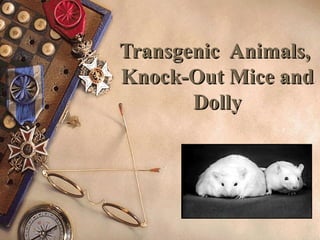
- 1. Transgenic Animals,Transgenic Animals, Knock-Out Mice andKnock-Out Mice and DollyDolly
- 2. Transgenic Animals: A Focus on Transgenic Mice Studies http://www.hku.hk/biochem/tgcentre/transcentre.html
- 3. Introduction Transgenic animals: – Animals which have been genetically engineered to contain one or more genes from an exogenous source. – Transgenes are integrated into the genome. – Transgenes can be transmitted through the germline to progeny. – First transgenic animal produced = “Founder Animal”
- 4. Introduction of foreign genes into intact organisms Procedure is basically the same regardless of which animal is involved. Integration usually occurs prior to DNA replication in the fertilized oocyte. – Majority of transgenic animals carry the gene in all of their cells, including the germ cells. Transmission to next generation requires germline integration. – Some integration events occur subsequent to DNA replication giving rise to mosaic animals which may or may not contain the transgene in its germline.
- 5. Procedure for Producing Transgenic Mice Three different breeding pairs of mice are required.
- 6. First Breeding Pair: – Fertile male + superovulated female • Fertile male = stud (changed regularly to ensure performance) • Superovulated female = immature female induced to superovulate – Pregnant mare’s serum (=FSH) on day 1 – Human Chorionic Gonadotropin (=LH) on day 3 • Mated on day 3 • Fertilized oocytes microinjected on day 4 with foreign DNA construct. • Microinjected oocytes are transferred to the oviducts of surrogate mothers at end of day 4.
- 7. Second breeding pair: – Sterile male + surrogate mother • Sterile male produced through vasectomy • Surrogate mother must mate to be suitable recipient of injected eggs • Mated on day 3 • Microinjected oocytes from first breeding pair are transferred to oviducts on day 4 • Embryos implant in uterine wall and are born 19 days later. • Southern blotting techniques confirm presence and copy number of transgenes.
- 9. Third breeding pair: – Foster parents • Fertile male + female mated to give birth on same day surrogate mother • Serves as foster parent if caesarian section is required for surrogate mother
- 11. Manipulation ofManipulation of Fertilized OocytesFertilized Oocytes Refer to transparencies and slides presented in class.
- 13. Flow Cytometry for the Analysis of Transgenic Mice
- 15. Integration of Transgene into One Chromosome Normally the transgene inserts into one chromosome giving rise to a heterozygote. – 50% probability of passing transgene onto offspring. Two heterozygous mice may be bred to obtain a homozygous line that contains the transgene on both chromosomes. – 100% probability of passing transgene onto offspring. Most transgenes are stably transmitted for many generations without detectable rearrangement.
- 16. Mechanisms of DNA Integration Linear molecules integrate more efficiently than circular molecules (~5x) Once in the oocyte, the linear molecules circularize. Usually all of the molecules that integrate are on the same chromosome and at the same site. Multiple copies are usually arranged in a tandem, head- to-tail array. The size of the DNA molecule (0.7 – 50Kb) is not an important parameter. The concentration and purity of the injected DNA is critical (1-3 µg/ml maximum).
- 17. Working Hypothesis of DNA Integration The ends of the injected linear DNA integrate at breaks that occur spontaneously in the chromosome. Other injected molecules which have circularized probably recombine with each other and the integrated copies to generate a tandem, head-to-tail array. Recombination is probably favored because of high local DNA concentration and special properties such as the absence of normal chromatin structure. The number of chromosomal breaks is presumably limiting explaining the low number of integration events and why different DNA molecules are usually integrated at the same site.
- 18. Gene Expression in Transgenic Mice In order to discriminate the products of the injected gene from those of an endogenous counterpart, the injected gene must be marked in some way. – Mini-genes where exons are deleted of cDNA where introns are absent. – Modification by insertion/deletion/mutagenesis of a few nucleotides (e.g. the gain or loss of a restriction endonuclease site). – Hybrid genes where foreign epitopes are expressed on transgenic products.
- 19. Tissue-Specific Gene Expression Generally, if a tissue-specific gene is expressed at all, then it is expressed appropriately, despite the fact that it has integrated at a different chromosomal location. – Trans-acting proteins involved in establishing tissue-specific expression are capable of finding their cognate sequences and activation transcription at various chromosomal locations. – Levels of expression vary between founder animals as chromosomal position can influence accessibility of the transgenes to these trans-acting transcription factors. – Some founders do not express the transgene at all owing to integration into heterochromatin domains where DNA is methylated heavily (silent).
- 20. Prokaryotic Sequences Must be Removed for Optimal Expression Prokaryotic sequences derived from the plasmid or bacteriophage vector used for replication of the transgene in bacteria can be inhibitory or “poisonous” for some transgenes. Therefore, the transgene fragment requires purification from contaminating vector sequence prior to microinjection.
- 21. Possible Reasons for Lack of Transgene Expression Integration in cis-acting silencer sequences (the negative counterpart of enhancer elements) might be sites for covalent modification of DNA (e.g. methylation) which might initiate condensation into an inactive chromatin configuration, or they might phase nucleosomes in an inappropriate manner. The inadvertent loss of certain regulatory sequences during the production of the constructs (e.g. topoisomerase-binding sites, nuclear matrix-attachment sites). Use of cDNA rather than genomic DNA. (Introns thought to contribute to stability of mRNA and may even contain enhancer sequences essential for tissue-specific expression. Flanking DNA may also contain regulatory sequences.)
- 22. Examples of Studies Utilizing Transgenic Mice The Oncomouse – c-myc oncogene + MMTV sequences breast cancer – Int-2 oncogene + viral promoter prostate cancer To obtain abnormal expression of genes to study their effects – Rat growth hormone + cadmium-inducible metallothionein promoter – Transgenic mouse was much larger, but also suffered complications with fertility and organ diseases
- 23. To study developmentally regulated genes http://www.ucihs.uci.edu/anatomy/calofpix1b.html
- 24. More Examples of Studies Utilizing Transgenic Mice “Pharm” animals (transgenic livestock) – Bioreactors whose cells have been engineered to synthesize marketable proteins – DNA constructs contain desired gene and appropriate regulatory sequences (tissue-specific promoters) – More economical than producing desired proteins in cell culture
- 25. Examples of Bioreactors Naked human Hb from pigs Human lactoferrin in cows’ milk Alpha-1-antitrypsin in sheep HGH in mouse urine (uroplakin promoters) Human antibodies in mice (H and L chain tgenics hybridomas) CfTCR in goats Tissue plasminogen activator (TPA) in goats Human antithrombin III in goats Malaria antigens in goats (vaccine) Alpha-glucosidase in rabbits (Pompe’s disease
- 29. Transgenic Pigs for the Production of Organs for Transplantation Pig organs are rejected acutely due to the presence of human antibodies to pig antigens. Once human antibodies are bound to pig organs, human complement is activated and triggers the complement cascade and organ destruction. Transgenic pigs with complement inhibitors have been produced and are gaining feasibility as a source of xenogeneic organs for transplantation.
- 34. What is a Knockout Mouse? A really good-looking mouse? A mouse in which a very specific endogenous gene has been altered in such a way that interferes with normal expression, i.e. it has been knocked out.
- 35. Why Produce KO Mice? To study effects of gene products, biochemical pathways, alternative (compensatory) pathways, and developmental pathways To recreate human diseases in animals to establish models to test the beneficial effects of drugs or gene therapy.
- 36. Procedure for Generating a KO Mouse Gene alteration in KO mice is targeted to very specific genes. DNA must integrate at precise positions in the genome. Integration of the altered gene takes place in embryonic stem cells ex vivo. Verification of exact location of integration occurs before the ESC is introduced into blastocysts to become part of the developing embryo.
- 37. Pluripotent ES Cells Pluripotent ES cells are undifferentiated early embryonic cells derived from the inner cell mass of mouse blastocysts. In vitro ES cells must be grown on a feeder layer of fibroblasts to prevent them from differentiating. Introduction of the transgene is achieved by electroporation of retroviral infection. The transgene must integrate via recombination, not randomly. Cells transfected successfully can be identified prior to injection into blastocysts.
- 38. Specific Gene Targeting in ES Cells Gene targeting can be achieved using gene constructs designed for homologous recombination. This technique can be used to either: – Knockout functional genes to study their contribution to different developmental or disease processes (null mutations) • Genes encoding β2m, MHC class I and II. CD2, Ii, TCR, Ig, IL-4, IL-2, FcεR, TAP1/2, RAG-2,and many more (>100)! – Replace a functional gene for a mutated/non- functional gene to restore wild type phenotype . • Gene encoding HGPRT in mice deficient for HGPRT (called Lesch-Nhyan syndrome in humans).
- 39. DNA Constructs for Recombination DNA vectors contain the gene of interest which has been interrupted with an antibiotic resistance gene (hygromycin resistance, or G418 resistance). To ensure targeted integration has occurred, the flanking DNA contains the thymidine kinase gene. If TK integrates (random insertion), then the transfected cells die when grown in selective media (gancyclovir).
- 41. Selection of Targeted ES Cells Gancyclovir resistant and G418 resistant ES cells grow into small clumps on top of feeder cells. The colonies of cells can be “picked” off and transferred to new wells (at 0.3 cells per well seeding density) containing feeder cells. When sufficient numbers of cells are obtained, they are: – Frozen for safe storage – Analyzed by Southern blotting or PCR to determine nature of integration event – Microinjected into the blastocoel cavity of blastocysts.
- 44. Dolly and theDolly and the Advancement ofAdvancement of Animal CloningAnimal Cloning
- 46. How Was Dolly Made?