
Recomendados
Más contenido relacionado
La actualidad más candente
La actualidad más candente (20)
Similar a Identifying Histological Structures in Tissue Samples Less Than 40 Characters
Similar a Identifying Histological Structures in Tissue Samples Less Than 40 Characters (20)
Más de heidiw
Último
Último (20)
Identifying Histological Structures in Tissue Samples Less Than 40 Characters
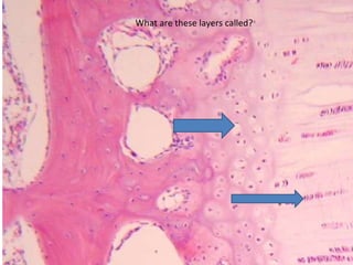
- 1. What are these layers called?
- 2. Identify the structures indicated
- 3. Identify the type of epithelium and what tissue you are looking at
- 4. Identify the structures indicated
- 5. Identify the structures indicated.
- 6. Identify the structure indicated (the connective tissue layer).
- 7. What type of muscle is this? Identify the structure indicated.
- 8. What type of muscle is this? Identify the structure indicated.
- 9. Identify the connective tissue structure.
- 10. Identify this type of muscle.
- 14. Identify this cortical tubule.
- 15. Identify this area of the kidney.
- 16. Where is this histological section found? What is the location of the pointed structure?
- 17. Characterize this tissue and name its location.
- 18. Identify the two layers of the Dermis and characterize their Connective Tissue.
- 19. Identify the structures pointed by arrows.
- 20. Identify the cartilage found and its location.
- 21. Name the primary cells of epithelim and name the layers of Muscularis Mucosae.
- 22. Identify location of the histological section and name its characterizing feature.
- 23. Name the glands found in this section.
- 24. Which region of the stomach is this and how can you tell?
- 25. What are the two cell types found here?
- 26. What is this structure?
- 27. What is this cell?
- 28. What is this layer?
- 29. What is this structure?
- 30. What is this layer?
- 34. IDENTIFY AND DESCRIBE THE CONTENTS OF THE CELLS IN LAYER “X”
- 35. IDENTIFY THE STRUCTURES THAT LINK THE CELLS IN THIS LAYER
- 37. A
- 39. Z
- 40. *
- 46. What is the layer identified by the arrow?
- 47. What cellular structure is this pointing at?
- 48. What is this structure?
- 49. What cell type is this?
- 50. What is the arrow pointing at?
- 51. *
- 53. *
- 56. What type of secretion occurs in the sebaceous gland?
- 58. Identify the structure of the arrow and the layer of B.
- 60. What type of cell is indicated?
- 67. What type of granules make up this layer?
- 68. What is the name of this cell?
- 69. What cell is this?
- 70. What cell is the blue arrow pointing to?
- 76. Identify the layer marked by the asterisk
- 77. Identify the cell marked by the arrow with the asterisk
- 78. Identify the layer marked by the asterisk *
- 79. A The lacuna at A gains a white appearance as it fills with what substance?
- 80. * What hormone is the cell (marked by the asterisk) regulated by?
- 81. What type of secretion does this gland undergo?
- 82. Identify this structure.
- 83. I Classify this gland.
- 84. * Identify this (*) structure.
- 85. * Identify this structure.
- 86. Identify the type of secretion
- 87. Identify this layer of IRS
- 88. Identify the type of epithelium
- 89. Identify the type of secretion
- 90. Identify granules contained in cells
- 91. 1)… 2)…
- 92. 1) …
- 94. Zone of…
- 95. 1) … 2) … Name the two layers and the structure that they make up.
- 96. Name the areas below: 1)… 2)…
Notas del editor
- Zone of resting cartilage (hyaline cartilage) Zone of proliferation
- Submucosa-plicae circularis Tunica muscularis – inner circular Simple columnar epithelium with goblet cell
- Regular Bronchile Simple ciliated columnar epithelium
- Hepatic arteriole Portal venule Bile duct
- Serous demilune Striated duct (interlobular duct)
- endomysium
- Cardiac muscle, glycogen halo
- Cardiac muscle, intercalated disk
- Perimysium
- Skeletal muscle (longitudinal section)
- 1. Medullary rays
- 2. PCT
- 3. Bowman’s space/urinary space
- 4. DCT
- 5. Area Cribosa
- Hyaline Cartilage found in Trachea. Location of the chondrocytes is in matrix lacunae.
- Dense Regular CT found in the tendon
- Papillary Layer and Reticular Layer of Dermis. Papillary Layer has loose irregular CT. Reticular Layer has dense irregular CT.
- The secretory portion of the sweat gland and adipocytes.
- Elastic cartilage in larynx.
- Large Intestine. Its epithelium is primarily composed of goblet cells secreting mucous. There are two layers in the muscularis mucosae. The inner circular and outer longitudinal.
- Brunner’s glands found in duodenum
- Pyloric region of the stomach. Notice the 1:3 (4) Pit to gland ratio.
- Zymogenic cells and parietal cells found in pits and glands of stomach
- Isogenic group
- Chondroblast
- Perichondrium
- Secretory portion of the sweat gland
- Papillary layer of dermis
- 1) Stratified squamous keratinized epithelium
- 2) dermis
- 3) basement membrane
- 4) stratum granulosum: keratohyalin granules
- 5) desmosomes
- 1) Periosteum
- 2) Cementing Line
- 3) Condrocyte
- 4) Isogenic group
- 5) Calcitonin
- Goblet cell
- Respiratory bronchiole
- Alveolar macrophage
- Cilia
- Regular bronchiole
- Tunica muscularis
- Goblet cell
- Crypt of ileum
- Parietal cell
- Brush border
- 1) Zymogen granules
- 2) Striated duct
- 3) Sinusoid
- 4) Islet of Langerhans, Serrous acini
- 5) Arteriole (Not cholangiole!)
- 1. Holocrine
- 2. Excretory duct of sweat gland
- 4. Tricohyaline granules and Glassy membrane
- 5. Dermal Papilla
- 6. Myoepithelial Cell
- 1. Dentin
- 2. Gingiva
- 3. Taste Bud
- 4. Pulp
- 5. Stellate Reticulum
- 6. Von Ebners Gland
- Keratohyaline granules
- Pneumocyte Type II
- Clara cells
- Fibrocyte
- What is this? Capillary
- Layers of elastic membrane
- Arteriole
- Lumen of elastic artery
- Lumen of large vein
- Lab 4---Compact Bone (low mag) Inner Circumferential Layer
- Osteocyte
- Zone of Proliferation
- Glycogen
- Calcitonin
- Lab 10---Salivary Gland Merocrine
- Image 5—Submandibular gland Striated Duct
- Image 8---Sublingular Mucous Acini with Serous Demilunes
- Image 16---kidney---label one of the arrows Intercalated duct
- In the Portal Space Hepatic Arteriole
- Holocrine secretion (sebaceous gland)
- Henle’s Layer (squamous and keratinized)
- Stratified cuboidal epithelium (duct of sweat gland)
- Merocrine secretion (pointing at secretory portion of a sweat gland)
- Tricohyline granules-acidophilic in Huxley’s layer
- Dentin Enamel Stellate Reticulum
- 1) Sinusoids
- Arteriole
- Cell death
- Fibrous layer Osteogenic layer Periosteum
- Epiphyseal Plate Epiphysis