Furcation involvement (Dr. Himanshu Shekhar)
•Descargar como PPT, PDF•
162 recomendaciones•12,595 vistas

detailed discussion about furcation.

Recomendados
Más contenido relacionado
La actualidad más candente
La actualidad más candente (20)
Destacado
Destacado (14)
Similar a Furcation involvement (Dr. Himanshu Shekhar)
Similar a Furcation involvement (Dr. Himanshu Shekhar) (20)
Último
Último (20)
Furcation involvement (Dr. Himanshu Shekhar)
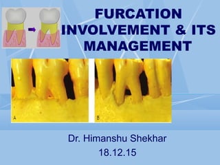
- 1. FURCATION INVOLVEMENT & ITS MANAGEMENT Dr. Himanshu Shekhar 18.12.15
- 2. Contents Introduction Etiologic Factors Diagnosis & Classification of Furcation Defects Local anatomic factors Root trunk length Root length Interradicular dimension Anatomy of Furcation Cervical Enamel Projections Anatomy of the bony lesions Pattern of attachment loss Other Dental Findings Treatment Therapeutic Classes of Furcation Defects Non-surgical Therapy Oral Hygiene Procedures Scaling & Root Planing Surgical Therapy Osseous Resection Regenerative Root Resection Hemi-section Root resection/Hemi-section Procedure Extraction Dental Implants Prognosis
- 3. Introduction The furcation is an area of complex anatomic morphology [Bower RC 1979] that may be difficult or impossible to debride by routine periodontal instrumentation. [Metzler DG 1991]
- 4. Introduction … The presence of furcation involvement is a sign of advanced periodontitis poor prognosis.
- 5. Definition
- 6. Parts of furcation: Root complex Root trunk Root cone Furcation entrance Furcation fornix Degree of seperation Divergence Coefficient of separation
- 10. Mandibular Molars 100% mesial roots 99% distal roots Maxillary Molars 94% mesiobuccal roots 31% distobuccal roots 17% palatal roots
- 11. Is larger than the second molar. The distal surface of the mesio-buccal root has a concavity which is about 0.3 mm deep (Bower 1979). This concavity gives the cross section of the mesio-buccal root an “hourglass” configuration .
- 12. The mesial furcation entrance is located about 3 mm from the CEJ, while the buccal is 3.5 mm and the distal entrance about 5 mm apical to the CEJ (Abrams & Trachtenberg 1974; Rosenberg 1988).
- 14. The mandibular first molar is larger than the second molar, which in turn is larger than the third molar. The cross section of the distal root is circular while the mesial root has an “hour-glass” shape. In addition, furrows and concavities often occur on the distal surface of the mesial root . The distal concavity of the mesial root is more pronounced than that of the distal root (Bower 1979a,b; Svärdström & Wennström 1988).
- 15. The lingual entrance is frequently found more apical to the CEJ (>4 mm) than the buccal entrance (>3 mm). Thus, the furcation fornix is inclined in the bucco-lingual direction. The buccal furcation entrance is often <0.75 mm wide while the lingual entrance is >0.75 mm in most cases (Bower 1979). The degree of separation and divergence between the roots decreases from the first to the third molar.
- 17. In about 40% of cases the maxillary first premolars have two root cones, one buccal and one palatal, and hence have a mesiodistal furcation. A concavity (about 0.5 mm deep) is often present in the furcation aspect of the buccal root. In many cases the furcation is located in the middle or in the apical third of the root complex . The mean distance between CEJ and the furcation entrance is about 8 mm. The width of the furcation entrance is about 0.7 mm.
- 18. Furcations may be present also in teeth which normally have only one root. In fact, two-rooted incisors , canines , and mandibular. premolars may exist. Occasionally three-rooted maxillary premolars and three-rooted mandibular molars can be found .
- 19. Bacterial plaque Inflammatory consequences Extent of attachment loss required to produce a furcation defect. Local anatomic factors Root trunk length Root morphology Local developmental anomalies Cervical enamel projections Enamel pearls Iatrogenic TFO Combined Origin
- 20. Local factors may affect the rate of plaque deposition or complicate the performance of oral hygiene procedures, thereby contributing to the development of periodontitis and attachment loss. Studies indicate that prevalence and severity of furcation involvement increase with age. [Hou GL JP 1978] Dental caries and pulpal death may also affect a tooth with furcation involvement or even the area of the furcation. All of these factors should be considered during the diagnosis, treatment planning, and therapy of the patient with furcation defects.
- 21. prevalence of furcation involvement In periodontal patients > 40 years old, all second molars have furcation involvement. Maxillary molars (distal) > mandibular molars > Maxillary Premolars (Larrato et al 1970) Highest frequency of involvement, distal furcation of maxillary first molar Svardstrom & Wennström 1996 Greater severity of furcation involvement in smokers Kerdvongbundit & Wikesjo 2000 Incidence of 35% in Mandibular Molars 90% in Maxillary Molars (Ross & Thompson 1980)
- 22. Clinical examination of the patient should allow the therapist to identify not only furcation defects but also many of the local anatomic factors that may affect the result of therapy (prognosis). Well-made dental radiographs, although not allowing a definitive classification of furcation involvement, provide additional information vital for treatment planning.
- 23. A key factor in both the development and the treatment of furcation involvement. The distance from the cementoenamel junction to the entrance of the furcation can vary extensively. Teeth may have very short root trunks, moderate root trunk length, or roots that may be fused to a point.
- 24. Root length is directly related to the quantity of attachment supporting the tooth. Teeth with long root trunks and short roots may have lost a majority of their support by the time that the furcation becomes affected. [Hermann DW 1983] Teeth with long roots and short-to-moderate root trunk length are more readily treated because sufficient attachment remains to meet functional demands.
- 25. The mesial root of most mandibular first and second molars and the mesio-buccal root of the maxillary first molar are typically curved to the distal side in the apical third. The curvature and fluting may increase the potential for root perforation during endodontic therapy or complicate post placement during restoration. [Langer B 1981][Langer B 1981] These anatomic features may also result in an increased incidence of vertical root fracture.
- 26. The degree of separation of the roots is also an important factor in treatment planning. Closely approximated or fused roots can preclude adequate instrumentation during scaling, root planing, and surgery. Teeth with widely separated roots present more treatment options and are more readily treated.
- 27. The presence of bifurcational ridges, a concavity in the dome, [Everett F 1958][Everett F 1958] and possible accessory canals [Gutmann JL 1978][Gutmann JL 1978] complicates not only scaling, root planing, and surgical therapy [Matia JB[Matia JB 1986]1986] but also periodontal maintenance. Odontoplasty to reduce or eliminate these ridges may be required during surgical therapy for an optimal result.
- 28. Cervical enamel projections (CEPs) are reported to occur on 8.6% to 28.6% of molars. [Larato DC 1975, Masters DH 1964][Larato DC 1975, Masters DH 1964] The prevalence is highest for mandibular and maxillary second molars. These projections can affect plaque removal, can complicate scaling and root planing, and may be a local factor in the development of gingivitis and periodontitis. CEPs should be removed to facilitate maintenance.
- 29. CEPs are flat, ectopic extensions of enamel that extend beyond the Normal contours of the CEJ. (Masters DH 1964)
- 30. • Predilection for maxillary third and second molars. • The prevalence is less than that of cervical enamel projections. • incidence of 2.6% (range 1.1–9.7%). Moskow & Canut (1990) Enamel pearls
- 31. Pattern of Attachment Loss Other Dental Findings
- 32. Horizontal bone loss can expose the furcation as thin facial/lingual plates of bone that may be totally lost during resorption. Alternatively, areas with thickened bony ledges may persist and predispose to the development of furcations with deep vertical components.
- 33. Complex multiwalled defects with deep, interradicular vertical components may be candidates for regenerative therapies. Alternatively, molars with advanced attachment loss on only one root may be treated by resective procedures.
- 34. The dental and periodontal condition of the adjacent teeth must be considered during treatment planning for furcation involvement.
- 37. This is an early lesion. The pocket is suprabony, involving the soft tissue. There is slight bone loss in the furcation area. Radiographic change is not usual since bone loss is minimal. A periodontal probe will detect root outline or may sink into a shallow V-shaped notch into the crestal area.
- 38. The level of bone loss allows for the insertion of the periodontal probe into the concavity of the root trunk.
- 39. In this, bone is destroyed in one or more aspects of the furcation, but a portion of the alveolar bone and periodontal ligament remain intact, permitting only partial penetration of the probe into the furca. Radiographs may or may not reveal this type of furcation.
- 40. The level of bone loss allows for the insertion of a periodontal probe into the furcation area between the roots.
- 41. In class III probe penetrates completely from one side to the other side characterized by severe bone destruction in the furcation area. It is clearly shown in the radiographs as a radiolucent area in between the roots, especially in the lower molars.
- 42. As in Class III, but the gingival tissues recede apically so that furcation is clearly visible.
- 43. GRADE II degree I VERTICAL COMPONENT OF >1MM BUT <3MM GRADE II degree II VERTICAL COMPONENT OF > 3 MM BUT NOT THROUGH & THROUGH
- 44. Incorporated a descriptive classification of furcation involvement as follows: GRADE I: Incipient GRADE II: Cul- de- sac GRADE III: Through and through
- 46. F 0 - No furcation involvementNo furcation involvement Hamp et al 1975 (horizontal component) F1- Probed 3mm in horizontal direction.F1- Probed 3mm in horizontal direction.
- 47. F2 -Probed deeper than 3mmProbed deeper than 3mm F3 - Through and throughThrough and through
- 48. Ramjford and Ash in 1979( horizontalRamjford and Ash in 1979( horizontal component)component) Classified similarly but instead of 3 mm increments, they utilized 2 mm increments. Class I: Beginning Involvement: The tissue destruction should not extend more than 2 mm [or not more than 1/3rd of the tooth width] into the furcation. Class II: Cul-de-sac Involvement: Tissue destruction extends deeper than 2 mm [or more than 1/3rd of the tooth width] into the furcation opening. Class III: Through and through. Tissue destruction extends throughout the entire length of furcation, so that an instrument can be passed between the roots and emerges on the other side of the tooth
- 50. VERTICAL CLASSIFICATION 1984 Vertical component of furcation is measured From floor of the furcation to the roof of the furcation
- 51. Hou et al. (1998): (horizontal &vertical component) In 1998 Hou et al. presented a new classification of furcation involvement based on -- root trunk , horizontal and vertical bone loss. The types of root trunk were classified on the basis of the ratio of the vertical dimension of the root trunk to root length as type A, B and C where Type A – Root trunk involving the cervical third of root length. Type B - Root trunks involving the cervical 2/3rd of the root length. Type C - Root trunks involving more than the cervical 2/3rd root length.
- 52. A thorough clinical examination is the key to diagnosis and treatment planning. Careful probing is required to determine the presence and extent of furcation involvement.
- 53. Transgingival sounding may further define the anatomy of the furcation defect. [Mealy BL 1994] The goal of this examination is to identify and classify the extent of furcation involvement.
- 54. Factors contributed to the dev. of the furcation defect or that could affect treatment outcome- (1) the morphology of the affected tooth, (2) the position of the tooth relative to adjacent teeth, (3) the local anatomy of the alveolar bone, (4) the configuration of any bony defects, and (5) the presence and extent of other dental diseases (e.g., caries, pulpal necrosis).
- 57. Naber’s curved 1 & 2 probes with Gradation 3,6,9,12 mm. No 23 explorer
- 58. The probe’s tip is initially applied on the root trunk just under the gingival margin in order to locate the furcation entrance.
- 59. Should include both peri-apical & bitewing. Sometimes superimposition of palatal root or thick bone may obscure furcation.
- 60. Slightest radiographic change in furcation area should be investigated clinically, especially if there is bone loss on adjacent roots. Diminished radio density in furcation area in which outlines of bony trabeculae are visible. o Whenever there is marked bone loss in relation to a single molar root, it may be assumed that the furcation is also involved.
- 61. Introduced subgingivally to visualize furcation. Consist of re-usable fiber optic endoscope which fits onto the periodontal probes & ultrasonic instruments that have been designed to accept it.
- 62. Thin fan of x- ray beam rotates around the patient to get multiple axial slice of the area of interest . These slices are merged to get the final image.
- 64. Pulpal pathosis may some times cause a lesion in the periodontal tissues of the furcation. Trauma from occlusion may cause inflammation and tissue destruction within the interradicular area of a multirooted tooth
- 67. Class I: Early Defects Incipient or early furcation defects (Class I) are amenable to conservative periodontal therapy. [Goldman HM 1958] Because the pocket is suprabony and has not entered the furcation, oral hygiene, scaling, and root planing are effective. [Gutmann JL 1978] Any thick overhanging margins of restorations, facial grooves, or CEPs should be eliminated by odontoplasty, recontouring, or replacement.
- 68. Class II Once a horizontal component to the furcation has developed (Class II), therapy becomes more complicated. Shallow horizontal involvement without significant vertical bone loss usually responds favorably to localized flap procedures with odontoplasty, osteoplasty, and ostectomy. Isolated deep Class II furcations may respond to flap procedures with osteoplasty and odontoplasty.
- 69. This reduces the dome of the furcation and alters gingival contours to facilitate the patient's plaque removal.
- 70. Classes II to IV: Advanced DefectsClasses II to IV: Advanced Defects Nonsurgical treatment is usually ineffective because the ability to instrument the tooth surfaces adequately is compromised. [Parashish AO 1993] Periodontal surgery, endodontic therapy, and restoration of the tooth may be required to retain the tooth.
- 71. Furcation involvement degree I Recommended therapy: Scaling and root planing, Furcation plasty. Furcation involvement degree II Recommended therapy: Furcation plasty, Tunnel preparation, Root resection, Tooth extraction & Guided tissue regeneration. Furcation involvement degree III Recommended therapy: Tunnel preparation, Root resection, Tooth extraction. Lindhe et al 2008
- 72. Oral hygiene procedures Scaling & Root Planing Kooher T., Plagmann H.C. (1999) modified sonic & ultrasonic scaler inserts to increase effectiveness of root surface instrumentation of molars with furcation involvement
- 73. Odontoplasty should eliminate any facial grooves or cervical enamel projections, enamel pearls. Root caries is another possibility if excessive tooth structure is removed (Goldman 1958, Fleischer et al 1989).
- 75. Odontoplasty + Osteoplasty when done together, termed furcationplasty. This reduces the dome of furcation and alters gingival contours to facilitate patients plaque removal. Hamp, Nyman and Lindhe (1975)
- 79. Regenerative therapy induce new attachment and reconstruction on molars with furcation defects. Many surgical procedures using a variety of grafting materials have been tested on teeth with different classes of furcation involvement. Furcation defects with deep two-walled or three- walled components may be suitable for reconstruction procedures.
- 80. BONE GRAFTS The furcation area is characterized by defects, the walls of which are primarily of tooth structure. The area is capable of holding a graft, it has little or no vascularity to support the graft, due to this reason the success of graft is limited in furcation defects (Sepe, Saunders) Grafts are indicated where destruction of furcation is only partial (grade I or II) or where deep vertical lesions have still left some bone on the inner aspects of the roots. (Bowers and Colleagues,2003)
- 81. Tsao et al have shown that the furcation defect is a graftable lesion. They found that lesions that were grafted had greater vertical fill than areas treated with open flap debridement alone.
- 83. GTR IN FURCATION DEFECTS BECKER ET AL 1988 – TOTAL OF 3.7 MM GAIN FOR VERTICAL DEFECTS PONTORIERO 1987,1988,1989,1992 – Grade ii and iii defects with Gore- tex membrane – 90% of grade ii and 25 to 35% of grde iii Defects were non probable - stated that grade iii furcation With less than 3mm entrance height can have complete closure METZER ET AL 1991 - Gtr has limited therapeutic modality For mesial and distal maxillary grade ii furcations. LEKOVIC ET AL 1989 In his re-entry study stated that – no diff in bone level between control and test but significant Improvements in the probing depth reduction of upto 4.09mm. SELVIG 1990 – Postulated – membrane potentiates or contributes To clot stability and protection by protecting the clot from disruptive Movements of the over lying flap. GOTTLOW & KARRING 1992 – 5 yr follow-up study and stated that – Attachment gain obtained could be maintained over a period up to 5 yrs OPENPROBING ATTACHMENT
- 84. LINDHE 2003 – REVIEW – 21 CLINICAL TRAILS (423 MANDIBULAR GRADE II FURCATION MOLARS) 1) No difference between bioresorbale and non resorbable membranes. 2) Gtr significantly improved the horizontal clinical attachment (cal-h) result over ofd ( 2.5mm vs 1.3mm). 3) Gtr significantly improved vertical attachment and reduction in pd. 4) Cal-h in maxillary furcation was only 1.6mm 5) Both the 1st and the 2nd mandibular molars were equal.
- 86. Conclusions: Overall, GTR was consistently more effective than OFD in reducing open horizontal furcation depths, horizontal and vertical attachment levels and pocket depths for mandibular or maxillary class II furcation defects. However, these improvements were modest, variable and there was only a limited number of studies available to appraise the effects, thus limiting general conclusions about the clinical benefit of GTR Future studies should aim to identify factors associated with achieving consistent and more pronounced benefits over open flap debridement.
- 88. Results: The meta-analysis showed that the GTR and GTR WITH OG groups obtained greater furcation closure rate, vertical/horizontal bone fill, and vertical/horizontal attachment level gain than the OFD group in mandibular molars. The GTR group obtained greater vertical/horizontal bone fill and vertical attachment level gain than the OFD group in maxillary molars. The GTR OG group achieved better clinical outcomes than the GTR group did in all the comparing outcomes in mandibular molars.
- 89. GTR with bone grafts in furcations:GTR with bone grafts in furcations: The predictability of connective tissue attachment gain is high using GTR for furcation involvement, but new bone formation is not predictable in the same area. (Schallhorn,Becker). Reports have shown that bone regeneration is increased by the use of bone grafts in combination with GTR for new bone formation. (Schallhorn and McClain). • The success is likely due to the fact that bone grafts facilitate space making and membrane placement.
- 90. GRADE II FURCATION IN MAXILLARYGRADE II FURCATION IN MAXILLARY MOLAR TREATED WITH BONE GRAFT &MOLAR TREATED WITH BONE GRAFT & RESORBABLE & NON RESORBABLERESORBABLE & NON RESORBABLE MEMBRABEMEMBRABE A) Grade II furcation in buccal aspect of maxillary molar B) DFDBA placed in the defect
- 91. D) Surgical reentry after 1 year of healing C) a. treated with non resorbable (Gore Tex ) membrane. b.Treated with resorbable vicryl mesh membrane
- 92. B) Teflon membrane sutured in position & supported by interproximal alveolar bone. A) Grade II furca in lingual aspect of mandibular first molar
- 93. D) Surgical reentry after 6 months of healing C) Flap sutured back in position
- 94. BIOLOGIC MEDIATORS • Enamel matrix derivative (EMD),Enamel matrix derivative (EMD), • Platelet-derived growth factor (PDGF),Platelet-derived growth factor (PDGF), • Platelet concentrates (PRP, PRF),Platelet concentrates (PRP, PRF), • Bone morphogenetic proteins (BMPs),Bone morphogenetic proteins (BMPs), • Fibroblast growth factor (FGF),Fibroblast growth factor (FGF),
- 95. Rossa C et al 2000: Regeneration of Class III furcation defects in dogs with bFGF + GTR. Concluded : bFGF in smaller doses – may enhance regenerative results in Class III furcation lesions , leading to greater filling with both mineralised & non- mineralised tissues. Nevins et al 2003: •Periodontal regeneration in humans (in intrabony defects & molar class II furcation ) – rhPDGF- BB & Allogenic bone •Concluded : use of purified rhPDGF-BB + Allograft robust periodontal regeneration in both Class II furcation & intrabony defect. •1st report of demonstrating periodontal regeneration histologically in human class II furcation defects.
- 96. PROCESS BY WHICH ONE OR MORE ROOTS OF A TOOTH ARE REMOVED AT THE LEVEL OF FURCATION WHILE LEAVING THE CROWN & REMAINING ROOTS IN FUNCTION
- 97. Modified from Basaraba.N ; Root amputation & Hemi section Dent clin North Am 13 (1) 121 ; 1969 INDICATIONS 1.Severe vertical bone loss on one Root of a multirooted tooth not amenable for Regeneration. 2. Furcation involvement not treatable / maintainable. 3. Periodontally involved abutment with hopeless one Root. 4. Vertical or horizontal Root fracture. 5. Endodontic therapy impossible on one Root of multirooted tooth. 1.Severe vertical bone loss on one Root of a multirooted tooth not amenable for Regeneration. 2. Furcation involvement not treatable / maintainable. 3. Periodontally involved abutment with hopeless one Root. 4. Vertical or horizontal Root fracture. 5. Endodontic therapy impossible on one Root of multirooted tooth. CONTRAINDICATIONS 1. Advanced bone loss around more than one Root with unfavorable crown-Root ratio 2. Fused roots that cannot be separated. 3. If remaining root(s) would be inadequate to serve as prosthetic abutment. 4. Poor socio-economic conditions 5. In the presence of inadequate oral hygiene. 1. Advanced bone loss around more than one Root with unfavorable crown-Root ratio 2. Fused roots that cannot be separated. 3. If remaining root(s) would be inadequate to serve as prosthetic abutment. 4. Poor socio-economic conditions 5. In the presence of inadequate oral hygiene.
- 98. 1. Remove the Root that has least amount of Remaining bone support & greatest amount of attachment loss. 2. Remove the Root that will eliminate the Furcation and produce an easily maintainable architecture. 3. Remove the Root with greatest number of anatomic problems like severe curvature, developmental grooves or accessory and multiple canals. 4. Remove the Root that is difficult for endodontist to treat. 5. Remove the Root that least complicates future periodontal maintenance. 6. Remove the Root that best contributes to the elimination of periodontal problems on adjacent teeth. The most commonly resected Root is distobuccal of Maxillary first molar due to less surface area and divergence The most commonly resected Root is distobuccal of Maxillary first molar due to less surface area and divergence
- 101. Resective treatment of multi-rooted teeth only root resection and tooth hemisection. (The Guidelines for Periodontal Therapy produced by the American Academy of Periodontology in 1992) ROOT AMPUTATION VITALVITAL NON VITALNON VITAL HEMISECTION & BICUSPIDIZATION
- 102. REMOVAL OF THE ROOT APICAL TO FURCATION WITH OUT REMOVAL OF THE CORRESPONDING CROWN STRUCTURE. May be indicated in multi-rooted teeth with advanced gradeII to grade IV furcation involvement.
- 103. Hemisection is the splitting of a two-rooted tooth into two separate portions. This process has been called bicuspidization or separation because it changes the molar into two separate roots. Molars with advanced bone loss in the interproximal and interradicular zones are not good candidates for hemisection.
- 104. After sectioning of the teeth, one or both roots can be retained. This decision is based on the extent and pattern of bony loss, root trunk and root length, ability to eliminate the osseous defect, and endodontic and restorative considerations. The anatomy of the mesial roots of mandibular molars often leads to their extraction and the retention of the distal root to facilitate both endodontic and restorative therapy.
- 106. HEMISECTION Is the surgical removal of a Root of multirooted tooth along with the corresponding portion of the crown. INDICATIONS 1. Vertical root fracture or root perforation during endodontic therapy 2. Endo dontically treated Mandibular molar with advanced bone loss on only one root. 3. Grossly decayed one portion of crown with calcified corresponding root. HEMISECTION Is the surgical removal of a Root of multirooted tooth along with the corresponding portion of the crown. INDICATIONS 1. Vertical root fracture or root perforation during endodontic therapy 2. Endo dontically treated Mandibular molar with advanced bone loss on only one root. 3. Grossly decayed one portion of crown with calcified corresponding root. BICUSPIDIZATION Is the splitting of a Mandibular molar & retaining both the fragments so as to change the molar into two separate units. INDICATIONS 1. Mandibular molars with Buccal & Lingual Class II or III Furcation involvements. BICUSPIDIZATION Is the splitting of a Mandibular molar & retaining both the fragments so as to change the molar into two separate units. INDICATIONS 1. Mandibular molars with Buccal & Lingual Class II or III Furcation involvements.
- 107. After appropriate local anesthesia, a full-thickness mucoperiosteal flap is elevated. With contra-angel hand piece & cross- cut bur sever the root where it joins the crown . Remove the root. With stone or diamond point smooth the resected root stump & contour the tooth to create easily cleansable area Clean the area & replace the flap , sutured & cover with periodontal pack . Root Resection Procedure The most common root resection involves the distobuccal root of the maxillary first molar, [Basten CH 1996].
- 108. Tunneling is a periodontal surgical procedure that creates access for patient cleaning, and maintenance within the furcal area of a molar tooth that has incurred severe attachment loss owing to periodontal disease.
- 109. Tunneling often fails because of decay in the furcation area (Lindhe, 1983). Hellden and colleagues (1989) concluded that teeth with tunnel preparations have a considerably better prognosis than that previously reported.
- 110. Potential development of root caries. Sensitivity Exposure to patent lateral canals that will require endodontic therapy in the future. Requirement that a patient should have good manual dexterity to maintain optimal oral hygiene.
- 111. The goal was to improve plaque control by eliminating the automatic niches within the furcation where bacteria can accumulate. ZOE: Kingsberg et al(1981) advocated the use of polymeric reinforced zinc oxide-Eugenol (IRM) & reported clinical success for up to 5 years. Kalkwart and Reinhardt(1988) reported in their clinics progressive bone loss around ZOE material and increased plaque retention.
- 112. SILVER AMALGUM: Van Swol et al. (1993) utilized amalgam restoration to fill grade-II furcation invasions. But on 1 year radiographic follow-up noted, radiolucency at the base of the restoration. RESIN IONOMER & GLASS IONOMER Dragoo (1997) demonstrated histologic evidence that both epithelium and connective tissue can adhere to the resin ionomer when placed in a subgingival environment. Reddy KP (2005) concluded, a glass ionomer restorative material may be effective as an occlusal barrier when treating maxillary molar grade III furcation defects.
- 113. The extraction of teeth with through-and-through furcation defects (Classes III and IV) and advanced attachment loss. This is particularly true for individuals who cannot or will not perform adequate plaque control, who have a high level of caries activity, who will not commit to a suitable maintenance program, or who have socioeconomic factors that may preclude more complex therapies. Some patients are reluctant to accept periodontal surgery. Although additional attachment loss may occur, such teeth may survive a significant number of years. [ Ross I 1978]
- 114. The advent of osseointegrated dental implants as an alternative abutment source has had a major impact on the retention of teeth with advanced furcation problems.
- 115. LASERs in Furcations A study to evaluate the influence of PhotodynamicPhotodynamic Therapy (PDT)Therapy (PDT) on bone loss in furcation areas in rats with experimentally-induced periodontal disease was conducted Concluded that within the parameters used in this study,Concluded that within the parameters used in this study, PDT may be an effective alternative for control of bone lossPDT may be an effective alternative for control of bone loss in furcation areas in periodontitisin furcation areas in periodontitis Photodynamic Therapy de Almeida JM 2008
- 117. Treated 20 Human Mandibular class ii Furcation Defects using PDL cells & observed Significant Regeneration & bone fill.
- 118. • Degree I could be successfully managed by nonsurgical mechanical debridement. • Tunneling Dental Caries Extraction • Resective surgeries Endodontic failures & Vertical root fractures • Complete furcation closure was not predictably achieved following GTR or the application of EMD in maxillary and mandibular molars.
- 119. Prognosis for individual teeth depend on: 1.Morphology of the bone deformity. 2.Root anatomy 3.Tooth morphology 4.Chronicity of the destructive process. 5.Clinical crown to clinical root ratio. 6.Mobility: Tooth mobility caused by inflammation and trauma from occlusion may be correctable, but mobility resulting from loss of alveolar bone alone is not likely to be corrected. 7.Patients age and general health 8.Therapists knowledge and skill.
- 120. Grade I & II Furcation ----- Good Prognosis. Grade III (early stage) ----- Fair Prognosis Grade III (advanced) & IV -- Poor Prognosis Prognosis is poor for short, tapered roots and relatively large crowns. Because of disproportionate crown to root ratio and reduced root surface available for periodontal support, the periodontium is more susceptible to injury by occlusal forces.
- 121. Molars with varying degrees of FI can be effectively treated and preserved similar to that of teeth with intact furcations. Effective management of furcation defects requires knowledge of the anatomic problems associated with FI and the application of treatment methods that are consistent with the nature and extent of the involvement. The keys to successful treatment of molar FI are Early diagnosis, thorough treatment planning, good oral hygiene by the patient, careful technical execution of the therapeutic modality and a well designed and implemented program of periodontal maintenance.
- 122. 1.CARRANZA 10th and 11th edition. 2.Lindhe –clinical periodontology. 3. Periodontology 2000, Vol. 9, 1995, 69-89 PERIODONTOLOGY 2000 “Management of furcation involvement” GIANFRANCCOA RNEVALREO, BERTOP ONTORIER&O M ARKUBS. HURZELER. 4. Jop 2011 Oct;82(10):1396-403. E pub 2011 Feb 2. 5. Journal of Clinical Periodontology Volume 36, Issue 7, pages 581– 588, July 2009.
- 123. AAAAAAAAAAAAAA
Notas del editor
- .[17,23]
- Root complex is the portion of a tooth that is located apical to the cemento-enamel junction (CEJ), i.e. the portion that normally is covered with a root cementum.
- is the portion of the root that is located apical to CEJ which is normally covered with cementum
- Loss of attachment on radicular flat surface or in furcation, is strongly correlated with subgingival plaque.
- The shorter the root trunk, the less attachment needs to be lost before the furcation is involved. Once the furcation is exposed, teeth with short root trunks may be more accessible to maintenance procedures, and the short root trunks may facilitate some surgical procedures. Alternatively, teeth with unusually long root trunks or fused roots may not be appropriate candidates for treatment once the furcation has been affected.
- The pattern of bone loss on other surfaces of the affected tooth and adjacent teeth must also be considered during treatment planning.
- The resolution of inflammation and subsequent repair of the periodontal ligament and bone are usually sufficient to restore periodontal health.
- These vertical bony deformities respond favorably to a variety of surgical procedures, including debridement with or without membranes and bone grafts.
- Hemisection is most likely to be performed on mandibular molars with buccal and lingual Class II or III furcation involvements.