exodontia / dental implant courses
•
51 recomendaciones•4,779 vistas

The Indian Dental Academy is the Leader in continuing dental education , training dentists in all aspects of dentistry and offering a wide range of dental certified courses in different formats.

Recomendados
Recomendados
Más contenido relacionado
La actualidad más candente
La actualidad más candente (20)
Destacado
Destacado (20)
Similar a exodontia / dental implant courses
Similar a exodontia / dental implant courses (20)
Más de Indian dental academy
Más de Indian dental academy (20)
Último
Último (20)
exodontia / dental implant courses
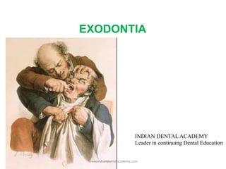
- 1. EXODONTIA INDIAN DENTAL ACADEMY Leader in continuing Dental Education www.indiandentalacademy.com
- 2. 1. Introduction 2. Definition 3. Indications 4. Contraindications 5. Presurgical assessment 6. Armamentarium 7. Mechanical principles of extraction 8. Methods of extraction 9. Extraction procedure. 10. Post extraction instructions 11. Sequence of extraction 12. Complications 13. Conclusion Contents: www.indiandentalacademy.com
- 3. Introduction: Extraction of the teeth is a procedure that combines the principles of surgery and principles of physics and mechanics. When these principles are applied correctly, a tooth can be easily removed from a alveolar process without untoward force or sequelae. www.indiandentalacademy.com
- 4. Definition According to Geoffrey Howe---- www.indiandentalacademy.com
- 5. 1.Carious teeth that is non restorable 2.Periodontally involved teeth 3.Teeth which can’t be restored endodontically 4.Prophylactic removal of teeth for radiation therapy 5.Teeth in the line of fracture 6.Fractured teeth which is non restorable 7.Teeth associated with pathologic lesions 8.Malposed teeth 9.Cracked teeth 10.Impacted teeth www.indiandentalacademy.com
- 6. 11. Supernumerary teeth 12. Orthodontic reasons 13. Prosthetic reasons 14. Retained deciduous teeth 15. Extraction of teeth for osteotomy. 16. Financial issues www.indiandentalacademy.com
- 7. Most feared side effect of radiotherapy is osteoradionecrosis. Should teeth be extracted ? Extraction may spare the patient, months or years of suffering from osteoradionecrosis www.indiandentalacademy.com
- 8. Woman's head undergoing radiotherapy treatment for basal cell carcinoma Attempt is made to remove a good portion of the alveolar process along with the teeth & achieve a primary soft tissue closure Traditionally 7 to 14 days between tooth extractions and radiotherapy has been suggested Radiotherapy should be delayed for 3 weeks if possible to ensure sufficient soft tissue healing www.indiandentalacademy.com
- 9. If tooth is grossly displaced, severely mobile, or grossly decayed – remove If tooth is non carious & appears secure in alveolar bone – retain www.indiandentalacademy.com
- 10. Contraindications for extraction Systemic contraindications Local contraindications Absolute Relative Absolute Relative www.indiandentalacademy.com
- 11. Systemic contraindications Absolute Relative Severe uncontrolled metabolic diseases ( Brittle diabetes & End stage renal disease with severe uremia) Uncontrolled leukemia & lymphoma Severe uncontrolled cardiac diseases (severe MI, unstable angina pectoris, malignant hypertension, recent MI, CVA Pregnancy Severe bleeding disorders like hemophilia or platelet disorder Pt. taking certain medications like corticosteroids, immunosuppressive agents, cancer chemotherapeutic agents www.indiandentalacademy.com
- 12. Local contraindications Absolute Relative Teeth located in malignant tumor Vascular lesions like A-V malformations & Hemangioma in the vicinity of the tooth H/O therapeutic radiation (Osteoradionecrosis) Severe Pericoronitis around impacted molars www.indiandentalacademy.com
- 13. Medical history Dental history (history of difficult extraction) Patient’s emotional maturity Clinical examination Radiographic examination www.indiandentalacademy.com
- 16. Radiographic examination Condition of the surrounding bone www.indiandentalacademy.com
- 22. Armamentarium: Extractionforceps Even though elevator can be used to luxate the tooth, it’s extraction forceps that does most of the work. Forceps have blades and handles joined by a hinge joint. The larger the ratio between the length of the handle and length of the blades, greater the leverage that can be exerted upon the root. www.indiandentalacademy.com
- 23. Ideally, the blade of the forceps should be sharp and also the whole of the inner surface of the forceps blades should fit the root surface. But because of variation in size & shape of tooth from individual to individual, it is not practicable. Hence a forceps is termed ideal if two point contact is achieved. www.indiandentalacademy.com
- 24. Expansion of the body socket by use of wedge shaped beaks of the forceps and the movement of the tooth itself. Removal of the tooth from the socket www.indiandentalacademy.com
- 26. How a forceps cause luxation of tooth and expansion of bony socket Apical pressure : Helps to wedge the forceps firmly in PDL space but results in minimal movement of the tooth Care should be taken so that beaks are not placed too apically and also not above the CEJ. Buccal force : results in expansion of buccal cortical plate. lingual force : cause expansion of lingual cortical plate. www.indiandentalacademy.com
- 27. Rotational force : cause rotation of tooth and this results in internal expansion of the tooth socket. This force in used in single, conical rooted tooth. Tractional force : used to deliver the tooth from the socket once adequate bone expansion is achieved. www.indiandentalacademy.com
- 28. Dental elevators Indications: To reflect mucoperiosteum To luxate & remove teeth which can’t be engaged by the beaks of forceps To remove fractured or carious root To loosen teeth prior to application of forceps To remove intraradicular bone www.indiandentalacademy.com
- 29. Rules when using elevators: Never use adjacent tooth as fulcrum unless that tooth is to be extracted also. Never use buccal plate at the gingival line as a fulcrum, except where odontectomy is performed Never use lingual plate at the gingival line as a fulcrum. Always use finger guards to protect the patient in case the elevator slips. Forces applied should be under control. When cutting through interseptal bone, take care not to engage the root of an adjacent tooth, thus inadvertently forcing it from its alveolus. www.indiandentalacademy.com
- 30. Classification of elevators: According to use:- 1. Elevators designed to remove the entire tooth( 1L- 1R). E.g. Cross bar elevator 2. Elevators designed to remove roots broken off at the gingival line(30-4-5) E.g. Apexo elevators 3. Elevators designed to remove roots broken off halfway to the apex (30-4-5, or 14L-14R, or 11L- 11R) E.g. Apexo elevators, Cryer elevator, cross bar elevator 4. Elevators designed to remove apical third of the root E.g. Apical fragment ejectors No. 1, 2 & 3 5. Elevators design to reflect mucoperiosteum E.g. Periosteal elevator According to Form:- 1. Straight: wedge type 2. Angular: Right & Left 3. Cross bar( handle at right angle to shank) www.indiandentalacademy.com
- 34. Mechanical principles of elevators LEVER & FULCRUM PRINCIPLE www.indiandentalacademy.com
- 37. Mechanical principles of extraction 1. Expansion of bony socket www.indiandentalacademy.com
- 38. 2. Use of Lever & fulcrum 3. Insertion of wedge or wedges B/W root & bony socket wall. www.indiandentalacademy.com
- 39. Methods of extraction Forceps extraction or Closed extraction Trans alveolar extraction or Open or surgical extraction www.indiandentalacademy.com
- 40. EXTRACTION PROCEDURE Over garment Mask Cap: hair covered Eye protection www.indiandentalacademy.com
- 41. Patient’s & operator’s position Administration of L.A Severing of gingival attachment from the tooth Positioning of Elevators & Forceps for extraction Role of other hand Compression of socket Pressure pack www.indiandentalacademy.com
- 42. Patient’s & operator’s position www.indiandentalacademy.com
- 44. Forceps always held with palm of hand above the handles of the forceps. Patient is inclined 15-20o for extraction in the lower left quadrant & 30 – 45o in the other 3 quadrants. Dentist stands behind the patient for extraction in the lower right quadrant & in front of the patient for all other extractions. www.indiandentalacademy.com
- 45. Forceps are usually held with the palm of the hand below the handles of the forceps The patient is usually inclined 30-45 degrees for all extractions The dentist normally stands behind the patient in all extractions www.indiandentalacademy.com
- 46. “You have to see well what you do in order to do well what you see” G.C Ingham www.indiandentalacademy.com
- 47. Position of Elevators & Forceps www.indiandentalacademy.com
- 48. Specific technique for removal of each tooth Maxillaryteeth: The left index finger of the surgeon should reflect the lip and cheek tissue Thumb should rest or palatal process. Stabilize the patients head and support alveolar process In this position of extraction surgeon can frequently feel the left palatal root of the molar becoming free in alveolar process. Right side index finger is positioned on the palate and thumb on buccal aspect. www.indiandentalacademy.com
- 54. Maxillary 2nd molar : anatomy is similar to the first molar except the roots are short and less diverged. Buccal roots are more lowerly fused and same techniques used as 1st molar. Maxillary 3rd molar has conical root extracted with no.210s forceps. Universal forceps for both right and left side. Erupted 3rd molar usually extracted with elevators. www.indiandentalacademy.com
- 55. Mandibular teeth: Index finger of left hand in buccal vestibule and second finger in lingual vestibule reflecting lip cheek and tongue The thumb of the left hand is placed below chin to support the mandible and minimize Temporomandibular joint pressure Bite block on contra lateral side also useful in limiting pressure on TMJ www.indiandentalacademy.com
- 56. strong buccal and lingual motion used to expand the socket and allow the teeth to be delivered in buccal-occlusal direction. Lingual bone is thinner in 2nd molar hence can more easily be removed with stronger lingual than buccal pressure. Erupted 3rd molar usually have fused conical roots. care should be taken not to use large amount of forces. www.indiandentalacademy.com
- 60. Removal of broken roots www.indiandentalacademy.com
- 64. Alveolar purchase technique Kruger recommended a unique closed method technique for removal of anterior teeth or root. A fractured tooth often can be grasped by root forceps, or anterior forceps. Alveolar purchase may be obtained by detaching labial gingival cuff. Then labial beak of forceps is placed under the tissue on the labial plate of the alveolar plate of the bone. The pressure on a forceps grasp the root along with labial alveolar bone By this technique the root can be delivered with labial alveolar bone easily. www.indiandentalacademy.com
- 65. Role of other hand www.indiandentalacademy.com
- 68. Compression of socket: Pressure pack: A B C www.indiandentalacademy.com
- 69. Debride only if necessary- Periapical pathology Debris Compress the expanded buccal and lingual plate Granulation tissue of gingiva should be curetted when required Bone margins should be smooth Hemorrhage should be controlled www.indiandentalacademy.com
- 70. Post extraction instructions 1. Patient should be advised to firmly bite on the gauze piece placed on the extraction socket for a minimum of half an hour after extraction. 2. Patient should be advised not to rinse his mouth vigorously for the next 24 hours. 3. To avoid any hot food or beverages for the next 24 hours. Patient should take cold food. 4. Advised to take soft diet on the day of extraction. 5. Not to suck from a straw on the day of extraction. 6. Warm saline rinses & gentle brushing should be advised from the next day. 7. Do not spit. 8. Take prescribed medication on time. www.indiandentalacademy.com
- 71. Sequence of extraction Max. post. except 1st molar Max. ant. Except canine Mand. post. Except 1st molar Mand. canine Mand. ant. Except canine Mand. 1st molar Max. canine Max. 1st molar www.indiandentalacademy.com
- 72. Procedure involves reflection of a mucoperiosteal flap, cutting of the bone obstructing the removal of the tooth & if required sectioning of the roots & then removal Forceps extraction of these teeth resulted in removal of bone & tooth instead of just tooth www.indiandentalacademy.com
- 73. Indications : 1. Attempts at forceps extraction have failed 2.Retained roots, especially those in close proximity to the maxillary sinus 3.History of difficult or attempted extractions. 4. Heavily restored tooth 5. Hypercementosed & ankylosed teeth 6. Geminated & dilacerated teeth 7. Teeth shown radiographically to have -Complicated root patterns or -Roots with conflicting lines of withdrawal www.indiandentalacademy.com
- 74. Heavy buccal plate suggests difficult forceps extraction Teeth exhibiting bruxism may have denser bone & stronger PDL attachment www.indiandentalacademy.com
- 75. procedure: Raising a flap Removal of bone Tooth division Removal of tooth Wound toilet Primary closure www.indiandentalacademy.com
- 78. Flap must be large enough to allow easy access without stretching Flap must be full thickness and include periosteum when reflected www.indiandentalacademy.com
- 79. Incisions that outline the flap must be made over intact bone The flap should avoid injury to local vital structures In the mandible vertical releasing incisions could damage the lingual nerve & the mental nerve www.indiandentalacademy.com
- 80. Releasing incisions should be used only when necessary When necessary, only single vertical incision used Healing time for a short flap is comparatively less to that for a long one www.indiandentalacademy.com
- 81. Types of mucoperiosteal flap www.indiandentalacademy.com
- 85. Maxillary bone much more porous than mandibular Mallet should follow with a sequence of taps Advantage • ease of autoclaving • no irrigation required www.indiandentalacademy.com
- 86. No: 8 rose head bur Rose head burs cut more efficiently & clog less than flat fissure burs www.indiandentalacademy.com
- 87. Rose head burs have 3 advantages over flat fissure burs for removal of bone 1. More easily cooled by water jet 2. Site of cutting more readily seen 3. Easier to control www.indiandentalacademy.com
- 88. Chisel and Mallet Quicker and cleaner removal of bone when used correctly No heat production Do not require irrigation No chances of bone necrosis Not pleasant for the patient Bur Slow removal of bone comparatively Heat production Require constant irrigation Chances of bone necrosis due to heat Comparatively pleasant www.indiandentalacademy.com
- 90. Particularly useful in cases where Lines of withdrawal of different roots conflict Minimize need to remove more bone www.indiandentalacademy.com
- 91. Teeth sectioned using either osteotome or powered hand piece Before splitting the tooth, point of application to elevator facilitate delivery of the roots must be made Separate the root mass of the lower molar from below upwards www.indiandentalacademy.com
- 92. Progress of healing & the amount of after-pain greatly influenced by the care with which postoperative socket cleaning is performed Judicious bone removal will speed healing by reducing the • amount of bone to be resorbed & remodeled • Volume of blood-clot which fills the socket www.indiandentalacademy.com
- 93. Surgical defect must be sealed from the oral environment by replacement of the flap The flap acts as a primary dressing & contains the osteogenic layer of periosteum which will help promote bone regeneration www.indiandentalacademy.com
- 94. 3 conditions must exist for a tooth to be left in the alveolar process • Root fragment must be small • Root deeply embedded in bone • Root must not be infected www.indiandentalacademy.com
- 95. Removal of root will cause excessive destruction of surrounding tissue Removal of root endangers vital structures Attempts of recovering the root can displace it into the maxillary sinus or tissue spaces www.indiandentalacademy.com
- 97. COMPLICATIONS OF TOOTH EXTRACTION ARE MANY & VARIED Some are unavoidable (occurs even when utmost care is exercised) Some are avoidable (if proper planning & surgical execution done) www.indiandentalacademy.com
- 102. Dry socket Causes: Preexisting infection Trauma to bone during extraction Decreased bleeding due to hemostatic effect of epinephrine or other vasoconstrictor injected with L.A Infection entering into the socket after the tooth has been removed Presence of dense bone General dehabilitation Loss of clot because of rinsing of the mouth or sucking the woundwww.indiandentalacademy.com
- 103. Etiology & pathogenesis of fibrinolytic alveolitis [ BIRN’S HYPOTHESIS] www.indiandentalacademy.com
- 105. Before undertaking the extraction of a tooth, one should thoroughly evaluate the problems involved and select the right anesthesia , radiographs and forceps. The right procedure is selected to yield the best result. Hurry always leads to worry Conclusion: www.indiandentalacademy.com
- 106. references Contemporary oral & Maxillofacial Surgery, 5th Edition James R. Hupp, Edward Ellis III, Myron R. Tucker The extraction of teeth, 3rd Edition Geoffrey Howe Minor oral surgery Geoffrey Howe, 3rd Edition Oral & Maxillofacial Surgery, Vol: 2 Daniel M. Laskin Oral & Maxillofacial Surgery, Vol: 1 W. Harry archer, 5th Edition www.indiandentalacademy.com
Notas del editor
- Extraction is the primary & original procedure of oral surgery so much that the dentistry for a layman implies extraction, while an apparently simple procedure, the process of extraction of teeth is like a serpentile path with many twist and turns.
- The ideal tooth extraction is the painless removal of the whole tooth, or tooth-root, with minimal trauma to the investing tissues, so that the wound heals uneventfully & no postoperative prosthetic complication is created.
- Malposed teeth causing soft tissue trauma or can’t be aligned by orthodontic treatment and prevents construction of opposing denture should be extracted.
- Many absolute contraindications are relative contraindications if we have requisite training & equipments. An absolute contraindication is a situation which makes a particular treatment or procedure absolutely inadvisable. In a baby, for example, aspirin is absolutely contraindicated because of the danger that aspirin will cause Reye syndrome. A relative contraindication is a condition which makes a particular treatment or procedure somewhat inadvisable but does not rule it out. For example, X-rays in pregnancy are relatively contraindicated (because of concern for the developing fetus) unless the X-rays are absolutely necessary. Reye syndrome is sudden (acute) brain damage (encephalopathy) and liver function problems of unknown cause. The syndrome has occurred with the use of aspirin to treat chickenpox or the flu in children. However, it has become very uncommon since aspirin is no longer recommended for routine use in children.
- There are a few absolute contraindications to the removal of teeth when it is necessary for the well-being of the patient . However there are a no. of instances where it may be judicious to delay extraction until local n systemic contraindications r corrected or modified. Diabetes- 1.peripheral circulation is reduced as a result of deposition of cholestrol into peripheral vessels. 2. high% of sugar in the body fluids helps in bacterial growth by supplying the organisms with a rich source of food. Recent MI- defer Rx for 6 months becoz 1. some pts. Are physiologically more stable after they have had a chance to recover from the infarction. 2. some of the more unstable postmyocardial infarction patients exclude themselves by having second infarction, the likelihood of which decreases with increasing time since initial infarct. 3. If symptomatic teeth can’t be palliated by use of antibiotics, then emergency Rx is indicated even in the relatively immediate postinfarction period. The operating room is the safest setting 4 these pts. Although suitably equipped & staffed ambulatory facility is satisfactory in most cases. Hemophillia: mild- 10 to 50% factor, major surgeries cause excessive bleeding. Moderate: 2 to 10% factor levels, mild trauma causes excessive bleeding. Severe: <2% factor level, spontaneous hemarthroses. aPTT & CT increased, Manage- whole blood, FFP & cryoprecipitate not used nowadays, freeze dried factor 8 & 9 concentrate, desmopressin acetate(vasopressin analogue), EACA & tranexemic acid (antifibrinolytic) block conversion of plasminogen to plasmin.
- Malignant tumor- spread of tumor & non healing wound in extn site.
- First evaluate number Then Curvature Shape Size length
- No.2 for mesial application & no.3 distal.
- Apexo elevators No.4(302), 301, no. 5(303) are particularly effective in the removal of roots #ed at gingival line.
- Lever 1- scissor, elevators. Lever 2- Nut cracker, Lever 3- tweezer.
- Apexo elevators are wedge elevators
- 2.Use of lever & fulcrum to force a tooth or root out of the socket along the path of least resistance. 3. Beaks of forceps act as wedge to expand alveolar bone & displace tooth in occlusal direction.
- Five general steps in closed extraction- 1. loosening of soft tissue attachment from the cervical portion of the tooth. 2.Luxation of the tooth with a dental elevator. 3. adaptation of the forceps to the tooth. 4. luxation of the tooth with the forceps. 5. removal of the tooth from the socket.
- Standing or sitting In front or behind patient Patient almost supine (back 10 to ground) for maxillary teeth Patient almost supine or semi-sitting (back 20 - 30 to ground) Be comfortable. Back straight. Head not bent forward blocking light.
- • Comfortable for both the patient and surgeon • Stand or sit during extraction • Maxillary occlusal plane 60° to the floor • Mandibular occlusal plane parallel to the floor In standing posn., 3 inch /8cm below the operator’s shoulder for maxillary extraction & 6 inch /16 cm below operator’s elbow. Force should be applied from the trunk. When extracting any tooth except the right mand. cheek teeth, the operator stands on the right side of the patient. For the removal of right mandibular cheek teeth , the operator stands behind the patient.
- Three fundamental requirement of good extraction- 1. adequate access & visualization of field of surgery 2. Unimpeded pathway for the removal of tooth 3. use of controlled force to luxate & remove the tooth. -Mouth props: bite block, side action mouth prop -Not usually required in awake patient
- Elevator shd be grasped in fingers & forced down the PDL memb. At angle of 45 degree to the long axis of the root. Tip of index finger rests against the alveolar bone for control of instrument.
- All maxillary anteriors & 2nd premolars are removed by upper universal forceps(150),
- Initial movement slow steady firm in labial direction Less vigorous palatal force used Followed by firm rotational movement Tooth delivered in labio-incisal direction with small amount of traction force.
- Longest tooth in mouth Root is oblong in cross section Difficult in extraction because of long root Often universal forceps used for extraction Beaks should be placed more apically as possible Initial movement is to buccal aspect with return pressure palataly As the bone expands & tooth mobilized, reposition the forceps more apically Small amount of rotational forces for expansion of socket Delivered in labio incisal direction
- Usually bifurcated palatal and labial root and furcated at middle of apical 3rd Thin roots: more chances of fracture Forceps used is upper universal (150A) forceps (upper premolar forceps) Careful control of forceps - single root are thin and bifurcated mild forces to luxate the tooth more force toward buccal direction rotation forces are avoided buccally delivered with slight tractional forces
- Single rooted teeth for entire root length Thick root with blunt end Forceps used is universal forceps no.150 Apical placement of beaks Strong movement in buccal and palatal direction With rotational and traction forces
- large and strong roots Buccal roots closes together ,palatal root diverges more palataly Buccal cortical plate thin Thick heavy palatal plate Radiograph to evaluate size shape and curvature of tooth Carefully evaluate the roots approximation to sinus Close proximity to sinus: surgical extraction preferred. No. 53R & 53L for all max. molars & some prefer No. 89 & No. 90, used preferrably for grossly decayed or large restorations. No. 210S forceps( universal forceps) for Max. 3rd molar.
- No. 151 lower universal forceps for mand. ant. & PM. Alternative NO. 151A or English style of Ashe forceps. No. 17 for mand. molars, No. 23 cowhorn forceps for GD. Usually erupted mand. 3rd molar have fused root, No. 222 forceps- short beaked, right angled forceps are used to extract this tooth.
- For extraction of primary teeth No. 150S & 151S forceps are used which are adaptation from upper & lower universal forceps respectively. Roots of primary teeth are long & delicate & subject to fracture.
- Mandible stabilised soft tissue reflected, No. 151 forceps are positioned. Forceps are seated apically as far as possible to displace center of rotation & to begin expansion of crestal bone, moderate buccal force applied followed by slight lingual force, tooth delivered with rotational & tractional force.
- No. 17 or 23 forceps, strong buccal then strong lingual force, tooth delivered in buccoocclusal direction with buccal & tractional forces.
- Use of apexo elevator no. 302(4) & 303 (5) as wedges for removal of a lower bicuspid root fractured at gingival line. 302 mesial application on both side & 303 distal application on both side.
- Technique for removal of apical 3rd root tip. B. slide the apical fragment ejector up the alveolus on the same side as the root flange. C. contact the flange & gently work the point B/W the flange & alveolus, moving flange away from tooth socket wall. D. rotate the tip of the instrument & work it b/w flange of the root tip & alveolus, gradually increasing the space b/w the root tip & the alveolus. E. slide the apical third root tip forceps up the side of the alveolus & grasp the flange. Rock the tip & withdraw.
- Removal of bicuspid root or lingual molar root in proximity to antrum. Root in situ. B & C. Williams apical pick inserted into the socket & intraradicular bone removed, if this bone is too dense to be removed round bur. D. williams apical pick reinserted, root engaged & removed with downward pressure. E & F. if root cannot be engaged with hook, drill a pathway along the lingual surface of the root with a small round bur, insert a straight no. 1 apical fragment ejector into this space & with wedging action move the root into the center of the socket, where it can be grasped with the apical fragment forceps & removed.
- To reflect the soft tissues To protect other teeth To stabilize patients head during extraction While extraction of mandibular teeth, to support the jaw To determine the alveolar bone expansion
- Helps in stabilization of the clot in the socket. 2. cause dislodging of the clot from the socket. Heat may cause vasodilatation & encourage bleeding from the socket. Also as the region may be numb, the pt. may not be able to sense the heat & it may produce burns. 4. hard food may traumatize the socket clot & cause bleeding. Create a negative pressure in the mouth which may dislodge the clot & causes bleeding. To maintain oral hygiene & to prevent infection. 7. repeated spitting can dislodge the clot.
- Periosteal elevator: #9 molt Full thickness Pointy end: initiate flap, elevate papilla, severe crestal PDL, sub-periosteal dissection Broad end: sub-periosteal dissection for much larger maxillofacial surgery flaps; not for exodontia
- If flap is too narrow at d base ,blood supply may b inadequate which may lead to flap necrosis.
- A- releasin incision is given with adequate bone width B- releasin incision is given too close to bone removel-delayed healing results
- Ash surgical burs( toller’s pattern) excellent can cut most dense mand. bone quickly & efficiently.
- A row of small holes are made with a bur and then joined together with either bur or chisel cuts.
- Maxillary tuberosity #- if attached to periosteum should take measures to ensure the survival of that bony fragment. If bony fragment can be dissected away from the tooth, & tooth removed in usual way. Tuberosity is then stabilised with mucosal sutures. If it can’t be dissected from tooth then- splint the tooth with adjacent teeth for 6 to 8 weeks. Teeth is then extracted with open technique.
- Alveolar osteitis, fibrinolytic alveolitis. Diagnosis is confirmed by gently passing a probe into extraction socket, bare bone is encountered which is extremely sensitive. Suppuration –ve, foul odour +ve, severe throbbing and radiating pain, third to fifth day of extraction, if untreated last for 7 to 14 days.
- Nitzin theory(1983)- bacterial enzymes cause dissolution of clot. It is possible that all these factors may play varying parts in the etiology. Bacteriologic examination- mixed infection(Alling) shows large no. of fusiform bacilli & vincent’s spirochetes.
- Primarily directed towards relieve of pain- local therapy: irrigation of socket with warm sterile saline or dilute solution of H2O2 to remove necrotic material & other debris, followed by application of either obtundant(eugenol) or topical anesthetic(benzocaine) & analgesics are prescribed. Reexamine after 24 hrs. if pain stopped no need to change dressing, otherwise repeat the procedure. Curretage should never be employed. Routine use of antibiotics not recommended. Use systemic antibiotics if required not topical. Pom pom containing whitehead’s varnish can be left in situ for 2 to 3 weeks, & socket found to be granulating when removed.