Microscope 2[1]/ orthodontic course by indian dental academy
•
2 recomendaciones•674 vistas

The document discusses optimal operating positions for endodontic surgery which involve coordinating the positions of the patient's head, the dental chair, the microscope, the surgeon, and the assistants. The key factors are ensuring comfort for all individuals while allowing the surgeon an unobstructed view of the surgical site through the microscope from an ergonomic position. Specific positioning recommendations are provided for different types of maxillary and mandibular surgeries.

Recomendados
Recomendados
Más contenido relacionado
La actualidad más candente
La actualidad más candente (20)
Similar a Microscope 2[1]/ orthodontic course by indian dental academy
Similar a Microscope 2[1]/ orthodontic course by indian dental academy (20)
Más de Indian dental academy
Más de Indian dental academy (20)
Último
Último (20)
Microscope 2[1]/ orthodontic course by indian dental academy
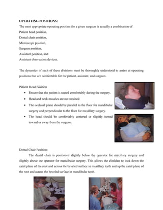
- 1. OPERATING POSITIONS: The most appropriate operating position for a given surgeon is actually a combination of Patient head position, Dental chair position, Microscope position, Surgeon position, Assistant position, and Assistant observation devices. The dynamics of each of these divisions must be thoroughly understood to arrive at operating positions that are comfortable for the patient, assistant, and surgeon. Patient Head Position • Ensure that the patient is seated comfortably during the surgery. • Head and neck muscles are not strained • The occlusal plane should be parallel to the floor for mandibular surgery and perpendicular to the floor for maxillary surgery. • The head should be comfortably centered or slightly turned toward or away from the surgeon. Dental Chair Position: The dental chair is positioned slightly below the operator for maxillary surgery and slightly above the operator for mandibular surgery. This allows the clinician to look down the axial plane of the root and across the beveled surface in maxillary teeth and up the axial plane of the root and across the beveled surface in mandibular teeth.
- 2. Microscope Position: Most endodontists prefer an operating microscope that is mounted to the ceiling. Friction couplings position the microscope and suspension arms in an infinite number of axes within three-dimensional space. Inserting a 135-degree inclined coupler between the mounting arm and the head of the microscope provides additional axis of movement and more versatility. The combination of microscope position and dental chair position places the microscope in three-dimensional space, which can be accessed by the surgeon. The microscope must be positioned to provide the' necessary visual access to perform the surgery while allowing for postural comfort for the surgeon and assistant. Surgeon Position: The surgeon should use an adjustable operator stool. The surgeon's thighs should be parallel to the floor so that the large muscle groups are at rest. The surgeon's arms should be relaxed and comfortable at his or her side. Specially designed surgical stools are available with arm supports that can be used to provide additional comfort and minimize fatigue. The surgeon should be facing the affected side of the patient. This mayor may not mean that the surgeon is seated on the affected side. Often the surgeon can accomplish the same result by having the patient turn slightly toward or away from him or her. a left-handed surgeon position himself or herself on the left side of the chair in all situations except during mandibular right surgeries, when the surgeon moves to the right side of the dental chair. A right-handed surgeon positions himself or herself on the right side of the dental chair in all situations except during mandibular left surgeries, when the surgeon moves to the left side of the dental chair. Assistant Position:
- 3. A well-designed microsurgery may use three dental assistants. The first assistant is primarily responsible for suctioning and is usually seated, although he or she may prefer to stand in some situations. The second assistant passes instruments and usually stands. This assistant is positioned next to the surgeon's dominant side to facilitate instrument passing. If a front delivery system is used, the second assistant can be positioned across from the surgeon and may pass instruments from the tray over the patient. The third assistant functions as a charge nurse and can leave the operatory to obtain additional instruments or materials if necessary. The third assistant is also in charge of video and photographic functions. The positions of the assistants may vary depending on their visual access and which observation devices are being used. Good communication is essential between the surgeon and the assistants. The first or suctioning assistant must let the surgeon know if he or she does not have good visual access to the surgical field. Positional adjustments may be necessary for both the surgeon and the assistants at times during the surgery depending on the location of the tooth being treated. Assistant Observation Devices: In most clinical situations, the assistant has a choice of three observation devices: articulating assistant binoculars, LCD screens, and high resolution monitors. SPECIFIC OPERATING POSITIONS: Operating position for maxilla anterior position: The occlusal plane is perpendicular to the floor Patient looking straight ahead Microscope angled down the axial plane of the roots
- 4. Dental chair position or surgical site low in relation to the microscope Maxillary left posterior position: Occlusal plane perpendicular to floor Patient turns slightly to right for premolar surgery and turns right to molar surgery Microscope angled down the axial plane of the roots Dental chair position or surgical site low in relation to the microscope Maxillary right posterior position: OPERATING POSITIONS FOR MANDIBLE: Anterior position: Occlusal plane parallel to the floor Patient looking straight ahead Microscope angled up the axial plane of the roots Dental chair position or surgical site slightly high in relation to microscope
- 5. Mandibular left posterior position: Occlusal plane parallel to the floor The patient is lying on the right side with head turned up slightly Microscope angled up the axial plane of the roots Dental chair position or surgical site low in relation to the microscope Mandibular Right Posterior Position: ADVANTAGES: DENTISTS Greater reliability better quality, diagnosis and therapy Greater comfort- upright posture Greater differentiation – better image PATIENTS Greater likelihood of successful treatment Faster healing – less traumatization Tooth conservation instead of tooth replacement Cost effective No scars- cosmetic benefits DISADVANTAGES
- 6. Costly equipment Patient cooperation is a must Needs a lot of surgeon’s skill and experience
- 7. Costly equipment Patient cooperation is a must Needs a lot of surgeon’s skill and experience