SlideShare una empresa de Scribd logo
1 de 115
Mixed Radiolucent- Radiopaque Lesions in
Jaws
By:
• INDIAN DENTALACADEMY
• Leader in continuing Dental Education
www.indiandentalacademy.com
Contents
• Definition
• Classification
• Clinical features
• Radiographic features
• Differential Diagnosis
• Treatment
www.indiandentalacademy.com
• Radiolucent — refers that portion of a
processed radiograph which appears as dark /
black.
• Radiopaque — refers that portion of processed
radiograph which appears as light / white.
• Some normal anatomic structures and disease states
can produce mixed radiolucent-radiopaque images on
radiographs.
www.indiandentalacademy.com
• Pathologic entity may commences as an osteolytic
lesion, which appears as radiolucency in radiograph.
• During its development, foci of calcified material may
form within the osteolytic area, when these foci
become large & sufficiently mineralized, they become
radiographically apparent.
• Thus mixed radiolucent-radiopaque condition
frequently represents an intermediate stage in
development of the lesion.
www.indiandentalacademy.com
Mixed Radiolucent-Radiopaque
Lesions Associated With Teeth
1. Calcifying crown of developing tooth
2. Tooth root with rarefying osteitis
3. Rarefying & Condensing osteitis
4. Chronic Osteomyelitis
5. PCOD
6. Cementoossifying fibroma
7. Cementoblastoma
8. Odontoma
9. Adenomatoid Odontogenic Tumor
10. Calcifying Odontogenic Cyst
11. Ameloblastic fibroodontoma
12. Calcifying Epithelial Odontogenic Tumorwww.indiandentalacademy.com
MixedRadiolucent-Radiopaque
LesionsAssociatedWithTeeth
www.indiandentalacademy.com
Calcifying crown of developing tooth
• Before calcification, permanent tooth buds may
appear in the periapical region of deciduous teeth.
• In early stages of development, tooth germs appear as
cyst like radiolucency.
• After few months undergo sufficient mineralization,
appear as periapical radiolucency with radiopaque
foci.
www.indiandentalacademy.com
• The cusp tips are first part
to calcify in developing
tooth.
• As soon as sufficient mineral
deposited in matrix of cusp
tip, developing tooth can
recognized as a radiolucency
with radiopaque foci.
www.indiandentalacademy.com
Tooth Root With Rarefying Osteitis
• Retained root & root tips are
abnormal radiopacities
commonly found in
edentulous region of the
jaws.
• Retained roots present in
jaws of every 1 of 4
edentulous persons.
• 80% of retained roots are in
posterior region of the jaws.
• 6% of all retained root
tips are associated with
radiolucent areas.
• Usually asymptomatic,
or c/o intermittent pain
or swelling.
• ill-defined radiolucency
with ragged margins.
www.indiandentalacademy.com
• The root canal may
become channel for
infection with a resulting
rarefying osteitis in
periapex, and production
of radiolucent-
radiopaque jaw lesions.
• Usually identifiable by the
shape of root, shadow of
root canal, PDL space and
surrounding lamina dura.
www.indiandentalacademy.com
Management
• Root tips should be removed.
• Surrounding soft tissue enucleate and bone
defect should curetted.
www.indiandentalacademy.com
Rarefying and Condensing Osteitis
• Frequently occurs at apex of a nonvital tooth / retained
root.
• Chronic infection acts as irritating factor and as stimulating
factor.
• Bone resorption occurs at apex; on other hand bone
apposition occurs at periphery of rarefying lesion.
• When chronic infection is present well-defined
homogenous radiopacity is seen more / less circumscribing
radiolucency around root end.
www.indiandentalacademy.com
Differential Diagnosis
• Chronic osteomyelitis
Treatment:
• Affected teeth should treat endodontically or
extraction
www.indiandentalacademy.com
Periapical Cementoosseous Dysplasia
• It is a localized change in normal bone metabolism that
results in replacement of normal cancellous bone with
fibrous tissue & cementum-like material and abnormal
bone.
• It is a reactive fibroosseous lesion; it arises from
periodontal ligament, where mature osteoblasts,
cementoblasts & precursor cells reside.
• This lesion usually identified during routine dental
radiographic examination.
www.indiandentalacademy.com
Clinical Features
• Typically it occurs in middle age mean age 39 years.
• Females > males (9:1)
• Involved teeth are vital; no H/O pain & swelling.
Radiographic features
• It usually lies at the apex of a tooth.
• Mainly in mandibular anteriors.
www.indiandentalacademy.com
Internal structure
• In early stage normal bone is
resorbed and replaced with
fibrous tissue that continuous
with PDL ( causing loss of lamina
dura).
• Radiographically – radiolucency
at apex of tooth.
• In mixed stage radiopaque
tissue appears in the radiolucent
structure.
• This material is amorphous, has
a round, oval shape; composed
of cementum /abnormal bone.www.indiandentalacademy.com
• In mature stage internal
aspect may be totally
radiopaque without any
obvious pattern.
• Lamina dura of involved
teeth is lost making PDL
space less apparent / widen
appearance.
• Small lesions do not cause
jaw expansion but large
lesions may cause expansion,
always a thin intact outer
cortex.
www.indiandentalacademy.com
Differential Diagnosis
• Periapical Abscess
• Periapical Granuloma
• Calcifying crown
• Rarefying and Condensing osteitis
• Odontoma
www.indiandentalacademy.com
Cementoossifying fibroma
• It is uncommon neoplastic condition - originate from
elements of periodontal ligament.
• Usually occurs as periapical lesions – round and well
marginated.
• It can occur at any age; commonly found in adults
mainly 2nd to 3rd decades.
• Definite female predilection.
www.indiandentalacademy.com
• Usually asymptomatic; displacement of teeth- early c/f
• 70-80% of lesions occur in mandible, primarily
premolar & molar region, superior to inferior alveolar
canal, in maxilla in canine fossa & zygomatic arch area.
• Juvenile ossifying fibroma is aggressive form; it occurs
in first 2 decades of life.
• Because of rapid growth in young patients deformity of
involved jaw may occur.
www.indiandentalacademy.com
Radiographic Features
• Most of the lesions are discovered
during routine dental examination.
• The borders of the lesion is well
defined.
• A thin radiolucent line representing
a fibrous capsule, may separate it
from surrounding bone.
• The internal structure is mixed
radiolucent-radiopaque density.
• Lesions produce more cementum
like material may contain solid,
amorphous radiopacities.
www.indiandentalacademy.com
Differential
Diagnosis
• Fibrous dysplasia
• P C O D
• C O C
• C E O T
• A O T
Management
• Surgical enucleation or
resection of the lesion
• Recurrence rate is 6%
www.indiandentalacademy.com
Cementoblastoma
• It is a slow growing mesenchymal neoplasm principally
composed cementum
• It manifest as bulbous growth around & attached to the
apex of tooth root
• Tooth is vital and painful
• Males > females
• Relatively young adults
• Mandible > maxilla, premolar & 1st molarwww.indiandentalacademy.com
• These are mixed radiolucent-radiopaque lesions in
which majority of the internal structure is radiopaque.
• This central radiopaque mass is surrounded by a
radiolucent band; indicating tumor is maturing from
central aspect to periphery.
• In most of the cases external resorption seen.
• Large lesions can cause expansion of mandible with
intact outer cortex.
www.indiandentalacademy.com
Differential Diagnosis
• Odontoma
• P C O D
• Hypercementosis
• Periapical sclerosing
osteitis
Treatment
• These are self-limiting
• Simple excision and
extraction of associated
tooth.
• Rarely recur after
enucleation.
www.indiandentalacademy.com
Chronic Osteomyelitis
• Osteomyelitis is an inflammation of bone and bone
marrow.
1. Suppurative : a. Acute
b. Chronic
2. Sclerosing: a. focal
b. diffuse
3. Chronic nonsuppurative sclerosing osteitis
www.indiandentalacademy.com
• It can occur at any age; common in first 3 decades of life.
• Proliferative periosteitis is common in children and
young adults.
• Mandible more frequently involved.
• Clinical examination shows signs of inflammation,
tenderness, pain, swelling, intraoral & extraoral draining
sinus tracts, fever and regional lymphadenopathy.
www.indiandentalacademy.com
• Denuded bone may protrude from open mucosal or
cutaneous ulcers.
• The sequestra are surrounded by sclerotic bone which
is relatively avascular.
• Within the bone, the haversian canals become blocked
with scar tissue, and the bone becomes surrounded by
thickened periosteum
• Drainage in chronic disease is intermittent and modest
in volume.
www.indiandentalacademy.com
Radiographic Features
• Acute osteomyelitis – No bony changes.
• Chronic osteomyelitis – radiographic images are
irregularly shaped radiolucency with ragged, poorly
defined borders.
• Sometimes lesions appear as mixed radiolucent-
radiopaque pattern.
www.indiandentalacademy.com
• Radiolucent areas consists of infected granulation
tissue, fibrosis or both.
• Radiopaque areas represent sclerosed, often nonvital
bone sequestra or both.
• Proliferative periostitis may show an alternating
radiolucent-radiopaque laminated appearance at
surface of affected bone (onion-skin appearance)
www.indiandentalacademy.com
Periapical changes
Sequestrum
www.indiandentalacademy.com
Garre’s
Osteomyelitis
Unusual appearance of the
mandible produced by
osteomyelitis.
www.indiandentalacademy.com
Differential Diagnosis
• Rarefying and condensing osteitis
• Fibrous dysplasia
• Paget’s disease
• Malignant tumors
www.indiandentalacademy.com
Management
Medical
• Systemic antibiotics
• Local antibiotics
- irrigation
- beads
Surgical
• Sequestrectomy
• Saucerization
• Decortication
• Resection
www.indiandentalacademy.com
Odontoma
• It is benign odontogenic tumor representing 67% of all
odontogenic tumors.
• Odontomas result from budding of extra odontogenic
epithelial cells from dental lamina.
• 3 types of odontomas present;
1. Compound
2. Complex
3. Compound-Complex
www.indiandentalacademy.com
• Compound odontoma –
comprises odontogenic tissues
laid down in a normal
relationship, resulting structures
will resemble to teeth.
• Complex odontoma – tooth
components are less well
organized; toothlike structures
are not formed.
• Compound-Complex – it contain
not only multiple tooth like
structures but also calcified
masses of dental tissue in
haphazard manner. www.indiandentalacademy.com
• 62% of compound variety occurs
in maxilla; predilection for incisor-
canine region, no gender bias.
• 70% of complex type occur in
mandible; located in 1st and 2nd
molar region, mostly in females.
• An odontoma frequently situated
between crown of an unerupted
tooth and crest of alveolar ridge,
blocks tooth eruption.
www.indiandentalacademy.com
• Intermediate stage compound odontoma appears
as a well-defined radiolucent lesion containing
varying no. of radiopaque shadows of developing
tooth.
• Degree of calcification & opacity varies from stage
to stage and lesion to lesion.
• Complex type appears as a well-defined
radiolucency with many radiopaque foci vary
greatly in size, shape & prominence.
www.indiandentalacademy.com
Differential
Diagnosis
• Cementoblastoma
• P C O D
• Cementoossifying
fibroma
Management
• Surgical enucleation
• Periodic
postoperative
examination
www.indiandentalacademy.com
Adenomatoid Odontogenic Tumor
• It is a uncommon and nonaggressive tumor of
odontogenic epithelium.
• It is 2 types - central and peripheral.
• The central tumor divided into follicular type
(associated with embedded tooth) & extra-follicular
type( with no embedded tooth).
• 73% of central lesions are follicular type
www.indiandentalacademy.com
• Age ranges from 5-50 years; 70% cases occur in 2nd
decade, average age is 16 years.
• It has female predilection (2:1)
• Commonly occur in maxilla; incisor-canine-premolar
• It is a slow growing & presents a gradually enlarging
painless swelling.
• Causes facial asymmetry, often associated with
missing tooth.
• It displace the teeth rather than root resorption.
www.indiandentalacademy.com
Radiographic Features
• It is a pericoronal cyst-
like radiolucency.
• In maturing stages,
sharply defined
radiopaque foci are seen
within radiolucency.
www.indiandentalacademy.com
Differential Diagnosis
• COC
• CEOT
• Odontoma
Management:
• Surgical enucleation.
www.indiandentalacademy.com
Calcifying Odontogenic Cyst
• It is also called as Gorlin’s cyst.
• It is uncommon, benign slow growing lesion.
• WHO categories as benign tumor.
• It have a wide age distribution that peaks at 10-19
years and second peak age is during 7th decade;
• Mean age is 36yrs
www.indiandentalacademy.com
Radiographic Features
• At least 75% occurs in bone
with equal distribution
between jaws.
• 75% lesions occur anterior to
the first molar, especially
incisor-canine region.
• Sometimes it manifests as a
pericoronal radiolucency.
• Periphery can vary from well
defined to irregular.
www.indiandentalacademy.com
• The internal aspect can
vary in appearance.
• It is cyst like radiolucency
containing quite distinct
radiopaque foci
• It can be completely
radiolucent to mixed R/L-
R/O.
www.indiandentalacademy.com
Differential Diagnosis
• AOT
• CEOT
• Odontoma
Treatment
• Surgical enucleation
• Recurrence is reported
www.indiandentalacademy.com
Ameloblastic fibroodontoma
• It is benign mixed odontogenic tumor that contain
cords & nets of odontogenic epithelium & some
calcified odontogenic tissue.
• Clinical features are similar to odontoma, often
associated with missing tooth / tooth has failed to
erupt.
• Occasionally tumor takes in position of a missing
tooth.
www.indiandentalacademy.com
Radiological features
• Most of the cases occur in
posterior aspect of mandible.
• The epicenter of lesion is
usually occlusal to a developing
tooth / towards the alveolar
crest.
• Lesion is well defined;
sometimes corticated.
• The internal structure is mixed
with the majority of the lesion
being radiolucent.
www.indiandentalacademy.com
• Small lesions may appear as
enlarged follicles with one / two
small discrete radiopacities.
• Large lesions may have a more
extensive calcified internal
structure.
• In some cases small calcifications
have around shape with
radiopaque enamel like margin.
www.indiandentalacademy.com
Differential Diagnosis
• Odontoma
• COC
• AOT
Management
 Surgical enucleation.
 No tendency to recur.
 Follow up is necessary
www.indiandentalacademy.com
Calcifying Epithelial Odontogenic
Tumor
• It is also called Pindborg tumor.
• It is less aggressive lesion; rarely may have an
extraosseous location.
• Males > females.
• Mean age is 42 years
• 68% tumors occur in mandible; especially molar regionwww.indiandentalacademy.com
Radiological Features
• It may have several radiographic appearance.
1. Pericoronal radiolucency,
2. Pericoronal radiolucency with radiopaque foci,
3. Mixed radiolucent-radiopaque lesion not associated with
an unerupted tooth,
4. A “driven snow” appearance,
5. Dense radiopacity (occasionally).www.indiandentalacademy.com
• More common in
mandible (2:1)
• 52% associated with
unerupted / impacted
tooth.
• Borders are well defined,
cyst like cortex, sometimes
irregular & ill defined
www.indiandentalacademy.com
• The internal aspect may
appear unilocular /
multilocular with numerous
scattered radiopaque foci.
• Characteristic & diagnostic
finding is radiopacities close
to crown of embedded
tooth.
• In addition small, thin
opaque trabeculae may cross
the radiolucency in many
directions
www.indiandentalacademy.com
Differential Diagnosis
• A O T
• C O C
Management
• Surgical resection is
recommended.
• Low recurrence rate.
• Follow up is mandatory
www.indiandentalacademy.com
MixedRadiolucent-RadiopaqueLesionsNot
NecessarilyContactingTeeth
www.indiandentalacademy.com
Osteoradionecrosis
• A disease condition of hard and soft tissue after
irradiation of the region.
• Usually develops after 12 months radiotherapy
completion.
• Asymptomatic in early stages.
• When ulceration occurs, tenderness and pain are
common complaint.
• Mixed radiolucent-radiopaque lesion in a full-blown
case.
www.indiandentalacademy.com
Irradiation
Narrowing of lumina ( principally of small arterioles)
Hypovascular, hypocellular, hypoxic tissue
Diminished capacity for normal repair
Breakdown of tissue and its overlying mucosa
Superficial infection of the denuded bone
Osteoradionecrosis
www.indiandentalacademy.com
• Mandible is more commonly affected than maxilla.
• This due to microanatomy & reduced vascularity of bone.
• Posterior mandible is more affected than anterior portion.
• Loss of mucosal covering & exposure of bone is hall mark of
ORN.
• Exposed bone becomes necrotic as a result of loss of
vascularity from periosteum.
• Intense pain may occur, with intermittent swelling & extra-
oral drainage.
www.indiandentalacademy.com
Radiological features
• The periphery is ill defined similar to osteomyelitis.
• If the lesion reaches the inferior border, irregular
resorption of bony cortex occurs.
• Internal structure appearance will be depends on range
of bone destruction and bone formation.
• It will be overall sclerotic to radiopaque appearance.
www.indiandentalacademy.com
Prevention
• Pt should undergo
thorough dental checkup
• Patient oral hygiene should
be assessed & motivate the
pt for good oral hygiene
after radiotherapy.
• Pt should undergo
extraction of grossly
carious teeth, periodontally
involved teeth at least 10-
14 days before
commencement of
radiotherapy.
• Restore if any decayed
tooth present & patient
must undergo thorough
oral prophylaxis.
• Teeth sockets should heal
completely prior to
radiation exposure.
• After radiation exposure, all
extractions, and invasive
procedures should be
avoided for a period of 3
months
www.indiandentalacademy.com
Management
• Both surgical and nonsurgical treatments have been
done.
• Hyperbaric oxygen therapy is effective treatment.
• Treatment is done in 3 stages;
• Stage I: 30 dives
• Stage II: sequestrectomy+ primary closure+ 60 dives
• Stage III: 30 dives, resection and remaining 30 dives
www.indiandentalacademy.com
Focal Cementoosseous Dysplasia
• It is a reactive lesion of
PDLO.
• It is PCOD like lesions occur
in the premolar-molar
region.
• These are asymptomatic
lesions occur in edentulous
areas.
• Females are commonly
affected.
• Average age is 37 yrs.
The lesion matures through 3
stages:
1. Early radiolucent
2. Middle mixed stage
3. Late radiopaque
www.indiandentalacademy.com
Radiological Features
• The distinctness of
margin varies;
radiopaque pattern
scattered throughout the
lesion.
• Radiolucent area is
surrounded by thin
radiopaque border.
• Often it appears as
ground glass appearance
www.indiandentalacademy.com
Management
• Periodic follow up is recommended.
• If lesion shows signs of serious enlargement
surgical intervention is indicated.
www.indiandentalacademy.com
Sailolith
• Sailoliths are stones found
within ducts of salivary
glands.
• Most common in
submandibular glands.
• Men > women
• Age ranges 3rd to 4th
decade.
• Usually occur single (70-
80%) but may occur
multiple
• Patients usually
asymptomatic but H/O
pain swelling in floor of
the mouth.
• This discomfort may
intensify at meal time.
www.indiandentalacademy.com
Radiographic Features
• Sailoliths usually
cylindrical
• Some stones are
homogenously
radiopaque and some
show multiple layers of
calcification.
Management
• Small stones may be
“milked out” through
duct orifice using
bimanual palpation.
• Large stones can remove
by lithotripsy or surgical
intervention.
www.indiandentalacademy.com
Phlebolith
• These are calcified thrombi found in veins, venulae or
sinusoidal vessels of hemangiomas.
• In head & neck phleboliths indicates presence of
hemangioma.
• Involved soft tissue swollen, throbbing or discolored by
presence of veins or soft tissue hemangioma
www.indiandentalacademy.com
Radiographic Features
• In cross section round or
oval in shape with smooth
periphery.
• Internal aspect maybe
homogenously radiopaque.
• It gives “bulls eye” or
target appearance.
www.indiandentalacademy.com
Rhinolith / Antrolith
• Hard calcified bodies or stones occur in the nose or
maxillary sinus.
• Pt is asymptomatic for long time but expanding mass
may impinge on mucosa which cause pain, congestion
and ulceration.
• Pt may develop a unilateral purulent rhinorrhea,
sinusitis, headache, fetor and fever
www.indiandentalacademy.com
• These stones have variety of
shapes & sizes
• Stones present as
homogenous or
heterogeneous radiopacities.
• Occasionally density will
exceed the surrounding
bone.
www.indiandentalacademy.com
Traumatic Myositis Ossificans
• It is characterised by ossification of muscle by single
acute traumatic episode or multiple minor traumatic
episodes.
• Exact mechanism for ossification is not clear.
• It usually manifests as firm painful mass in the injured
muscle within 1-4 weeks.
• In facial muscles masseter and temporalis muscle
frequently involved.
www.indiandentalacademy.com
Radiographic Features
• It may appear as a feather type of calcification in
muscle following ossification of hematoma along
dissected muscle bundle.
• Calcification may be seen within 2-3 weeks after
trauma.
Treatment:
• Surgical excision
www.indiandentalacademy.com
Fibrous dysplasia
• It is a hamartomatous fibroosseous lesion.
• Etiology- Unknown
• It is a bone lesion that produces lysis of bone with
fibrous proliferation as a replacement (radiolucent) in its
early stage.
www.indiandentalacademy.com
• Monostotic type most common & frequently involves
the jaws and skull ( older adults).
• Mandibular lesions are usually solitary.
• Maxillary lesions may involve neighboring bones
(Craniofacial FD).
• Polyostotic form usually seen in children and young
adults ( < 6 years).
• Polyostotic form is static when skeletal growth stops,
but proliferation may continue.
www.indiandentalacademy.com
• Early lesions are spherical radiolucency
• Margins are ragged and poorly defined, merging into
normal bone.
• Lesions are usually situated deep within jaw bone.
• Maturing changes produce ground glass appearance
may commence at periphery of the lesion.
• Gross displacement of mandibular canal is seen.
www.indiandentalacademy.com
• Fibrous dysplasia
(intermediate stage)
A) Smoky or mottled
appearance(also seen in
B).
• B)Radiograph showing
one lesion in maxilla &
one in mandible.
www.indiandentalacademy.com
Differential Diagnosis
• Chronic osteomyelitis
• Paget’s disease
• Osteogenic sarcoma
www.indiandentalacademy.com
Paget’s Disease (Osteitis Deformins)
• First described in England by Sir James Paget in 1876.
• Etiology: Unclear; genetics and undetected slow-acting
virus may play roles.
• Usually old age people are affected.
• It is disorder of bone metabolism.
www.indiandentalacademy.com
• Osteoclastic resorptive process is overactive, with a
compensatory increase in osteoblastic action
attempting to maintain strong bones.
• Progresses so slowly that annual radiographs show little
change.
• The vertebrae, pelvis, skull, tibia, femur and humerus
are most likely to be involved
• Patient may develop arthritis like symptoms & bowed
legs with a waddling gait and may complain of an
increasing hat size, which is due to skull expansion.
www.indiandentalacademy.com
• Maxilla>Mandible
• Involved bones are thickened, and the foramina are often
constricted.
• Consequently pressure is induced on the structures that pass
through the foramina, causing neurologic signs such as
deafness and diminishing vision.
• Calcified areas interspersed with radiolucent areas producing
the cotton-wool appearance.
• Teeth in the involved jaw may demonstrate spreading,
migration, diminution of the laminadura, and
(characteristically) hypercementosis.
www.indiandentalacademy.com
Differential Diagnosis
• Florid Cementoosseous
dysplasia
• Fibrous dysplasia
Management
• In early stages, drug
therapy with
diphosphonates or
calcitonin arrests
osteoclastic activity.
• Anti-inflammatory agents
for pain relief.
• Edentulous pts may
require frequent
adjustment or continued
fabrication of new
dentures.
www.indiandentalacademy.com
Osteogenic Sarcoma
• Arises from primitive
undifferentiated cells and
from malignant
transformation of osteoblasts.
• Etiology – unknown.
• Irradiated bone is more
prone to become osteogenic
sarcoma.
• Age: 3rd - 4th decade.
• Jaw lesions have less
tendency to metastasize.
• Prognosis is better for jaw
lesions.
• Metastasizes almost
exclusively through
hematogenous spread.
• Intermittent local pain,
swelling.
www.indiandentalacademy.com
Radiographic features
• Three different
radiographic images:
1. Totally radiolucent
2. Mixed radiolucent-
radiopaque
3. Completely radiopaque
• The classic sun-burst
appearance may be seen in
the latter two types.
• The lesion may have
ragged, ill-defined borders.
• In some lesions sequestra
are formed.
• If the tumor invades the
periosteum, many thin,
irregular spicules of new
bone may develop
outward and perpendicular
to the surface of the
lesion. This produces the
sun-burst effect.www.indiandentalacademy.com
• Sometimes, two triangular radiopacities project from
the cortex and mark the lateral extremities of the
lesion. These are referred to as Codman’s triangles.
• Rarely, it causes periosteal deposition of bone in an
“onion-skin” pattern.
• In some cases a bandlike widening involving complete
length of PDL space on one or both sides of the root &
involvement of the mandibular canal with widening of
the canal can be seen.
• Cemental resorption may be seen.
www.indiandentalacademy.com
Differential Diagnosis
• Chondrosarcoma
• Fibrosarcoma
• Osteoblastic metastatic
carcinoma
• Fibrous dysplasia
Management
• Resection with large
border of normal bone
• Radiation &
chemotherapy used for
control metastatic
spread.
www.indiandentalacademy.com
Osteoblastic metastatic carcinoma
• Occasionally metastatic
tumors to the jawbone
from primary lesions in
the prostate or breast
may be seen.
• It may appear as entirely
radiolucent, mixed
radiolucent-radiopaque,
or entirely radiopaque.
• Less aggressive– more
circumscribed.
• When small nests of
sclerotic foci are
disseminated throughout
the jawbone, salt & pepper
pattern may be seen.
• H/o surgery or symptoms of
a primary tumor indicate
metastatic disease.
www.indiandentalacademy.com
PA view cotton-wool
appearance in skull & large
bilateral radiolucent lesions
seen in mandible
Lateral view showing
radiolucent & radiopaque
areas
www.indiandentalacademy.com
Differential Diagnosis
• Chondrosarcoma
• Ossifying subperiosteal hematoma
www.indiandentalacademy.com
Chondrosarcoma
• It is malignant tumor of cartilaginous origin.
• It can occur at any age; common in adults.
• Male and females are equally affected.
• Maxilla more frequently involved than mandible
• Pt may have firm to hard swelling with long duration.
• Enlargement cause pain headache and deformity
www.indiandentalacademy.com
Radiographic Features
• Maxillary lesions occur in anterior maxilla
• Mandibular lesions occur In coronoid process, condylar
head and neck; occasionally symphysis region.
• Lesions are round, ovoid, or lobulated
• Borders are well defined and corticated some times meld
with adjacent normal bone.
• Aggressive lesions have infiltrative, ill-defined, and
nonocorticated borders.
www.indiandentalacademy.com
• Internal structure usually exhibit some form of
calcification within center of the lesion which gives
mixed radiolucent-radiopaque appearance.
• This diffuse calcification may be superimposed on a
bony background that resembles granular / ground-
glass appearance.
• Lesions are rarely completely radiolucent.
• In mandibular cases the inferior border / alveolar
process grossly expanded with intact cortical covering.
www.indiandentalacademy.com
• Radiograph of a surgical specimen.
• Radiopaque foci can be seen in the exophytic mass
www.indiandentalacademy.com
Differential Diagnosis
• Osteogenic sarcoma
• Fibrous dysplasia
Management
• Surgical resection is
mandatory
• It is quite radioresistent;
radiation therapy can used
as palliative procedure
with large inoperable
tumors.
• At least 60% of cases have
recurrences within 5 years.
www.indiandentalacademy.com
Desmoblastic Ameloblastoma
• It is histologic variant of ameloblastoma.
• Distinctive with a moderately cellular fibrous connective
tissue with an abundance of collagen.
• The tumor epithelium consists of small, ovoid, or follicle
shaped islands & narrow cords.
• Maxilla>Mandible.
• Anterior region is commonly affected.
www.indiandentalacademy.com
Edentulous premolar
region of maxilla
Canine and premolar
region of mandible
www.indiandentalacademy.com
Rarities
www.indiandentalacademy.com
• Eruption sequestrum
• Ameloblastic fibrodentinoma
• Calcifying hyperplastic dental
follicle
• Central odontogenic fibroma
• Ameloblastoma
• Cystic odontoma
• Lymphoma of bone
• Juvenile ossifying fibroma
• Odontodysplasia
• Odontogenic myxoma
• Central hemangioma
• Ewing’s sarcoma
• Osteoblastoma
• Osteiod osteoma
RARITIES
www.indiandentalacademy.com
Foreign Bodies
• Usually root canal filling
materials.
• Extruded gutta percha,
silver points, sealers or
retrograde amalgam
and filled root canal
www.indiandentalacademy.com
Florid Cementoosseous Dysplasia
• It appears as widespread form of
periapical cemental dysplasia.
• It has poor vascular supply, more
susceptible for infections.
• Females > Males
• Mean age 42 years
• Mandible > Maxilla
• Mostly posterior to cuspids
www.indiandentalacademy.com
Lymphoma of Bone
www.indiandentalacademy.com
Cystic odontoma
www.indiandentalacademy.com
References
• Differential Diagnosis Wood & Goaz 5th Edition
• Dental Radiology White & Pharow 5th Edition
• Oral Pathology Shafer 5th Edition
• Oral pathology Neville & Dam 2nd Edition
• Oral Radiographic Diagnosis Stafne’s 5th Edition
www.indiandentalacademy.com
THANK YOU
www.indiandentalacademy.com

Más contenido relacionado

La actualidad más candente

Radiographic assessment of dental caries
Radiographic assessment of dental cariesRadiographic assessment of dental caries
Radiographic assessment of dental cariesDrMohamedEkram
 
28.regional odontodysplasia
28.regional odontodysplasia28.regional odontodysplasia
28.regional odontodysplasiaNehal Vithlani
 
Soft tissue calcification orofacial region
Soft tissue calcification orofacial regionSoft tissue calcification orofacial region
Soft tissue calcification orofacial regionDr. Monali Prajapati
 
Multilocular radiolucencies
Multilocular radiolucenciesMultilocular radiolucencies
Multilocular radiolucenciesSelva Arockiam
 
Radiopaque Lesions
Radiopaque LesionsRadiopaque Lesions
Radiopaque LesionsMaryam Arbab
 
unilocular and multilocular radiolucencies
unilocular and multilocular radiolucenciesunilocular and multilocular radiolucencies
unilocular and multilocular radiolucenciesDr Sourav Malhotra
 
Radiographic Differential Diagnosis 2008
Radiographic Differential Diagnosis 2008Radiographic Differential Diagnosis 2008
Radiographic Differential Diagnosis 2008IAU Dent
 
Pulp therapy for primary and young teeth
Pulp therapy for primary and young teethPulp therapy for primary and young teeth
Pulp therapy for primary and young teethSaeed Bajafar
 
Radiographic Differential Diagnosis 2009
Radiographic Differential Diagnosis 2009Radiographic Differential Diagnosis 2009
Radiographic Differential Diagnosis 2009IAU Dent
 
Oral White lesions
Oral White lesionsOral White lesions
Oral White lesionsIAU Dent
 
Diseases of bone manifested in the jaws
Diseases of bone manifested in the jawsDiseases of bone manifested in the jaws
Diseases of bone manifested in the jawsIAU Dent
 
Multilocular radiolucencies/ oral surgery courses  
Multilocular radiolucencies/ oral surgery courses  Multilocular radiolucencies/ oral surgery courses  
Multilocular radiolucencies/ oral surgery courses  Indian dental academy
 
Radiopacities of jaws
Radiopacities of jawsRadiopacities of jaws
Radiopacities of jawsSwati Kalra
 
Endodontic radiography
Endodontic radiographyEndodontic radiography
Endodontic radiographyEdward Kaliisa
 

La actualidad más candente (20)

Radiographic assessment of dental caries
Radiographic assessment of dental cariesRadiographic assessment of dental caries
Radiographic assessment of dental caries
 
28.regional odontodysplasia
28.regional odontodysplasia28.regional odontodysplasia
28.regional odontodysplasia
 
Soft tissue calcification orofacial region
Soft tissue calcification orofacial regionSoft tissue calcification orofacial region
Soft tissue calcification orofacial region
 
Periapical radiolucencies
Periapical radiolucencies Periapical radiolucencies
Periapical radiolucencies
 
Multilocular radiolucencies
Multilocular radiolucenciesMultilocular radiolucencies
Multilocular radiolucencies
 
Radiopaque Lesions
Radiopaque LesionsRadiopaque Lesions
Radiopaque Lesions
 
Odontogenic cysts
Odontogenic  cystsOdontogenic  cysts
Odontogenic cysts
 
unilocular and multilocular radiolucencies
unilocular and multilocular radiolucenciesunilocular and multilocular radiolucencies
unilocular and multilocular radiolucencies
 
Jaw bone disaese
Jaw bone disaeseJaw bone disaese
Jaw bone disaese
 
Radiographic Differential Diagnosis 2008
Radiographic Differential Diagnosis 2008Radiographic Differential Diagnosis 2008
Radiographic Differential Diagnosis 2008
 
Pulp therapy for primary and young teeth
Pulp therapy for primary and young teethPulp therapy for primary and young teeth
Pulp therapy for primary and young teeth
 
Radiographic Differential Diagnosis 2009
Radiographic Differential Diagnosis 2009Radiographic Differential Diagnosis 2009
Radiographic Differential Diagnosis 2009
 
Oral White lesions
Oral White lesionsOral White lesions
Oral White lesions
 
Diseases of bone manifested in the jaws
Diseases of bone manifested in the jawsDiseases of bone manifested in the jaws
Diseases of bone manifested in the jaws
 
Multilocular radiolucencies/ oral surgery courses  
Multilocular radiolucencies/ oral surgery courses  Multilocular radiolucencies/ oral surgery courses  
Multilocular radiolucencies/ oral surgery courses  
 
Odontogenic tumor
Odontogenic tumorOdontogenic tumor
Odontogenic tumor
 
Radiopacities of jaws
Radiopacities of jawsRadiopacities of jaws
Radiopacities of jaws
 
Fibro Osseous Lesions
Fibro Osseous LesionsFibro Osseous Lesions
Fibro Osseous Lesions
 
Endodontic radiography
Endodontic radiographyEndodontic radiography
Endodontic radiography
 
red and white lesions of oral cavity
red and white lesions of oral cavityred and white lesions of oral cavity
red and white lesions of oral cavity
 

Similar a mixed radiolucent and radiopaque lesions / oral surgery courses

Mixed radiopaque & radiolucent lesions
Mixed radiopaque & radiolucent lesionsMixed radiopaque & radiolucent lesions
Mixed radiopaque & radiolucent lesionsDr. Samarth Johari
 
diseases of bone.pptx
diseases of bone.pptxdiseases of bone.pptx
diseases of bone.pptxHetvi23
 
Solitary cyst like radiolucencies not contacting teeth/ dental courses
Solitary cyst like radiolucencies not contacting teeth/ dental coursesSolitary cyst like radiolucencies not contacting teeth/ dental courses
Solitary cyst like radiolucencies not contacting teeth/ dental coursesIndian dental academy
 
Odontogenic tumors vi/certified fixed orthodontic courses by Indian dental ac...
Odontogenic tumors vi/certified fixed orthodontic courses by Indian dental ac...Odontogenic tumors vi/certified fixed orthodontic courses by Indian dental ac...
Odontogenic tumors vi/certified fixed orthodontic courses by Indian dental ac...Indian dental academy
 
radiopacity lesions : radiology lecture.
radiopacity lesions : radiology lecture.radiopacity lesions : radiology lecture.
radiopacity lesions : radiology lecture.ShafieMohamed2003
 
Management of jaw tumors
Management of jaw tumorsManagement of jaw tumors
Management of jaw tumorsUjwal Gautam
 
Odontogenic tumors v / dental implant courses by Indian dental academy 
Odontogenic tumors v / dental implant courses by Indian dental academy Odontogenic tumors v / dental implant courses by Indian dental academy 
Odontogenic tumors v / dental implant courses by Indian dental academy Indian dental academy
 
differential diagnosis
differential diagnosisdifferential diagnosis
differential diagnosisZafeena Zaham
 
Periapical radiopacities/ dental implant courses
Periapical radiopacities/ dental implant coursesPeriapical radiopacities/ dental implant courses
Periapical radiopacities/ dental implant coursesIndian dental academy
 
Cysts of oral and maxillofacial region by dr. maryam salman
Cysts of oral and maxillofacial region by dr. maryam salmanCysts of oral and maxillofacial region by dr. maryam salman
Cysts of oral and maxillofacial region by dr. maryam salmanDr.Maryam Salman
 
Skull pathologies on Xrays
Skull pathologies on XraysSkull pathologies on Xrays
Skull pathologies on Xraysdypradio
 
Bone Lesions Oral Pathology
Bone Lesions Oral PathologyBone Lesions Oral Pathology
Bone Lesions Oral PathologySana Rasheed
 
Radiographic interpretation
Radiographic interpretationRadiographic interpretation
Radiographic interpretationmoix rafiq
 
Periapical radiolucencies./ oral surgery courses
Periapical radiolucencies./ oral surgery courses Periapical radiolucencies./ oral surgery courses
Periapical radiolucencies./ oral surgery courses Indian dental academy
 
radiographicinterpretation.MS-Ceu.pptx
radiographicinterpretation.MS-Ceu.pptxradiographicinterpretation.MS-Ceu.pptx
radiographicinterpretation.MS-Ceu.pptxDrCarlosIICapitan
 

Similar a mixed radiolucent and radiopaque lesions / oral surgery courses (20)

Mixed radiopaque & radiolucent lesions
Mixed radiopaque & radiolucent lesionsMixed radiopaque & radiolucent lesions
Mixed radiopaque & radiolucent lesions
 
Solitary radiolucencies with ragged & poorly defined borders
Solitary radiolucencies with ragged & poorly defined bordersSolitary radiolucencies with ragged & poorly defined borders
Solitary radiolucencies with ragged & poorly defined borders
 
diseases of bone.pptx
diseases of bone.pptxdiseases of bone.pptx
diseases of bone.pptx
 
Solitary cyst like radiolucencies not contacting teeth/ dental courses
Solitary cyst like radiolucencies not contacting teeth/ dental coursesSolitary cyst like radiolucencies not contacting teeth/ dental courses
Solitary cyst like radiolucencies not contacting teeth/ dental courses
 
Odontogenic tumors vi/certified fixed orthodontic courses by Indian dental ac...
Odontogenic tumors vi/certified fixed orthodontic courses by Indian dental ac...Odontogenic tumors vi/certified fixed orthodontic courses by Indian dental ac...
Odontogenic tumors vi/certified fixed orthodontic courses by Indian dental ac...
 
radiopacity lesions : radiology lecture.
radiopacity lesions : radiology lecture.radiopacity lesions : radiology lecture.
radiopacity lesions : radiology lecture.
 
Cysts of jaws1
Cysts of jaws1Cysts of jaws1
Cysts of jaws1
 
ODONTOGENIC TUMORS.pptx
ODONTOGENIC TUMORS.pptxODONTOGENIC TUMORS.pptx
ODONTOGENIC TUMORS.pptx
 
Management of jaw tumors
Management of jaw tumorsManagement of jaw tumors
Management of jaw tumors
 
Odontogenic tumors v / dental implant courses by Indian dental academy 
Odontogenic tumors v / dental implant courses by Indian dental academy Odontogenic tumors v / dental implant courses by Indian dental academy 
Odontogenic tumors v / dental implant courses by Indian dental academy 
 
differential diagnosis
differential diagnosisdifferential diagnosis
differential diagnosis
 
Presentation
PresentationPresentation
Presentation
 
Periapical radiopacities/ dental implant courses
Periapical radiopacities/ dental implant coursesPeriapical radiopacities/ dental implant courses
Periapical radiopacities/ dental implant courses
 
Cysts of oral and maxillofacial region by dr. maryam salman
Cysts of oral and maxillofacial region by dr. maryam salmanCysts of oral and maxillofacial region by dr. maryam salman
Cysts of oral and maxillofacial region by dr. maryam salman
 
Skull pathologies on Xrays
Skull pathologies on XraysSkull pathologies on Xrays
Skull pathologies on Xrays
 
Bone Lesions Oral Pathology
Bone Lesions Oral PathologyBone Lesions Oral Pathology
Bone Lesions Oral Pathology
 
Radiographic interpretation
Radiographic interpretationRadiographic interpretation
Radiographic interpretation
 
Periapical radiolucencies./ oral surgery courses
Periapical radiolucencies./ oral surgery courses Periapical radiolucencies./ oral surgery courses
Periapical radiolucencies./ oral surgery courses
 
radiographicinterpretation.MS-Ceu.pptx
radiographicinterpretation.MS-Ceu.pptxradiographicinterpretation.MS-Ceu.pptx
radiographicinterpretation.MS-Ceu.pptx
 
Fibro osseous lesions
Fibro osseous lesionsFibro osseous lesions
Fibro osseous lesions
 

Más de Indian dental academy

Indian Dentist - relocate to united kingdom
Indian Dentist - relocate to united kingdomIndian Dentist - relocate to united kingdom
Indian Dentist - relocate to united kingdomIndian dental academy
 
1ST, 2ND AND 3RD ORDER BENDS IN STANDARD EDGEWISE APPLIANCE SYSTEM /Fixed ort...
1ST, 2ND AND 3RD ORDER BENDS IN STANDARD EDGEWISE APPLIANCE SYSTEM /Fixed ort...1ST, 2ND AND 3RD ORDER BENDS IN STANDARD EDGEWISE APPLIANCE SYSTEM /Fixed ort...
1ST, 2ND AND 3RD ORDER BENDS IN STANDARD EDGEWISE APPLIANCE SYSTEM /Fixed ort...Indian dental academy
 
Invisalign -invisible aligners course in india
Invisalign -invisible aligners course in india Invisalign -invisible aligners course in india
Invisalign -invisible aligners course in india Indian dental academy
 
Invisible aligners for your orthodontics pratice
Invisible aligners for your orthodontics praticeInvisible aligners for your orthodontics pratice
Invisible aligners for your orthodontics praticeIndian dental academy
 
Development of muscles of mastication / dental implant courses
Development of muscles of mastication / dental implant coursesDevelopment of muscles of mastication / dental implant courses
Development of muscles of mastication / dental implant coursesIndian dental academy
 
Corticosteriods uses in dentistry/ oral surgery courses  
Corticosteriods uses in dentistry/ oral surgery courses  Corticosteriods uses in dentistry/ oral surgery courses  
Corticosteriods uses in dentistry/ oral surgery courses  Indian dental academy
 
Cytotoxicity of silicone materials used in maxillofacial prosthesis / dental ...
Cytotoxicity of silicone materials used in maxillofacial prosthesis / dental ...Cytotoxicity of silicone materials used in maxillofacial prosthesis / dental ...
Cytotoxicity of silicone materials used in maxillofacial prosthesis / dental ...Indian dental academy
 
Diagnosis and treatment planning in completely endntulous arches/dental courses
Diagnosis and treatment planning in completely endntulous arches/dental coursesDiagnosis and treatment planning in completely endntulous arches/dental courses
Diagnosis and treatment planning in completely endntulous arches/dental coursesIndian dental academy
 
Properties of Denture base materials /rotary endodontic courses
Properties of Denture base materials /rotary endodontic coursesProperties of Denture base materials /rotary endodontic courses
Properties of Denture base materials /rotary endodontic coursesIndian dental academy
 
Use of modified tooth forms in complete denture occlusion / dental implant...
Use of modified  tooth forms  in  complete denture occlusion / dental implant...Use of modified  tooth forms  in  complete denture occlusion / dental implant...
Use of modified tooth forms in complete denture occlusion / dental implant...Indian dental academy
 
Dental luting cements / oral surgery courses  
Dental   luting cements / oral surgery courses  Dental   luting cements / oral surgery courses  
Dental luting cements / oral surgery courses  Indian dental academy
 
Dental casting alloys/ oral surgery courses  
Dental casting alloys/ oral surgery courses  Dental casting alloys/ oral surgery courses  
Dental casting alloys/ oral surgery courses  Indian dental academy
 
Dental casting investment materials/endodontic courses
Dental casting investment materials/endodontic coursesDental casting investment materials/endodontic courses
Dental casting investment materials/endodontic coursesIndian dental academy
 
Dental casting waxes/ oral surgery courses  
Dental casting waxes/ oral surgery courses  Dental casting waxes/ oral surgery courses  
Dental casting waxes/ oral surgery courses  Indian dental academy
 
Dental ceramics/prosthodontic courses
Dental ceramics/prosthodontic coursesDental ceramics/prosthodontic courses
Dental ceramics/prosthodontic coursesIndian dental academy
 
Dental implant/ oral surgery courses  
Dental implant/ oral surgery courses  Dental implant/ oral surgery courses  
Dental implant/ oral surgery courses  Indian dental academy
 
Dental perspective/cosmetic dentistry courses
Dental perspective/cosmetic dentistry coursesDental perspective/cosmetic dentistry courses
Dental perspective/cosmetic dentistry coursesIndian dental academy
 
Dental tissues and their replacements/ oral surgery courses  
Dental tissues and their replacements/ oral surgery courses  Dental tissues and their replacements/ oral surgery courses  
Dental tissues and their replacements/ oral surgery courses  Indian dental academy
 

Más de Indian dental academy (20)

Indian Dentist - relocate to united kingdom
Indian Dentist - relocate to united kingdomIndian Dentist - relocate to united kingdom
Indian Dentist - relocate to united kingdom
 
1ST, 2ND AND 3RD ORDER BENDS IN STANDARD EDGEWISE APPLIANCE SYSTEM /Fixed ort...
1ST, 2ND AND 3RD ORDER BENDS IN STANDARD EDGEWISE APPLIANCE SYSTEM /Fixed ort...1ST, 2ND AND 3RD ORDER BENDS IN STANDARD EDGEWISE APPLIANCE SYSTEM /Fixed ort...
1ST, 2ND AND 3RD ORDER BENDS IN STANDARD EDGEWISE APPLIANCE SYSTEM /Fixed ort...
 
Invisalign -invisible aligners course in india
Invisalign -invisible aligners course in india Invisalign -invisible aligners course in india
Invisalign -invisible aligners course in india
 
Invisible aligners for your orthodontics pratice
Invisible aligners for your orthodontics praticeInvisible aligners for your orthodontics pratice
Invisible aligners for your orthodontics pratice
 
online fixed orthodontics course
online fixed orthodontics courseonline fixed orthodontics course
online fixed orthodontics course
 
online orthodontics course
online orthodontics courseonline orthodontics course
online orthodontics course
 
Development of muscles of mastication / dental implant courses
Development of muscles of mastication / dental implant coursesDevelopment of muscles of mastication / dental implant courses
Development of muscles of mastication / dental implant courses
 
Corticosteriods uses in dentistry/ oral surgery courses  
Corticosteriods uses in dentistry/ oral surgery courses  Corticosteriods uses in dentistry/ oral surgery courses  
Corticosteriods uses in dentistry/ oral surgery courses  
 
Cytotoxicity of silicone materials used in maxillofacial prosthesis / dental ...
Cytotoxicity of silicone materials used in maxillofacial prosthesis / dental ...Cytotoxicity of silicone materials used in maxillofacial prosthesis / dental ...
Cytotoxicity of silicone materials used in maxillofacial prosthesis / dental ...
 
Diagnosis and treatment planning in completely endntulous arches/dental courses
Diagnosis and treatment planning in completely endntulous arches/dental coursesDiagnosis and treatment planning in completely endntulous arches/dental courses
Diagnosis and treatment planning in completely endntulous arches/dental courses
 
Properties of Denture base materials /rotary endodontic courses
Properties of Denture base materials /rotary endodontic coursesProperties of Denture base materials /rotary endodontic courses
Properties of Denture base materials /rotary endodontic courses
 
Use of modified tooth forms in complete denture occlusion / dental implant...
Use of modified  tooth forms  in  complete denture occlusion / dental implant...Use of modified  tooth forms  in  complete denture occlusion / dental implant...
Use of modified tooth forms in complete denture occlusion / dental implant...
 
Dental luting cements / oral surgery courses  
Dental   luting cements / oral surgery courses  Dental   luting cements / oral surgery courses  
Dental luting cements / oral surgery courses  
 
Dental casting alloys/ oral surgery courses  
Dental casting alloys/ oral surgery courses  Dental casting alloys/ oral surgery courses  
Dental casting alloys/ oral surgery courses  
 
Dental casting investment materials/endodontic courses
Dental casting investment materials/endodontic coursesDental casting investment materials/endodontic courses
Dental casting investment materials/endodontic courses
 
Dental casting waxes/ oral surgery courses  
Dental casting waxes/ oral surgery courses  Dental casting waxes/ oral surgery courses  
Dental casting waxes/ oral surgery courses  
 
Dental ceramics/prosthodontic courses
Dental ceramics/prosthodontic coursesDental ceramics/prosthodontic courses
Dental ceramics/prosthodontic courses
 
Dental implant/ oral surgery courses  
Dental implant/ oral surgery courses  Dental implant/ oral surgery courses  
Dental implant/ oral surgery courses  
 
Dental perspective/cosmetic dentistry courses
Dental perspective/cosmetic dentistry coursesDental perspective/cosmetic dentistry courses
Dental perspective/cosmetic dentistry courses
 
Dental tissues and their replacements/ oral surgery courses  
Dental tissues and their replacements/ oral surgery courses  Dental tissues and their replacements/ oral surgery courses  
Dental tissues and their replacements/ oral surgery courses  
 

Último

Exploring_the_Narrative_Style_of_Amitav_Ghoshs_Gun_Island.pptx
Exploring_the_Narrative_Style_of_Amitav_Ghoshs_Gun_Island.pptxExploring_the_Narrative_Style_of_Amitav_Ghoshs_Gun_Island.pptx
Exploring_the_Narrative_Style_of_Amitav_Ghoshs_Gun_Island.pptxPooja Bhuva
 
Google Gemini An AI Revolution in Education.pptx
Google Gemini An AI Revolution in Education.pptxGoogle Gemini An AI Revolution in Education.pptx
Google Gemini An AI Revolution in Education.pptxDr. Sarita Anand
 
How to Create and Manage Wizard in Odoo 17
How to Create and Manage Wizard in Odoo 17How to Create and Manage Wizard in Odoo 17
How to Create and Manage Wizard in Odoo 17Celine George
 
Key note speaker Neum_Admir Softic_ENG.pdf
Key note speaker Neum_Admir Softic_ENG.pdfKey note speaker Neum_Admir Softic_ENG.pdf
Key note speaker Neum_Admir Softic_ENG.pdfAdmir Softic
 
FSB Advising Checklist - Orientation 2024
FSB Advising Checklist - Orientation 2024FSB Advising Checklist - Orientation 2024
FSB Advising Checklist - Orientation 2024Elizabeth Walsh
 
Understanding Accommodations and Modifications
Understanding  Accommodations and ModificationsUnderstanding  Accommodations and Modifications
Understanding Accommodations and ModificationsMJDuyan
 
How to Manage Global Discount in Odoo 17 POS
How to Manage Global Discount in Odoo 17 POSHow to Manage Global Discount in Odoo 17 POS
How to Manage Global Discount in Odoo 17 POSCeline George
 
How to Give a Domain for a Field in Odoo 17
How to Give a Domain for a Field in Odoo 17How to Give a Domain for a Field in Odoo 17
How to Give a Domain for a Field in Odoo 17Celine George
 
Food safety_Challenges food safety laboratories_.pdf
Food safety_Challenges food safety laboratories_.pdfFood safety_Challenges food safety laboratories_.pdf
Food safety_Challenges food safety laboratories_.pdfSherif Taha
 
On National Teacher Day, meet the 2024-25 Kenan Fellows
On National Teacher Day, meet the 2024-25 Kenan FellowsOn National Teacher Day, meet the 2024-25 Kenan Fellows
On National Teacher Day, meet the 2024-25 Kenan FellowsMebane Rash
 
Holdier Curriculum Vitae (April 2024).pdf
Holdier Curriculum Vitae (April 2024).pdfHoldier Curriculum Vitae (April 2024).pdf
Holdier Curriculum Vitae (April 2024).pdfagholdier
 
COMMUNICATING NEGATIVE NEWS - APPROACHES .pptx
COMMUNICATING NEGATIVE NEWS - APPROACHES .pptxCOMMUNICATING NEGATIVE NEWS - APPROACHES .pptx
COMMUNICATING NEGATIVE NEWS - APPROACHES .pptxannathomasp01
 
ICT Role in 21st Century Education & its Challenges.pptx
ICT Role in 21st Century Education & its Challenges.pptxICT Role in 21st Century Education & its Challenges.pptx
ICT Role in 21st Century Education & its Challenges.pptxAreebaZafar22
 
Python Notes for mca i year students osmania university.docx
Python Notes for mca i year students osmania university.docxPython Notes for mca i year students osmania university.docx
Python Notes for mca i year students osmania university.docxRamakrishna Reddy Bijjam
 
SOC 101 Demonstration of Learning Presentation
SOC 101 Demonstration of Learning PresentationSOC 101 Demonstration of Learning Presentation
SOC 101 Demonstration of Learning Presentationcamerronhm
 
How to setup Pycharm environment for Odoo 17.pptx
How to setup Pycharm environment for Odoo 17.pptxHow to setup Pycharm environment for Odoo 17.pptx
How to setup Pycharm environment for Odoo 17.pptxCeline George
 
Single or Multiple melodic lines structure
Single or Multiple melodic lines structureSingle or Multiple melodic lines structure
Single or Multiple melodic lines structuredhanjurrannsibayan2
 
General Principles of Intellectual Property: Concepts of Intellectual Proper...
General Principles of Intellectual Property: Concepts of Intellectual  Proper...General Principles of Intellectual Property: Concepts of Intellectual  Proper...
General Principles of Intellectual Property: Concepts of Intellectual Proper...Poonam Aher Patil
 
ICT role in 21st century education and it's challenges.
ICT role in 21st century education and it's challenges.ICT role in 21st century education and it's challenges.
ICT role in 21st century education and it's challenges.MaryamAhmad92
 
Jual Obat Aborsi Hongkong ( Asli No.1 ) 085657271886 Obat Penggugur Kandungan...
Jual Obat Aborsi Hongkong ( Asli No.1 ) 085657271886 Obat Penggugur Kandungan...Jual Obat Aborsi Hongkong ( Asli No.1 ) 085657271886 Obat Penggugur Kandungan...
Jual Obat Aborsi Hongkong ( Asli No.1 ) 085657271886 Obat Penggugur Kandungan...ZurliaSoop
 

Último (20)

Exploring_the_Narrative_Style_of_Amitav_Ghoshs_Gun_Island.pptx
Exploring_the_Narrative_Style_of_Amitav_Ghoshs_Gun_Island.pptxExploring_the_Narrative_Style_of_Amitav_Ghoshs_Gun_Island.pptx
Exploring_the_Narrative_Style_of_Amitav_Ghoshs_Gun_Island.pptx
 
Google Gemini An AI Revolution in Education.pptx
Google Gemini An AI Revolution in Education.pptxGoogle Gemini An AI Revolution in Education.pptx
Google Gemini An AI Revolution in Education.pptx
 
How to Create and Manage Wizard in Odoo 17
How to Create and Manage Wizard in Odoo 17How to Create and Manage Wizard in Odoo 17
How to Create and Manage Wizard in Odoo 17
 
Key note speaker Neum_Admir Softic_ENG.pdf
Key note speaker Neum_Admir Softic_ENG.pdfKey note speaker Neum_Admir Softic_ENG.pdf
Key note speaker Neum_Admir Softic_ENG.pdf
 
FSB Advising Checklist - Orientation 2024
FSB Advising Checklist - Orientation 2024FSB Advising Checklist - Orientation 2024
FSB Advising Checklist - Orientation 2024
 
Understanding Accommodations and Modifications
Understanding  Accommodations and ModificationsUnderstanding  Accommodations and Modifications
Understanding Accommodations and Modifications
 
How to Manage Global Discount in Odoo 17 POS
How to Manage Global Discount in Odoo 17 POSHow to Manage Global Discount in Odoo 17 POS
How to Manage Global Discount in Odoo 17 POS
 
How to Give a Domain for a Field in Odoo 17
How to Give a Domain for a Field in Odoo 17How to Give a Domain for a Field in Odoo 17
How to Give a Domain for a Field in Odoo 17
 
Food safety_Challenges food safety laboratories_.pdf
Food safety_Challenges food safety laboratories_.pdfFood safety_Challenges food safety laboratories_.pdf
Food safety_Challenges food safety laboratories_.pdf
 
On National Teacher Day, meet the 2024-25 Kenan Fellows
On National Teacher Day, meet the 2024-25 Kenan FellowsOn National Teacher Day, meet the 2024-25 Kenan Fellows
On National Teacher Day, meet the 2024-25 Kenan Fellows
 
Holdier Curriculum Vitae (April 2024).pdf
Holdier Curriculum Vitae (April 2024).pdfHoldier Curriculum Vitae (April 2024).pdf
Holdier Curriculum Vitae (April 2024).pdf
 
COMMUNICATING NEGATIVE NEWS - APPROACHES .pptx
COMMUNICATING NEGATIVE NEWS - APPROACHES .pptxCOMMUNICATING NEGATIVE NEWS - APPROACHES .pptx
COMMUNICATING NEGATIVE NEWS - APPROACHES .pptx
 
ICT Role in 21st Century Education & its Challenges.pptx
ICT Role in 21st Century Education & its Challenges.pptxICT Role in 21st Century Education & its Challenges.pptx
ICT Role in 21st Century Education & its Challenges.pptx
 
Python Notes for mca i year students osmania university.docx
Python Notes for mca i year students osmania university.docxPython Notes for mca i year students osmania university.docx
Python Notes for mca i year students osmania university.docx
 
SOC 101 Demonstration of Learning Presentation
SOC 101 Demonstration of Learning PresentationSOC 101 Demonstration of Learning Presentation
SOC 101 Demonstration of Learning Presentation
 
How to setup Pycharm environment for Odoo 17.pptx
How to setup Pycharm environment for Odoo 17.pptxHow to setup Pycharm environment for Odoo 17.pptx
How to setup Pycharm environment for Odoo 17.pptx
 
Single or Multiple melodic lines structure
Single or Multiple melodic lines structureSingle or Multiple melodic lines structure
Single or Multiple melodic lines structure
 
General Principles of Intellectual Property: Concepts of Intellectual Proper...
General Principles of Intellectual Property: Concepts of Intellectual  Proper...General Principles of Intellectual Property: Concepts of Intellectual  Proper...
General Principles of Intellectual Property: Concepts of Intellectual Proper...
 
ICT role in 21st century education and it's challenges.
ICT role in 21st century education and it's challenges.ICT role in 21st century education and it's challenges.
ICT role in 21st century education and it's challenges.
 
Jual Obat Aborsi Hongkong ( Asli No.1 ) 085657271886 Obat Penggugur Kandungan...
Jual Obat Aborsi Hongkong ( Asli No.1 ) 085657271886 Obat Penggugur Kandungan...Jual Obat Aborsi Hongkong ( Asli No.1 ) 085657271886 Obat Penggugur Kandungan...
Jual Obat Aborsi Hongkong ( Asli No.1 ) 085657271886 Obat Penggugur Kandungan...
 

mixed radiolucent and radiopaque lesions / oral surgery courses

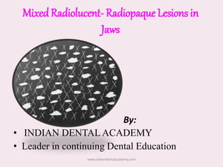
  • 1. Mixed Radiolucent- Radiopaque Lesions in Jaws By: • INDIAN DENTALACADEMY • Leader in continuing Dental Education www.indiandentalacademy.com
  • 2. Contents • Definition • Classification • Clinical features • Radiographic features • Differential Diagnosis • Treatment www.indiandentalacademy.com
  • 3. • Radiolucent — refers that portion of a processed radiograph which appears as dark / black. • Radiopaque — refers that portion of processed radiograph which appears as light / white. • Some normal anatomic structures and disease states can produce mixed radiolucent-radiopaque images on radiographs. www.indiandentalacademy.com
  • 4. • Pathologic entity may commences as an osteolytic lesion, which appears as radiolucency in radiograph. • During its development, foci of calcified material may form within the osteolytic area, when these foci become large & sufficiently mineralized, they become radiographically apparent. • Thus mixed radiolucent-radiopaque condition frequently represents an intermediate stage in development of the lesion. www.indiandentalacademy.com
  • 5. Mixed Radiolucent-Radiopaque Lesions Associated With Teeth 1. Calcifying crown of developing tooth 2. Tooth root with rarefying osteitis 3. Rarefying & Condensing osteitis 4. Chronic Osteomyelitis 5. PCOD 6. Cementoossifying fibroma 7. Cementoblastoma 8. Odontoma 9. Adenomatoid Odontogenic Tumor 10. Calcifying Odontogenic Cyst 11. Ameloblastic fibroodontoma 12. Calcifying Epithelial Odontogenic Tumorwww.indiandentalacademy.com
  • 7. Calcifying crown of developing tooth • Before calcification, permanent tooth buds may appear in the periapical region of deciduous teeth. • In early stages of development, tooth germs appear as cyst like radiolucency. • After few months undergo sufficient mineralization, appear as periapical radiolucency with radiopaque foci. www.indiandentalacademy.com
  • 8. • The cusp tips are first part to calcify in developing tooth. • As soon as sufficient mineral deposited in matrix of cusp tip, developing tooth can recognized as a radiolucency with radiopaque foci. www.indiandentalacademy.com
  • 9. Tooth Root With Rarefying Osteitis • Retained root & root tips are abnormal radiopacities commonly found in edentulous region of the jaws. • Retained roots present in jaws of every 1 of 4 edentulous persons. • 80% of retained roots are in posterior region of the jaws. • 6% of all retained root tips are associated with radiolucent areas. • Usually asymptomatic, or c/o intermittent pain or swelling. • ill-defined radiolucency with ragged margins. www.indiandentalacademy.com
  • 10. • The root canal may become channel for infection with a resulting rarefying osteitis in periapex, and production of radiolucent- radiopaque jaw lesions. • Usually identifiable by the shape of root, shadow of root canal, PDL space and surrounding lamina dura. www.indiandentalacademy.com
  • 11. Management • Root tips should be removed. • Surrounding soft tissue enucleate and bone defect should curetted. www.indiandentalacademy.com
  • 12. Rarefying and Condensing Osteitis • Frequently occurs at apex of a nonvital tooth / retained root. • Chronic infection acts as irritating factor and as stimulating factor. • Bone resorption occurs at apex; on other hand bone apposition occurs at periphery of rarefying lesion. • When chronic infection is present well-defined homogenous radiopacity is seen more / less circumscribing radiolucency around root end. www.indiandentalacademy.com
  • 14. Differential Diagnosis • Chronic osteomyelitis Treatment: • Affected teeth should treat endodontically or extraction www.indiandentalacademy.com
  • 15. Periapical Cementoosseous Dysplasia • It is a localized change in normal bone metabolism that results in replacement of normal cancellous bone with fibrous tissue & cementum-like material and abnormal bone. • It is a reactive fibroosseous lesion; it arises from periodontal ligament, where mature osteoblasts, cementoblasts & precursor cells reside. • This lesion usually identified during routine dental radiographic examination. www.indiandentalacademy.com
  • 16. Clinical Features • Typically it occurs in middle age mean age 39 years. • Females > males (9:1) • Involved teeth are vital; no H/O pain & swelling. Radiographic features • It usually lies at the apex of a tooth. • Mainly in mandibular anteriors. www.indiandentalacademy.com
  • 17. Internal structure • In early stage normal bone is resorbed and replaced with fibrous tissue that continuous with PDL ( causing loss of lamina dura). • Radiographically – radiolucency at apex of tooth. • In mixed stage radiopaque tissue appears in the radiolucent structure. • This material is amorphous, has a round, oval shape; composed of cementum /abnormal bone.www.indiandentalacademy.com
  • 18. • In mature stage internal aspect may be totally radiopaque without any obvious pattern. • Lamina dura of involved teeth is lost making PDL space less apparent / widen appearance. • Small lesions do not cause jaw expansion but large lesions may cause expansion, always a thin intact outer cortex. www.indiandentalacademy.com
  • 19. Differential Diagnosis • Periapical Abscess • Periapical Granuloma • Calcifying crown • Rarefying and Condensing osteitis • Odontoma www.indiandentalacademy.com
  • 20. Cementoossifying fibroma • It is uncommon neoplastic condition - originate from elements of periodontal ligament. • Usually occurs as periapical lesions – round and well marginated. • It can occur at any age; commonly found in adults mainly 2nd to 3rd decades. • Definite female predilection. www.indiandentalacademy.com
  • 21. • Usually asymptomatic; displacement of teeth- early c/f • 70-80% of lesions occur in mandible, primarily premolar & molar region, superior to inferior alveolar canal, in maxilla in canine fossa & zygomatic arch area. • Juvenile ossifying fibroma is aggressive form; it occurs in first 2 decades of life. • Because of rapid growth in young patients deformity of involved jaw may occur. www.indiandentalacademy.com
  • 22. Radiographic Features • Most of the lesions are discovered during routine dental examination. • The borders of the lesion is well defined. • A thin radiolucent line representing a fibrous capsule, may separate it from surrounding bone. • The internal structure is mixed radiolucent-radiopaque density. • Lesions produce more cementum like material may contain solid, amorphous radiopacities. www.indiandentalacademy.com
  • 24. Differential Diagnosis • Fibrous dysplasia • P C O D • C O C • C E O T • A O T Management • Surgical enucleation or resection of the lesion • Recurrence rate is 6% www.indiandentalacademy.com
  • 25. Cementoblastoma • It is a slow growing mesenchymal neoplasm principally composed cementum • It manifest as bulbous growth around & attached to the apex of tooth root • Tooth is vital and painful • Males > females • Relatively young adults • Mandible > maxilla, premolar & 1st molarwww.indiandentalacademy.com
  • 26. • These are mixed radiolucent-radiopaque lesions in which majority of the internal structure is radiopaque. • This central radiopaque mass is surrounded by a radiolucent band; indicating tumor is maturing from central aspect to periphery. • In most of the cases external resorption seen. • Large lesions can cause expansion of mandible with intact outer cortex. www.indiandentalacademy.com
  • 28. Differential Diagnosis • Odontoma • P C O D • Hypercementosis • Periapical sclerosing osteitis Treatment • These are self-limiting • Simple excision and extraction of associated tooth. • Rarely recur after enucleation. www.indiandentalacademy.com
  • 29. Chronic Osteomyelitis • Osteomyelitis is an inflammation of bone and bone marrow. 1. Suppurative : a. Acute b. Chronic 2. Sclerosing: a. focal b. diffuse 3. Chronic nonsuppurative sclerosing osteitis www.indiandentalacademy.com
  • 30. • It can occur at any age; common in first 3 decades of life. • Proliferative periosteitis is common in children and young adults. • Mandible more frequently involved. • Clinical examination shows signs of inflammation, tenderness, pain, swelling, intraoral & extraoral draining sinus tracts, fever and regional lymphadenopathy. www.indiandentalacademy.com
  • 31. • Denuded bone may protrude from open mucosal or cutaneous ulcers. • The sequestra are surrounded by sclerotic bone which is relatively avascular. • Within the bone, the haversian canals become blocked with scar tissue, and the bone becomes surrounded by thickened periosteum • Drainage in chronic disease is intermittent and modest in volume. www.indiandentalacademy.com
  • 32. Radiographic Features • Acute osteomyelitis – No bony changes. • Chronic osteomyelitis – radiographic images are irregularly shaped radiolucency with ragged, poorly defined borders. • Sometimes lesions appear as mixed radiolucent- radiopaque pattern. www.indiandentalacademy.com
  • 33. • Radiolucent areas consists of infected granulation tissue, fibrosis or both. • Radiopaque areas represent sclerosed, often nonvital bone sequestra or both. • Proliferative periostitis may show an alternating radiolucent-radiopaque laminated appearance at surface of affected bone (onion-skin appearance) www.indiandentalacademy.com
  • 35. Garre’s Osteomyelitis Unusual appearance of the mandible produced by osteomyelitis. www.indiandentalacademy.com
  • 36. Differential Diagnosis • Rarefying and condensing osteitis • Fibrous dysplasia • Paget’s disease • Malignant tumors www.indiandentalacademy.com
  • 37. Management Medical • Systemic antibiotics • Local antibiotics - irrigation - beads Surgical • Sequestrectomy • Saucerization • Decortication • Resection www.indiandentalacademy.com
  • 38. Odontoma • It is benign odontogenic tumor representing 67% of all odontogenic tumors. • Odontomas result from budding of extra odontogenic epithelial cells from dental lamina. • 3 types of odontomas present; 1. Compound 2. Complex 3. Compound-Complex www.indiandentalacademy.com
  • 39. • Compound odontoma – comprises odontogenic tissues laid down in a normal relationship, resulting structures will resemble to teeth. • Complex odontoma – tooth components are less well organized; toothlike structures are not formed. • Compound-Complex – it contain not only multiple tooth like structures but also calcified masses of dental tissue in haphazard manner. www.indiandentalacademy.com
  • 40. • 62% of compound variety occurs in maxilla; predilection for incisor- canine region, no gender bias. • 70% of complex type occur in mandible; located in 1st and 2nd molar region, mostly in females. • An odontoma frequently situated between crown of an unerupted tooth and crest of alveolar ridge, blocks tooth eruption. www.indiandentalacademy.com
  • 41. • Intermediate stage compound odontoma appears as a well-defined radiolucent lesion containing varying no. of radiopaque shadows of developing tooth. • Degree of calcification & opacity varies from stage to stage and lesion to lesion. • Complex type appears as a well-defined radiolucency with many radiopaque foci vary greatly in size, shape & prominence. www.indiandentalacademy.com
  • 42. Differential Diagnosis • Cementoblastoma • P C O D • Cementoossifying fibroma Management • Surgical enucleation • Periodic postoperative examination www.indiandentalacademy.com
  • 43. Adenomatoid Odontogenic Tumor • It is a uncommon and nonaggressive tumor of odontogenic epithelium. • It is 2 types - central and peripheral. • The central tumor divided into follicular type (associated with embedded tooth) & extra-follicular type( with no embedded tooth). • 73% of central lesions are follicular type www.indiandentalacademy.com
  • 44. • Age ranges from 5-50 years; 70% cases occur in 2nd decade, average age is 16 years. • It has female predilection (2:1) • Commonly occur in maxilla; incisor-canine-premolar • It is a slow growing & presents a gradually enlarging painless swelling. • Causes facial asymmetry, often associated with missing tooth. • It displace the teeth rather than root resorption. www.indiandentalacademy.com
  • 45. Radiographic Features • It is a pericoronal cyst- like radiolucency. • In maturing stages, sharply defined radiopaque foci are seen within radiolucency. www.indiandentalacademy.com
  • 46. Differential Diagnosis • COC • CEOT • Odontoma Management: • Surgical enucleation. www.indiandentalacademy.com
  • 47. Calcifying Odontogenic Cyst • It is also called as Gorlin’s cyst. • It is uncommon, benign slow growing lesion. • WHO categories as benign tumor. • It have a wide age distribution that peaks at 10-19 years and second peak age is during 7th decade; • Mean age is 36yrs www.indiandentalacademy.com
  • 48. Radiographic Features • At least 75% occurs in bone with equal distribution between jaws. • 75% lesions occur anterior to the first molar, especially incisor-canine region. • Sometimes it manifests as a pericoronal radiolucency. • Periphery can vary from well defined to irregular. www.indiandentalacademy.com
  • 49. • The internal aspect can vary in appearance. • It is cyst like radiolucency containing quite distinct radiopaque foci • It can be completely radiolucent to mixed R/L- R/O. www.indiandentalacademy.com
  • 50. Differential Diagnosis • AOT • CEOT • Odontoma Treatment • Surgical enucleation • Recurrence is reported www.indiandentalacademy.com
  • 51. Ameloblastic fibroodontoma • It is benign mixed odontogenic tumor that contain cords & nets of odontogenic epithelium & some calcified odontogenic tissue. • Clinical features are similar to odontoma, often associated with missing tooth / tooth has failed to erupt. • Occasionally tumor takes in position of a missing tooth. www.indiandentalacademy.com
  • 52. Radiological features • Most of the cases occur in posterior aspect of mandible. • The epicenter of lesion is usually occlusal to a developing tooth / towards the alveolar crest. • Lesion is well defined; sometimes corticated. • The internal structure is mixed with the majority of the lesion being radiolucent. www.indiandentalacademy.com
  • 53. • Small lesions may appear as enlarged follicles with one / two small discrete radiopacities. • Large lesions may have a more extensive calcified internal structure. • In some cases small calcifications have around shape with radiopaque enamel like margin. www.indiandentalacademy.com
  • 54. Differential Diagnosis • Odontoma • COC • AOT Management  Surgical enucleation.  No tendency to recur.  Follow up is necessary www.indiandentalacademy.com
  • 55. Calcifying Epithelial Odontogenic Tumor • It is also called Pindborg tumor. • It is less aggressive lesion; rarely may have an extraosseous location. • Males > females. • Mean age is 42 years • 68% tumors occur in mandible; especially molar regionwww.indiandentalacademy.com
  • 56. Radiological Features • It may have several radiographic appearance. 1. Pericoronal radiolucency, 2. Pericoronal radiolucency with radiopaque foci, 3. Mixed radiolucent-radiopaque lesion not associated with an unerupted tooth, 4. A “driven snow” appearance, 5. Dense radiopacity (occasionally).www.indiandentalacademy.com
  • 57. • More common in mandible (2:1) • 52% associated with unerupted / impacted tooth. • Borders are well defined, cyst like cortex, sometimes irregular & ill defined www.indiandentalacademy.com
  • 58. • The internal aspect may appear unilocular / multilocular with numerous scattered radiopaque foci. • Characteristic & diagnostic finding is radiopacities close to crown of embedded tooth. • In addition small, thin opaque trabeculae may cross the radiolucency in many directions www.indiandentalacademy.com
  • 59. Differential Diagnosis • A O T • C O C Management • Surgical resection is recommended. • Low recurrence rate. • Follow up is mandatory www.indiandentalacademy.com
  • 61. Osteoradionecrosis • A disease condition of hard and soft tissue after irradiation of the region. • Usually develops after 12 months radiotherapy completion. • Asymptomatic in early stages. • When ulceration occurs, tenderness and pain are common complaint. • Mixed radiolucent-radiopaque lesion in a full-blown case. www.indiandentalacademy.com
  • 62. Irradiation Narrowing of lumina ( principally of small arterioles) Hypovascular, hypocellular, hypoxic tissue Diminished capacity for normal repair Breakdown of tissue and its overlying mucosa Superficial infection of the denuded bone Osteoradionecrosis www.indiandentalacademy.com
  • 63. • Mandible is more commonly affected than maxilla. • This due to microanatomy & reduced vascularity of bone. • Posterior mandible is more affected than anterior portion. • Loss of mucosal covering & exposure of bone is hall mark of ORN. • Exposed bone becomes necrotic as a result of loss of vascularity from periosteum. • Intense pain may occur, with intermittent swelling & extra- oral drainage. www.indiandentalacademy.com
  • 64. Radiological features • The periphery is ill defined similar to osteomyelitis. • If the lesion reaches the inferior border, irregular resorption of bony cortex occurs. • Internal structure appearance will be depends on range of bone destruction and bone formation. • It will be overall sclerotic to radiopaque appearance. www.indiandentalacademy.com
  • 66. Prevention • Pt should undergo thorough dental checkup • Patient oral hygiene should be assessed & motivate the pt for good oral hygiene after radiotherapy. • Pt should undergo extraction of grossly carious teeth, periodontally involved teeth at least 10- 14 days before commencement of radiotherapy. • Restore if any decayed tooth present & patient must undergo thorough oral prophylaxis. • Teeth sockets should heal completely prior to radiation exposure. • After radiation exposure, all extractions, and invasive procedures should be avoided for a period of 3 months www.indiandentalacademy.com
  • 67. Management • Both surgical and nonsurgical treatments have been done. • Hyperbaric oxygen therapy is effective treatment. • Treatment is done in 3 stages; • Stage I: 30 dives • Stage II: sequestrectomy+ primary closure+ 60 dives • Stage III: 30 dives, resection and remaining 30 dives www.indiandentalacademy.com
  • 68. Focal Cementoosseous Dysplasia • It is a reactive lesion of PDLO. • It is PCOD like lesions occur in the premolar-molar region. • These are asymptomatic lesions occur in edentulous areas. • Females are commonly affected. • Average age is 37 yrs. The lesion matures through 3 stages: 1. Early radiolucent 2. Middle mixed stage 3. Late radiopaque www.indiandentalacademy.com
  • 69. Radiological Features • The distinctness of margin varies; radiopaque pattern scattered throughout the lesion. • Radiolucent area is surrounded by thin radiopaque border. • Often it appears as ground glass appearance www.indiandentalacademy.com
  • 70. Management • Periodic follow up is recommended. • If lesion shows signs of serious enlargement surgical intervention is indicated. www.indiandentalacademy.com
  • 71. Sailolith • Sailoliths are stones found within ducts of salivary glands. • Most common in submandibular glands. • Men > women • Age ranges 3rd to 4th decade. • Usually occur single (70- 80%) but may occur multiple • Patients usually asymptomatic but H/O pain swelling in floor of the mouth. • This discomfort may intensify at meal time. www.indiandentalacademy.com
  • 72. Radiographic Features • Sailoliths usually cylindrical • Some stones are homogenously radiopaque and some show multiple layers of calcification. Management • Small stones may be “milked out” through duct orifice using bimanual palpation. • Large stones can remove by lithotripsy or surgical intervention. www.indiandentalacademy.com
  • 74. Phlebolith • These are calcified thrombi found in veins, venulae or sinusoidal vessels of hemangiomas. • In head & neck phleboliths indicates presence of hemangioma. • Involved soft tissue swollen, throbbing or discolored by presence of veins or soft tissue hemangioma www.indiandentalacademy.com
  • 75. Radiographic Features • In cross section round or oval in shape with smooth periphery. • Internal aspect maybe homogenously radiopaque. • It gives “bulls eye” or target appearance. www.indiandentalacademy.com
  • 76. Rhinolith / Antrolith • Hard calcified bodies or stones occur in the nose or maxillary sinus. • Pt is asymptomatic for long time but expanding mass may impinge on mucosa which cause pain, congestion and ulceration. • Pt may develop a unilateral purulent rhinorrhea, sinusitis, headache, fetor and fever www.indiandentalacademy.com
  • 77. • These stones have variety of shapes & sizes • Stones present as homogenous or heterogeneous radiopacities. • Occasionally density will exceed the surrounding bone. www.indiandentalacademy.com
  • 78. Traumatic Myositis Ossificans • It is characterised by ossification of muscle by single acute traumatic episode or multiple minor traumatic episodes. • Exact mechanism for ossification is not clear. • It usually manifests as firm painful mass in the injured muscle within 1-4 weeks. • In facial muscles masseter and temporalis muscle frequently involved. www.indiandentalacademy.com
  • 79. Radiographic Features • It may appear as a feather type of calcification in muscle following ossification of hematoma along dissected muscle bundle. • Calcification may be seen within 2-3 weeks after trauma. Treatment: • Surgical excision www.indiandentalacademy.com
  • 81. Fibrous dysplasia • It is a hamartomatous fibroosseous lesion. • Etiology- Unknown • It is a bone lesion that produces lysis of bone with fibrous proliferation as a replacement (radiolucent) in its early stage. www.indiandentalacademy.com
  • 82. • Monostotic type most common & frequently involves the jaws and skull ( older adults). • Mandibular lesions are usually solitary. • Maxillary lesions may involve neighboring bones (Craniofacial FD). • Polyostotic form usually seen in children and young adults ( < 6 years). • Polyostotic form is static when skeletal growth stops, but proliferation may continue. www.indiandentalacademy.com
  • 83. • Early lesions are spherical radiolucency • Margins are ragged and poorly defined, merging into normal bone. • Lesions are usually situated deep within jaw bone. • Maturing changes produce ground glass appearance may commence at periphery of the lesion. • Gross displacement of mandibular canal is seen. www.indiandentalacademy.com
  • 84. • Fibrous dysplasia (intermediate stage) A) Smoky or mottled appearance(also seen in B). • B)Radiograph showing one lesion in maxilla & one in mandible. www.indiandentalacademy.com
  • 85. Differential Diagnosis • Chronic osteomyelitis • Paget’s disease • Osteogenic sarcoma www.indiandentalacademy.com
  • 86. Paget’s Disease (Osteitis Deformins) • First described in England by Sir James Paget in 1876. • Etiology: Unclear; genetics and undetected slow-acting virus may play roles. • Usually old age people are affected. • It is disorder of bone metabolism. www.indiandentalacademy.com
  • 87. • Osteoclastic resorptive process is overactive, with a compensatory increase in osteoblastic action attempting to maintain strong bones. • Progresses so slowly that annual radiographs show little change. • The vertebrae, pelvis, skull, tibia, femur and humerus are most likely to be involved • Patient may develop arthritis like symptoms & bowed legs with a waddling gait and may complain of an increasing hat size, which is due to skull expansion. www.indiandentalacademy.com
  • 88. • Maxilla>Mandible • Involved bones are thickened, and the foramina are often constricted. • Consequently pressure is induced on the structures that pass through the foramina, causing neurologic signs such as deafness and diminishing vision. • Calcified areas interspersed with radiolucent areas producing the cotton-wool appearance. • Teeth in the involved jaw may demonstrate spreading, migration, diminution of the laminadura, and (characteristically) hypercementosis. www.indiandentalacademy.com
  • 90. Differential Diagnosis • Florid Cementoosseous dysplasia • Fibrous dysplasia Management • In early stages, drug therapy with diphosphonates or calcitonin arrests osteoclastic activity. • Anti-inflammatory agents for pain relief. • Edentulous pts may require frequent adjustment or continued fabrication of new dentures. www.indiandentalacademy.com
  • 91. Osteogenic Sarcoma • Arises from primitive undifferentiated cells and from malignant transformation of osteoblasts. • Etiology – unknown. • Irradiated bone is more prone to become osteogenic sarcoma. • Age: 3rd - 4th decade. • Jaw lesions have less tendency to metastasize. • Prognosis is better for jaw lesions. • Metastasizes almost exclusively through hematogenous spread. • Intermittent local pain, swelling. www.indiandentalacademy.com
  • 92. Radiographic features • Three different radiographic images: 1. Totally radiolucent 2. Mixed radiolucent- radiopaque 3. Completely radiopaque • The classic sun-burst appearance may be seen in the latter two types. • The lesion may have ragged, ill-defined borders. • In some lesions sequestra are formed. • If the tumor invades the periosteum, many thin, irregular spicules of new bone may develop outward and perpendicular to the surface of the lesion. This produces the sun-burst effect.www.indiandentalacademy.com
  • 93. • Sometimes, two triangular radiopacities project from the cortex and mark the lateral extremities of the lesion. These are referred to as Codman’s triangles. • Rarely, it causes periosteal deposition of bone in an “onion-skin” pattern. • In some cases a bandlike widening involving complete length of PDL space on one or both sides of the root & involvement of the mandibular canal with widening of the canal can be seen. • Cemental resorption may be seen. www.indiandentalacademy.com
  • 95. Differential Diagnosis • Chondrosarcoma • Fibrosarcoma • Osteoblastic metastatic carcinoma • Fibrous dysplasia Management • Resection with large border of normal bone • Radiation & chemotherapy used for control metastatic spread. www.indiandentalacademy.com
  • 96. Osteoblastic metastatic carcinoma • Occasionally metastatic tumors to the jawbone from primary lesions in the prostate or breast may be seen. • It may appear as entirely radiolucent, mixed radiolucent-radiopaque, or entirely radiopaque. • Less aggressive– more circumscribed. • When small nests of sclerotic foci are disseminated throughout the jawbone, salt & pepper pattern may be seen. • H/o surgery or symptoms of a primary tumor indicate metastatic disease. www.indiandentalacademy.com
  • 98. PA view cotton-wool appearance in skull & large bilateral radiolucent lesions seen in mandible Lateral view showing radiolucent & radiopaque areas www.indiandentalacademy.com
  • 99. Differential Diagnosis • Chondrosarcoma • Ossifying subperiosteal hematoma www.indiandentalacademy.com
  • 100. Chondrosarcoma • It is malignant tumor of cartilaginous origin. • It can occur at any age; common in adults. • Male and females are equally affected. • Maxilla more frequently involved than mandible • Pt may have firm to hard swelling with long duration. • Enlargement cause pain headache and deformity www.indiandentalacademy.com
  • 101. Radiographic Features • Maxillary lesions occur in anterior maxilla • Mandibular lesions occur In coronoid process, condylar head and neck; occasionally symphysis region. • Lesions are round, ovoid, or lobulated • Borders are well defined and corticated some times meld with adjacent normal bone. • Aggressive lesions have infiltrative, ill-defined, and nonocorticated borders. www.indiandentalacademy.com
  • 102. • Internal structure usually exhibit some form of calcification within center of the lesion which gives mixed radiolucent-radiopaque appearance. • This diffuse calcification may be superimposed on a bony background that resembles granular / ground- glass appearance. • Lesions are rarely completely radiolucent. • In mandibular cases the inferior border / alveolar process grossly expanded with intact cortical covering. www.indiandentalacademy.com
  • 103. • Radiograph of a surgical specimen. • Radiopaque foci can be seen in the exophytic mass www.indiandentalacademy.com
  • 104. Differential Diagnosis • Osteogenic sarcoma • Fibrous dysplasia Management • Surgical resection is mandatory • It is quite radioresistent; radiation therapy can used as palliative procedure with large inoperable tumors. • At least 60% of cases have recurrences within 5 years. www.indiandentalacademy.com
  • 105. Desmoblastic Ameloblastoma • It is histologic variant of ameloblastoma. • Distinctive with a moderately cellular fibrous connective tissue with an abundance of collagen. • The tumor epithelium consists of small, ovoid, or follicle shaped islands & narrow cords. • Maxilla>Mandible. • Anterior region is commonly affected. www.indiandentalacademy.com
  • 106. Edentulous premolar region of maxilla Canine and premolar region of mandible www.indiandentalacademy.com
  • 108. • Eruption sequestrum • Ameloblastic fibrodentinoma • Calcifying hyperplastic dental follicle • Central odontogenic fibroma • Ameloblastoma • Cystic odontoma • Lymphoma of bone • Juvenile ossifying fibroma • Odontodysplasia • Odontogenic myxoma • Central hemangioma • Ewing’s sarcoma • Osteoblastoma • Osteiod osteoma RARITIES www.indiandentalacademy.com
  • 109. Foreign Bodies • Usually root canal filling materials. • Extruded gutta percha, silver points, sealers or retrograde amalgam and filled root canal www.indiandentalacademy.com
  • 110. Florid Cementoosseous Dysplasia • It appears as widespread form of periapical cemental dysplasia. • It has poor vascular supply, more susceptible for infections. • Females > Males • Mean age 42 years • Mandible > Maxilla • Mostly posterior to cuspids www.indiandentalacademy.com
  • 114. References • Differential Diagnosis Wood & Goaz 5th Edition • Dental Radiology White & Pharow 5th Edition • Oral Pathology Shafer 5th Edition • Oral pathology Neville & Dam 2nd Edition • Oral Radiographic Diagnosis Stafne’s 5th Edition www.indiandentalacademy.com