Trigeminal Nerve Anatomy and Branches
•
24 recomendaciones•1,360 vistas

The document discusses the trigeminal nerve, which is the 5th cranial nerve. It has three main branches - the ophthalmic, maxillary, and mandibular nerves. Each branch innervates different parts of the face and has its own set of branches. The trigeminal ganglion contains the cell bodies of the sensory fibers of the trigeminal nerve. Common disturbances of the trigeminal nerve include trigeminal neuralgia, which causes sudden, severe facial pain, and trigeminal nerve injuries. The document provides detailed information on the anatomy and branches of the trigeminal nerve.

Recomendados
Más contenido relacionado
La actualidad más candente
La actualidad más candente (20)
Destacado
Destacado (18)
Similar a Trigeminal Nerve Anatomy and Branches
Similar a Trigeminal Nerve Anatomy and Branches (20)
Más de Indian dental academy
Más de Indian dental academy (20)
Último
Último (20)
Trigeminal Nerve Anatomy and Branches
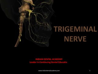
- 1. TRIGEMINAL NERVE 1 INDIAN DENTAL ACADEMY Leader in Continuing Dental Education www.indiandentalacademy.com
- 2. Contents • Introduction • Cranial nerves • Trigeminal ganglion • Trigeminal nerve Opthalmic branch Maxillary branch Mandibular branch • Disturbances associated with trigeminal nerve Trigeminal neuralgia Paratrigeminal syndrome Sphenopalatine neuralgia Auriculotemporal syndrome • Nerve blocks associated with trigeminal nerve branches • Clinical examination of trigeminal nerve • Trigeminal nerve injuries • Conclusion • References 2www.indiandentalacademy.com
- 3. NERVOUS SYSTEM Central nervous system Peripheral nervous system Brain and spinal cord Autonomic and somatic nervous system I N T R O D U C T I O N B.D. Chaurasia 4th edition 3www.indiandentalacademy.com
- 4. Structure of nerve Axon Fasciculi Nerve fibre Nerve Endoneurium Perineurium Epineurium I N T R O D U C T I O N Peter. L. Williams, grays anatomy, 38th edition,1995 4www.indiandentalacademy.com
- 5. C R A N I A L N E R V E S Motor nerves: III IV VI XI XII Sensory nerves I II VIII Mixed nerves V VII IX X Associated with parasympathetic ganglion III VII IX X Peter. L. Williams, grays anatomy, 38th edition,1995 5www.indiandentalacademy.com
- 6. T R I G E M I N A L G A N G A L I O N Trigeminal gangalion Gasserian/semilunar Sensory cell bodies of three branches of trigeminal Lies in a depression on the petrous apex, within a dural fold called the meckal cave Cunningham’s manual of practical anatomy, volume 3, 3rd edition 6www.indiandentalacademy.com
- 8. T R I G E M I N A L N E R V E Trigeminal nerve Tres meaning three and geminus meaning twin Largest cranial nerve Mixed nerve Sensory to greater part of the scalp, teeth and the oral and nasal cavity Motor supply is to muscles of mastication and extraocular muscles 5th cranial nerve Cunningham’s manual of practical anatomy, volume 3, 3rd edition 8www.indiandentalacademy.com
- 9. T R I G E M I N A L N E R V E MOTOR ROOT SENSORY ROOT TWO ROOTS Within pons and medulla oblongata Semilunar gangalion Foramen ovale along with mandibular nerve MOTOR ROOT Muscles of mastication Mylohyoid Anterior belly of digastric Tensor tympani Tensor veli palatine Cunningham’s manual of practical anatomy, volume 3, 3rd edition 9www.indiandentalacademy.com
- 10. T R I G E M I N A L N E R V E SENSORY ROOT Fibres arise in trigeminal ganglion Opthalmic nerve Leaving from concave surface of ganglion Cunningham’s manual of practical anatomy, volume 3, 3rd edition 10 Maxillary nerve Mandibular nerve www.indiandentalacademy.com
- 11. B R A N C H E S Main divisions Opthalmic Wholly Sensory division Superior and smallest division Leaves cranium through Superior orbital fissure 3 branches Lacrimal Frontal Nasocilliary Cunningham’s manual of practical anatomy, volume 3, 3rd edition 11www.indiandentalacademy.com
- 12. O P T H A L M I C N E R V E Peter. L. Williams, grays anatomy, 38th edition,1995 12www.indiandentalacademy.com
- 13. O P T H A L M I C N E R V E Lacrimal nerve Smallest branch Ends in the skin of upper eyelid Lacrimal gland and conjunctiva Pierces orbital septum Peter. L. Williams, grays anatomy, 38th edition,1995 13www.indiandentalacademy.com
- 14. O P T H A L M I C N E R V E Frontal nerve Largest branch Skin of forhead and scalp Mucous membrane of frontal sinus and pericranium Enter orbit through superior orbital fissure Supraorbital branch Supratochlear branch Upper eyelid Lower part of forhead Peter. L. Williams, grays anatomy, 38th edition,1995 14www.indiandentalacademy.com
- 15. O P T H A L M I C N E R V E Nasocilliary nerve Branches in orbit In nasal cavity Supply mucous membrane of nose Branches in nasal cavity Long ciliary nerve Supply iris , cornea Posterior ethmoi nerve Posterior ethmoid,sphenoidal air cells Anterior ethmoidal nerve Anterior ethmoid, frontal paranasal Nasal conchae, anterior nasal wall Tip and ala of nose On face supply lacrimal gland. Both eyelids and skin over nasal bridge Branches on face B.D. Chaurasia 4th edition 15www.indiandentalacademy.com
- 16. M A X I L L A R Y N E R V E Maxillary nerve Wholly sensoryBegins at trigeminal ganglion Enter orbit through inferior orbital fissure and appears on face through infra orbital foramen Leaves through foramen rotundum Peter. L. Williams, grays anatomy, 38th edition,1995 16www.indiandentalacademy.com
- 17. M A X I L L A R Y N E R V E Maxillary nerve branches Cranium Pterygopalatine fossa Infraorbital canal Face Cranium Pterygopalatine fossa Infraorbital canal Face Peter. L. Williams, grays anatomy, 38th edition,1995 17www.indiandentalacademy.com
- 18. M A X I L L A R Y N E R V E Middle superior alveolar nerve Anterior superior alveolar nerve Posterior superior alveolar nerve Peter. L. Williams, grays anatomy, 38th edition,1995 18www.indiandentalacademy.com
- 19. M A N D I B U L A R N E R V E Largest division Large sensory root, small motor root Mandibular nerve Motor root medial to sensory Main trunk 2-3mm Small anterior and large posterior branch B.D. Chaurasia 4th edition 19www.indiandentalacademy.com
- 20. M A N D I B U L A R N E R V E Branches of mandibular nerve Peter. L. Williams, grays anatomy, 38th edition,1995 20www.indiandentalacademy.com
- 21. M A N D I B U L A R N E R V E Meningeal branch Nerve to medial pterygoid Branches of undivided nerve Enter skull through foramen spinosum Supply dura of middle and anterior cranial fossa Slender branch Supply deep part of muscle Also tensor tympani and tensor velli palati muscle B.D. Chaurasia 4th edition 21www.indiandentalacademy.com
- 22. M A N D I B U L A R N E R V E Buccal nerve Anterior division Passes between two heads of lateral pterygoid Emerge under ramus and anterior border of masseter Supply skin over anterior part of buccinator and mucous membrane lining buccal surface of gums B.D. Chaurasia 4th edition 22www.indiandentalacademy.com
- 23. M A N D I B U L A R N E R V E Massetric nerve Passes through mandibular notch Deep temporal nerve Nerve to lateral pterygoid Supply masseter muscle Passes above upper head of lateral pterygoid Turn above infratemporal crest and sink into temporalis 2 branches supplying each muscle head B.D. Chaurasia 4th edition 23www.indiandentalacademy.com
- 24. M A N D I B U L A R N E R V E Branches of posterior division Auriculotemporal nerve Traverse upper part of parotid gland Auricular branch Articular branch Cunningham’s manual of practical anatomy, volume 3, 3rd edition 24 Temporal branch www.indiandentalacademy.com
- 25. M A N D I B U L A R N E R V E Lingual nerve Lies between ramus and medial pterygoid. Antriomedial to inferior alveolar nerve Base of tongue behind 3rd molar Medial to submandibular duct Peter. L. Williams, grays anatomy, 38th edition,1995 25www.indiandentalacademy.com
- 26. M A N D I B U L A R N E R V E Carries sensation from Anterior two third of the tongue Oral mucosa on the floor of the oral cavity Lingual gingiva associated with lower teeth Peter. L. Williams, grays anatomy, 38th edition,1995 26www.indiandentalacademy.com
- 27. M A N D I B U L A R N E R V E Inferior alveolar nerve Large terminal branch of posterior division Originates deep to the lateral pterygoid muscle in association with lingual nerve Passes between sphenomandibular ligament and ramus B.D. Chaurasia 4th edition 27www.indiandentalacademy.com
- 28. M A N D I B U L A R N E R V E Inferior alveolar nerve Mylohyoid branch which supply mylohyoid muscle Enter the mandibular foramen Branches into mental nerve and incisive nerve Peter. L. Williams, grays anatomy, 38th edition,1995 28www.indiandentalacademy.com
- 30. g a n g l i o n Ganglion associated with trigeminal nerve: B.D. Chaurasia 4th edition 30www.indiandentalacademy.com
- 31. g a n g l i o n Ciliary ganglion Sensory root Sympathetic root Plexus around opthalmic artery Nasocilliary nerve Parasympathetic root Occulomotor nerve Peter. L. Williams, grays anatomy, 38th edition,1995 31www.indiandentalacademy.com
- 32. g a n g l i o n Only structurally related to maxillary nerve Sphenopalatine/Pterygopalatine gangalion B.D. Chaurasia 4th edition 32 Sensory root Sympathetic root Plexus around internal carotid artery Maxillary nerve Secrotomotor root Greater petrosal nerve www.indiandentalacademy.com
- 33. g a n g l i o n Nasal Sphenopalatine/Pterygop alatine gangalion Palatine Orbital B.D. Chaurasia 4th edition 33 Pharyngeal www.indiandentalacademy.com
- 34. g a n g l i o n Otic ganglion Parasympathetic ganglion Topographically – Mandibular nerve Functionally – Glossopharyngeal nerve Situated in infratemporal fossa below foramen ovale B.D. Chaurasia 4th edition 34www.indiandentalacademy.com
- 35. g a n g l i o n Otic ganglion B.D. Chaurasia 4th edition 35 Sensory root Sympathetic root Plexus around middle meningeal artery Auriculotemporal nerve Secrotomotor root Lesser petrosal nerve www.indiandentalacademy.com
- 36. g a n g l i o n Submandibular ganglion Lies superficial to hyoglossus muscle in submandibular region Topographically – lingual branch of mandibular nerve Fumctionally – Facial nerve Peter. L. Williams, grays anatomy, 38th edition,1995 36www.indiandentalacademy.com
- 37. D I S T U R B A N C E S A S S O C I A T E D Trigeminal neuralgia Fothergill’s disease/Tic douolourex Sudden, unilateral, lancinating, recurrent pain Right side more commonly effected Cranial nerve functional anatomy, Stanley Monkhouse 37 Attack last from few seconds to several minutes Presence of trigger zones www.indiandentalacademy.com
- 38. D I S T U R B A N C E S A S S O C I A T E D Etiology Cranial nerve functional anatomy, Stanley Monkhouse 38 Intracranial tumours Intravascular abnormalities Multiple sclerosis Post traumatic neuralgia Viral etiologyDental etiology www.indiandentalacademy.com
- 39. D I S T U R B A N C E S A S S O C I A T E D Pathophysiology Cranial nerve functional anatomy, Stanley Monkhouse 39www.indiandentalacademy.com
- 40. D I S T U R B A N C E S A S S O C I A T E D Bilateral cases Maxillary division Mandibular division Opthalmic division All 3 divisions Both maxi and mandibular 3 % 66% 49% 16% 19% 1% Cranial nerve functional anatomy, Stanley Monkhouse 40www.indiandentalacademy.com
- 41. D I S T U R B A N C E S A S S O C I A T E D Treatment Peripheral neurectomy Alcohol injection into peripheral nerve area Injection of boiling water into ganglion Surgical sectioning of trigeminal sensory root Cranial nerve functional anatomy, Stanley Monkhouse 41 Microsurgical decompression of trigeminal root www.indiandentalacademy.com
- 42. D I S T U R B A N C E S A S S O C I A T E D Paratrigeminal syndrome/Raeder’s syndrome Middle aged males Severe hedache pain in area distibution of trigeminal nerve Signs of ocular sympathetic paralysis No sweating on the affected side Cranial nerve functional anatomy, Stanley Monkhouse 42www.indiandentalacademy.com
- 43. D I S T U R B A N C E S A S S O C I A T E D Sphenopalatine neuralgia Lower half headache/Sludders headache Vasoconstriction of vessels supplying nasal mucosa Deviated nasal septum Vasodialation of internal maxillary artery Cranial nerve functional anatomy, Stanley Monkhouse 43www.indiandentalacademy.com
- 44. D I S T U R B A N C E S A S S O C I A T E D Sphenopalatine neuralgia Unilateral paroxysm of intense pain for 15 min to several hours Alarm clock hedache Male > female 1st episode occur before 40 years of age Cranial nerve functional anatomy, Stanley Monkhouse 44 Sneezing, discharge and epiphoraNo trigger zones www.indiandentalacademy.com
- 45. D I S T U R B A N C E S A S S O C I A T E D Treatment Alcohol injection into the ganglion Surgical correction of septal defects Cranial nerve functional anatomy, Stanley Monkhouse 45 Drugs like ergotamine and methysergideResection of ganglion www.indiandentalacademy.com
- 46. D I S T U R B A N C E S A S S O C I A T E D Auriculotemporal syndrome Frey syndrome Due to reinnervation of sweat glands by parasympathetic salivary fibres Mainly after some surgical operation like removal of parotid gland Sweating and flushing of the involved side during eating Cranial nerve functional anatomy, Stanley Monkhouse 46www.indiandentalacademy.com
- 47. N E R V E B L O C K S Posterior superior alveolar nerve block Tuberosity block/zygomatic block Pulp of maxillary 3rd molar, 2nd and 1st molar (except MB root of 1st molar), buccal periosteum and bone Malamad SF. Handbook of local anesthesia. 6th ed. Missouri: Elsevier;201347www.indiandentalacademy.com
- 48. N E R V E B L O C K S Middle superior alveolar nerve block Pulps of maxillary 1st and 2nd premolar, MB root of 1st molar, buccal periosteum and bone Malamad SF. Handbook of local anesthesia. 6th ed. Missouri: Elsevier;201348www.indiandentalacademy.com
- 49. N E R V E B L O C K S Anterior superior alveolar nerve block Infraorbital nerve block Pulp of maxillary central incisor to the canine. In 72% of cases pulp of maxillary premolars and MB root of 1st molar. Buccal periosteum and bone of these teeth. Lower eyelid, lateral aspect of nose, upper lip. Malamad SF. Handbook of local anesthesia. 6th ed. Missouri: Elsevier;201349www.indiandentalacademy.com
- 50. N E R V E B L O C K S Greater palatine nerve block Anterior palatine nerve block Posterior portion of hard palate and overlying tissues, anteriorly till the 1st PM and medially till the midline. Malamad SF. Handbook of local anesthesia. 6th ed. Missouri: Elsevier;201350www.indiandentalacademy.com
- 51. N E R V E B L O C K S Nasopalatine nerve block Incisive nerve block Anterior portion of hard palate and overlying tissues, mesial from the right 1st PM to the mesial of the left 1st PM. Malamad SF. Handbook of local anesthesia. 6th ed. Missouri: Elsevier;201351www.indiandentalacademy.com
- 52. N E R V E B L O C K S Maxillary nerve block Second division block/V2 nerve block Pulpal anaesthesia of maxillary teeth on the side of the block. Buccal periodontium and bone overlying these teeth. Soft tissue and bone of the hard palate and part of the soft palate medial to midline. Skin of lower eyelid, side of nose, cheek and upper lip. Malamad SF. Handbook of local anesthesia. 6th ed. Missouri: Elsevier;201352www.indiandentalacademy.com
- 53. N E R V E B L O C K S Inferior alveolar nerve block Mandibular block Mandibular teeth to the midline. Body of the mandible, inferior portion of ramus. Buccal mucoperiosteum, mucous membrane anterior to mental foramen. Anterior 2/3rd of tongue and floor of oral cavity. Lingual soft tissues and periosteum. Malamad SF. Handbook of local anesthesia. 6th ed. Missouri: Elsevier;201353www.indiandentalacademy.com
- 54. N E R V E B L O C K S Mandibular nerve bolck 3rd division nerve block/Gow Gates technique Mandibular teeth to the midline. Body of the mandible, inferior portion of ramus. Buccal mucoperiosteum, mucous membrane on the side of injection. Anterior 2/3rd of tongue and floor of oral cavity. Lingual soft tissues and periosteum. Skin over zygoma and temporal region. Malamad SF. Handbook of local anesthesia. 6th ed. Missouri: Elsevier;201354www.indiandentalacademy.com
- 55. N E R V E B L O C K S Vazirani akinosi tecnique Closed mouth mandibular nerve block Mandibular teeth to the midline. Body of the mandible, inferior portion of ramus. Buccal mucoperiosteum, mucous membrane anterior to mental foramen. Anterior 2/3rd of tongue and floor of oral cavity. Lingual soft tissues and periosteum. Malamad SF. Handbook of local anesthesia. 6th ed. Missouri: Elsevier;201355www.indiandentalacademy.com
- 56. N E R V E B L O C K S Incisive nerve block Buccal mucous membrane anterior to mental foramen till midline. Lower lips. Pulp of premolars, canines and incisors. Malamad SF. Handbook of local anesthesia. 6th ed. Missouri: Elsevier;201356www.indiandentalacademy.com
- 57. C L I N I C A L E X A M I N A T I O N Clinical examination of trigeminal nerve Use sterile sharp item on forehead, cheek and jaw. Sensory function Motor function While cleching of teeth, firmnes of masseter will be felt on both sides Trigeminal Nerve Injuries. Michael Miloro;2013 57www.indiandentalacademy.com
- 58. T R I G E M I N A L N E R V E I N J U R I E S Trigeminal nerve injuries Neuropraxia Axontmesis Neurotmesis Types Trigeminal Nerve Injuries. Michael Miloro;2013 58www.indiandentalacademy.com
- 59. T R I G E M I N A L N E R V E I N J U R I E S Trigeminal nerve injuries Trigeminal Nerve Injuries. Michael Miloro;2013 59www.indiandentalacademy.com
- 60. T R I G E M I N A L N E R V E I N J U R I E S Causes of Trigeminal nerve injuries LA injections Cosmetic surgery Orthognathic surgery Maxillofacial trauma Tumours and oncologic surgery Salivary gland surgery Trigeminal Nerve Injuries. Michael Miloro;2013 60www.indiandentalacademy.com
- 61. T R I G E M I N A L N E R V E I N J U R I E S Extraction of mandibular 3rd molar Endodontic treatment Trigeminal Nerve Injuries. Michael Miloro;2013 61www.indiandentalacademy.com
- 63. REFERENCES: B.D. Chaurasia 4th edition Cunningham’s manual of practical anatomy, volume 3, 3rd edition Tubbs RS, Loukas M, May WR, Cohen-Gadol AA. A variation of the infraorbital nerve: its potential clinical consequence especially in the treatment of trigeminal neuralgia: case report. Neurosurgery. 2010 Sep; 67 Malamad SF. Handbook of local anesthesia. 6th ed. Missouri: Elsevier;2013 Peter. L. Williams, grays anatomy, 38th edition,1995 Trigeminal Nerve Injuries. Michael Miloro;2013 Cranial nerve functional anatomy, Stanley Monkhouse 63www.indiandentalacademy.com