SlideShare una empresa de Scribd logo
1 de 115
Dr. Joe M Das
DIABETIC FOOT
- Bahara, Mills et al. International Wound Journal 2009.
The theme – Diabetes
education and prevention
(2009 – 2013)
Statistics
 60-70% of those with diabetes will develop
peripheral neuropathy, or lose sensation in their
feet.
 Up to 25% of those with diabetes will develop a
foot ulcer.
 More than half of all foot ulcers (wounds) will
become infected, requiring hospitalization and 1 in
5 will require an amputation.
 After a major amputation, 50% of patient will have
their other limb amputated within 2 years.
The History of Diabetes
 1552 BC: the first known mention of diabetes
– found on the Ebers Papyrus
 Egyptian physician Hesy-Ra of the 3rd
Dynasty makes
 Lists remedies to combat the
‘passing of too much urine’
Wood Carving of
Hesy-Ra
The History.. Contd…
 250 BC: Diabetes described as ‘the
melting down of flesh and limbs into
urine’
 By Greek physician Aretaeus of
Cappodocia who gives the first
complete medical description of
diabetes
 1425 AD: Diabetes first appears in the
English language as the Middle
English word ‘diabete’.
 16th Century: Swiss physician Phillipus Aureolus
considered the ‘Martin Luther of Medicine’ –
identifies diabetes as a serious general disorder.
1776: English physician Mathew Dobson of Liverpool
 evaporates two quarts of urine from a patient with
diabetes.
 The resulting residue is granulated and smells and
tastes like sugar, conclusively establishing the presence
of ‘saccharine materials’ as a diagnosis of diabetes.
The History… Contd…
Phillipus Aureolus
 1869
 German medical student
 Paul Langerhans
 The islet cells of the pancreas but is unable to explain
their function.
 The find is dubbed the ‘islets of Langerhans.”
The History Continues..
 1871: French physician Apollinaire Bouchardat
notices the disappearance of glycosuria in his
diabetes patients during food rationing of
food under the Siege of Paris in the Franco-
Prussian War, and formulates individualized
diets to treat the condition.
 1889: Scientists Oskar Minkowski and Joseph
von Mering of the University of Strasbourg,
France demonstrate how removing a dog’s
pancreas produces diabetes.
The History Continues..
Apollinaire Bouchardat
Oskar Minkowski
 1901: American pathologist Eugene Opie of
John Hopkins University in Baltimore
establishes a connection between the
failure of the islets of Langerhans in the
pancreas and the occurrence of diabetes.
 1913: Prof. John J.R. Macleod writes a
monograph on diabetes entitled
‘Diabetes: Its Pathological Physiology.’
The History Continues..
 Dec. 1916: Boston pathologist Elliott Joslin
 compiles 1,000 of his own cases and creates the
textbook The Treatment of Diabetes
Mellitus.
 In it he reports that ‘the mortality of patients
was approximately 20 per cent lower than for
the previous year’, due to ‘the introduction of
fasting and the emphasis on regular
exercise.’
 1919: Dr. Frederick Allen of the Rockefeller
Institute in New York publishes
 “Total Dietary Regulations in the
Treatment of Diabetes” that introduces a
therapy of strict dieting – dubbed the
‘starvation treatment’ –- as a way to manage
diabetes
The History Continues..
Dr. Frederick Allen
Elliott Joslin
Oct. 31, 1920
Sir Frederick Grant Banting
 conceives of the idea of insulin after reading an
article in The Journal Surgery, Gynecology and
Obstetrics by Moses Barron, by an American
pathologist, titled ‘The Relation of Islets of
Langerhans to Diabetes with Special Reference to
Cases of Pancreatic Lithiasis.’
 With the support of Prof.. Macleod of the
University of Toronto, and the assistance of
 Best, a medical student he starts his research
using a variety of different extracts on
depancreatized dogs.
The History Continues..
 Summer 1921: Banting’s work leads to the discovery of insulin. On July
30, Dog 410 is the first to receive the extract. On August 4 the extract
is called ‘Isletin’ for the first time.
 Nov. 14, 1921 Dr. Banting and Charles Best deliver a preliminary report
of their research to the Journal Club of the University of Toronto,
Department of Physiology.
 Nov. 17, 1921: Banting and Best discover that extract from cattle foetal
pancreas lowers blood sugar levels of depancreatized dogs, leading
them toward plentiful, cheap sources for insulin. Experiments begin
to test the long-term effectiveness of insulin treatment.
 Dec. 30, 1921 :Banting, Macleod, Best and Dr. James Bertram
Collip, a biochemist present the results of their research at a session
of the American Physiological Society at Yale University. The paper
initially generates little interest.
 The paper – ‘The Internal Secretion of the Pancreas’ – is published two
months later in the prestigious Journal of Laboratory and Clinical
Medicine
More Of Banting & Best…
Oct. 25, 1923
 Banting and Macleod
 The Nobel Prize in Physiology or Medicine.
 Banting shares his award with Best
 Macleod shares his with Collip.
The Nobel Prize Goes To….
Banting & Macleod
Banting's and Best's laboratory, where
insulin was discovered.
 11th January 1922
 Leonard Thompson, 14,
 A ‘charity patient’ at the Toronto General Hospital,
 First person to receive and injection of insulin to treat Type 1
diabetes.
 Thompson lives another 13 years before dying of pneumonia
at age 27.
The History Continues..
 May 3, 1922: The word ‘insulin’ is used in public for the
first time when Macleod presents the paper ‘The Effect
Produced on Diabetes by the Extracts of Pancreas’ to the
 May 30, 1922: Pharmaceutical manufacturer Eli Lilly & Co.
of Indianapolis and the University of Toronto enter a deal
for the mass production of insulin.
The History Continues..
 1966
 First pancreas transplant
 University of Manitoba
The History Continues..
 July 7, 1989: Her Majesty Queen Elizabeth The Queen Mother
kindles the
 Flame of Hope at Banting House National Historic Site – ‘The
Birthplace of Insulin’ – in London, Ontario.
 As a symbol of hope, the flame will burn until a cure for
diabetes is found.
 March 1999
 Scientists conduct the first
successful islet transplant at the
University of Alberta Hospital.
 The Edmonton Protocol.
isolating islets from a cadaveric
donor pancreas using a mixture
of enzymes called Liberase
The History Continues..
 Dec. 20, 2006: The United Nations recognizes diabetes as a
global threat and designates World Diabetes Day,
November 14 – in honour of Frederick Banting’s birthday –
as a UN Day to be observed every year starting in 2007.
Pathogenesis
 Chronic hyperglycaemia
 Nerve microvascular dysfunction
 ↑ free radical formation (due to ↓ NADPH)
 Polyol pathway hyperactivity (Glucose → ↑sorbitol
→ ↓ myoinositol → ↓ Na-K ATPase)
 PKC hyperactivity
 Nonenzymatic
 Abnormalities of nerve growth
 Pathology:
 Axonal loss distally, with a “dying back”
phenomenon
 A reduction in myelinated fiber density and
 Focal areas of demyelination on teased fiber
preparations.
Neuropathy
Motor Sensory Autonomic
↓ nociception
↓ Proprioception,
Unawareness
of foot position
A-V Shunt* open
Permanent
Increase foot
Blood flow
Bulging foot veins,
Warm foot
Reduced
sweating
Dry skin
Fissures and
cracks
Muscle wasting
Foot weakness
Postural deviation
Deformities, stress
and shear pressures
Trauma
Stress on bones & joints
Plantar pressure
Callus formation
InfectionUlcer
Symmetrical neuropathies Asymmetrical neuropathies
Distal sensory and sensory-
motor neuropathy
Mononeuropathy
Large-fiber type Mononeuropathy multiplex
Small-fiber type Radiculopathies
Distal small-fiber neuropathy Lumbar plexopathy or
radiculoplexopathy
“Insulin neuropathy” CIDP
CIDP
 Distal Symmetrical Neuropathy
 The commonest neuropathic syndrome
 “Diabetic neuropathy”
 A “length-related” pattern of sensory loss
 Toes → Feet → Leg (stocking pattern)
 Later motor
 Autonomic neuropathy is less common
 Autonomic neuropathy:
 ↓ sweating → dry skin that is likely to crack easily →
risk of infection.
 Warm because of the arterio-venous shunting →
Distension of foot veins that fail to collapse even
when the foot is elevated
 A gangrenous toe in a foot that has bounding
arterial pulses may be seen as there is impairment of
the nutritive capillary circulation because of
arteriovenous shunting.
 The oxygen tension of the blood in these veins is
typically raised.
 Neuropathic edema, which is resistant to treatment
with diuretics but may respond to treatment with
ephedrine
 High blood sugar expedites
artherosclerosis giving peripheral
vascular disease (reduction of blood
supply to the foot).
 The delivery of essential nutrients
and oxygen to the foot is
compromised leading to anaerobic
infections and tissue necrosis.
Peripheral arterial disease
Artherosclerosis
narrows or blocks
the arterial lumen
Foot ischaemia
Foot ulcer
Necrosis/ Gangrene
Infection
Artheroma plaque
narrowing the arterial
lumen
Ischaemic toes due to
artherosclerosis
Peripheral Arterial Disease
Clinical features (Contd..)
 Ulcers
 ↓ vibration sense
 Glove and stocking distribution of sensory loss
 +ve Romberg’s sign; ↓ ankle tendon reflex
 Wasting of small muscles of feet → Unopposed
pulling of long extensors and flexors → Clawing
of toes → Elevated plantar pressure points at the
metatarsal heads→ Callus formation & foot
ulceration
Charcot arthropathy
 A progressive condition of the musculoskeletal system
that is characterized by joint dislocations, pathologic
fractures, and debilitating deformities.
Charcot Foot
 More dramatic – less common 1%
 Severe non-infective bony collapse with secondary
ulceration
 Two theories
 Neurotraumatic
 Neurovascular
Charcot Foot
 Neurotraumatic
 Decreased sensation + repetitive trauma = joint and
bone collapse
 Neurovascular
 Increased blood flow → increased osteoclast activity →
osteopenia → Bony collapse
 Glycolization of ligaments → brittle and fail →
Joint collapse
Charcot’s Foot-- Classification
Eichenholtz
1 – acute inflammatory process
Often mistaken for infection
2 – coalescing phase
3 - consolidation
Location
Forefoot, midfoot (most
common) , hindfoot
Atrophic or hypertrophic
•Radiographic finding
•Little treatment implication
Wagner’s classification
Depth of penetration
Extent of tissue necrosis
Treatment given
Grade 1
 No open lesion, skin intact
 Potential site for ulceration like prominent
metatarsal heads, hammer toes, Charcot’s
deformity
 Treatment:
 Education & prevention
 Self foot care & proper shoes
 Antibiotics for cellulitis
Grade 2
 Superficial ulcer – extension to subcutaneous
adipose tissue
 Treatment:
 Wound care + antibiotics
 Radiological evaluation
 Modified weight bearing bone resection
Grade 3
 Ulcer penetrating joint, deep abscess, OM,
tendon sheath infection
 No digital gangrene
 Treatment:
 Hospitalisation
 Debridement and IV antibiotics
 Joint and bone resection
Grade 4
 Some part of the foot gangrenous
 Most part salvagable
 Treatment:
 Similar to that for grade 3 + Local amputation
Grade 5
 Extensive gangrene
 Limb not salvagable
 Treatment:
 Major amputation
University of Texas Diabetic Wound Classification
 Stages
 Stage A: No infection or ischemia
 Stage B: Infection present
 Stage C: Ischemia present
 Stage D: Infection and ischemia present
 Grading
 Grade 0: Epithelialized wound
 Grade 1: Superficial wound
 Grade 2: Wound penetrates to tendon or capsule
 Grade 3: Wound penetrates to bone or joint
Depth
Classification
Definition Treatment
0 At-risk foot, no
ulceration
Patient education,
accommodative
footwear, regular
clinical examination
1 Superficial ulceration,
not infected
Offloading with total
contact cast (TCC),
walking brace, or
special footwear
2 Deep ulceration
exposing tendons or
joints
Surgical debridement,
wound care, offloading,
culture-specific
antibiotics
3 Extensive ulceration
or abscess
Debridement or partial
amputation, offloading,
culture-specific
antibiotics
Ischemia
Classification
Definition Treatment
A Not ischemic
B Ischemia without
gangrene
Noninvasive vascular
testing, vascular
consultation if symptomatic
C Partial (forefoot)
gangrene
Vascular consultation
D Complete foot
gangrene
Major extremity
amputation, vascular
consultation
International Working Group on Diabetic Foot
Classification System
 On the basis of the scientific literature and expert
opinion, five categories were identified,
which were considered the most relevant items for
research projects in diabetic foot ulcers:
 Perfusion
 Extent/size
 Depth/ tissue loss
 Infection
 Sensation
PEDIS Classification System
History taking
 Age
 Mode of onset
 Bare foot walking
 Paraesthesia
 Claudication pain
 Fever
 H/o DM – Duration, controlled/uncontrolled
 Treatment history
 H/o impotency
 Diabetics present to the surgeon in a number of scenarios:
 with any foot and ankle problem and diabetes as an
incidental part of the past medical history - important to
assess how much diabetes increases the risk of any
proposed surgical or non-surgical treatment
 with an acutely inflamed where the main differential
diagnosis is between infection and Charcot arthropathy
 with an ulcer which may or may not be complicated by
deep infection or ischemia
 with a deformity which has led to recurrent ulceration, or
is felt to be a risk factor for ulceration - important to assess
the relative risks of corrective surgery and continuing
protective care
Whatever the initial presentation, it is important to ask
about
 what type of diabetes does the patient have (type 1 or type 2)
 how is it managed - diet, oral hypoglycaemics, insulin - and
what agents and regime
 who normally looks after the patient's diabetes - GP/practice
nurse/hospital diabetic clinic and therefore who is likely to
have the patient's diabetic records
 how well controlled is the diabetes - review the patient's
glucose monitoring record and ask when the last HbA1c test
was done and what was the result and the previous trend
 does the patient have any of the major complications of
diabetes
 cardiovascular disease
 renal disease
 eye disease
 has the patient had an ulcer or Charcot arthropathy before - the
single biggest risk factor for either of these conditions is to have
had them before
Clinical Examination
 Nourishment, pallor
 A) Limb affected
B) Type of lesion:
 Ulcer
 Gangrene
 Cellulitis / abscess
Ulcer: “Malperforant ulcer”
 Site
 Size, shape, margin
 Floor: Slough ±
 Edge: Punched out
 Surrounding skin
 Base: May be bone
 Infected – foul smell
 Local anesthesia of skin
Gangrene:
 Usually wet
 Forefoot → Leg
 Edema
 Foul smell
 Skip lesion
 No line of demarcation
 Bone may be exposed
Cellulitis / abscess:
 Diffuse tender
 Swelling ±
Regional examination:
Deformity of foot
High arching
Local anaesthesia
Regional pulsations: Posterior tibial & Dorsalis
pedis
Lymphadenopathy
Local thrombophlebitis
Local fracture
Nylon monofilament test
 For identifying loss of
protective sensation
 Failure to feel the
filament at four of 10
sites is 97 percent
sensitive and 83
percent specific
Procedure of NMT
 10 gram Semmes-Weinstein nylon monofilament
 The patient must not watch
 Test the monofilament on the patients hand so that
he/she knows what to anticipate.
 Apply the monofilament perpendicular to the
 Apply sufficient force to cause the filament to bend or
buckle, using a smooth, not a jabbing motion.
 Apply the filament along the perimeter and NOT
ON an ulcer site, callus, scar or necrotic tissue
• Apply a vibrating 128 Hz tuning fork to the bony
prominence of the big toe
• If the patient cannot feel the vibration, gradually
move the fork upwards
• The sensitivity of this test for demonstrating a deficit
is ~53%
• A biothesiometer is a portable device that measures
the vibration perception threshold. A vibration
threshold of more than 25V has a sensitivity of 83%.
Tuning fork
test
Either an abnormal 10g monofilament test or a vibration
threshold of more than 25V predicts foot ulceration with a
sensitivity of 100% , hence the rationale for combining these
two tests in clinical practice.
Tuning Fork (vibration)
Biothesiometer
 To measure simply and accurately the
threshold of appreciation of vibration in
human subjects.
 An "electrical tuning fork" whose amplitude
may be set to any predetermined level or whose
amplitude may be gradually increased until the
threshold of vibratory sensation is reached.
 Conversely, the amplitude may be lowered
until the vibration is no longer discernible.
 To detect neurological changes that are not
disclosed with a tuning fork.
Thermal Exam
 Taking temperature readings on both the plantar and dorsal surfaces of the foot
 may be a good indicator of foot condition. A wide difference of temperatures
between left and
 right feet can indicate possible infection, injury or circulatory problems. Done with
gloves and
 the patient sitting comfortably, shoes and socks off.
 a. Dorsal Temp: Take a reading on the dorsum of the foot near the dorsalis pedis
artery.
 Plantar Temp: Take a reading on the plantar aspect of the foot in the medial arch
area.
Skin & Nail Exam:
 Done with gloves and the patient sitting comfortably, shoes and socks off.
 a. Corns & Calluses: Metatarsal heads, Dorsal aspect of 5th toe, Lateral aspect of 1st
toe, Heel, Plantar surface
 b. Skin appearance: Shiny, thin, fragile
 c. Hair: Normal hair growth / no hair growth
 d. Skin Features: Bruises, Cracks/breaks in skin, Soggy skin, Dry skin, Blisters,
 Discoloration, Ulcerations, Previous surgeries, Tinea Pedis (Fungus)
 e. Nails: Ingrown, Improperly cut, Sharp, Long, Absent
Footwear assessment
 Examine the inside of the shoes
 Look at the type of footwear
 Look at the fit of the footwear
 Are there any areas of increased pressure?
 Are there any accommodations made for any
deformities if already present?
 Is the footwear appropriate in size & shape for the
patients feet?
 Would the patient benefit from corrective footwear?
 From inserts?
Evaluation of Diabetic foot
 Urine – sugar, acetone
 FBS, PPBS
 Local –
 Radiology – deformity, osteomyelitis, #,
infection
 Doppler & Duplex
 Transcutaneous oximetry
 Mapping of pressure areas by foot prints
 C&S
 MRI, MRA
 Angiography
Transcutaneous oximetry
 Application of heat to skin → Localised hyperemia →
If O2 present in excess of skin requirement (depends
on oxygen delivery & blood flow) → O2 diffuses across
skin → Measured by electode
 TcPO2 - 40 – 70 mm Hg (normal)
 TcPO2 <20 mm Hg – Limb threatening ischaemia
MRI foot
 Most specific
 Differentiates between bony destruction from OM and
that from Charcot’s arthropathy
 Iridium – labelled white cell studies
Risk Stratification
Guidelines
Interventions in diabetic ulcer
Offloading
Debridement
Wound dressing
Antibiotics
Revascularisation
Limited amputation
Offloading
Offloading Devices
 Offloading devices are devices used to decrease the
pressure over a wound and protect wounds, thereby
giving the wound a good chance of healing.
 Although many offloading modalities are currently
utilized, only small numbers of case series exist
describing the frequency and rate of wound healing
associated with these devices.
 Reduction of pressure and shear forces on the foot
may be the single most important-and most
neglected-aspect of treating neuropathic ulceration.
 Off-loading therapy is a key part of the treatment
plan for diabetic foot ulcers.
 The goal is to off-load (reduce) the pressure at the
ulcer while keeping the patient ambulatory.
Offloading Devices Contd
Total Contact Casts (TCCs):
 Most common modality for offloading used by diabetic
foot specialists. This type of offloading was first described
by Milroy Paul in treating cases of neuropathic foot
wounds and became more popular by Dr.Paul Brand at
the Hansen's disease Center in Carville, Louisiana
 This modality of offloading is known as total contact cast
(TCC) due to the technique used to make it. It employs a
well-moulded, minimally padded cast that maintains
contact with the entire planter aspect of the foot and lower
leg.
 TCCs have been shown to reduce pressure at the site of
ulceration by 84 - 92%(7). It is also effective in treating a
majority of non-infected, non-ischemic plantar diabetic
foot wounds, with healing rates ranging from 72% - 100%
over a course of 5 - 7% weeks(8)
TCC
Gait modification
 Total contact casts
(TCCs) – gold standard
Advantages of TCCs
 Offloading the foot(mainly the anterior aspect)
 Protect the foot from infection
 Help in reduce or control edema
Disadvantages of TCCs
 Adds pressure on posterior aspect of foot hence not useful in
case of heal ulcers
 Technically difficult to apply, need special skills.
 Consuming time on its application & Improper cast application
can cause skin irritation.
 Improper cast application can cause frank ulceration
 Loss of flexibility of daily wound assessment.
 Difficulty in daily life activity such as bathing without wetting
the cast
 Users may have difficulty in sleeping comfortably.
 Some designs of TCCs may affect gait stability
Contraindications for use of TCCs
 For wounds with ischemia
 For infected wounds
 For wounds with osteomylitis
Removable Cast Walkers
Advantages of the Removable Cast Walkers
 Easily removed for self inspection of the wound
 Easily removed for local application of topical
therapies
 Easier to do activities such as bathing, sleeping
comfortably.
 Can be used for infected wounds
 Can be used for superficial ulcers.
 The amount of pressure reduction for certain RCW is equivalent to
TCCs
 Although some RCWs reduce the pressure as well as TCCs other data
showed that healing with TCCs is more readily achieved than healing
with other modalities.
 Because patients can remove RCWs easily, the best feature of this
device is potentially the most hazardous point.
 In a recently conducted study evaluated the activity of patients with
diabetic foot ulcers and their adherence to their offloading regime.
This study suggests that it is less than 30% of their total daily activity
 Due to the disadvantages of the TCCs it has been suggested to make
an alternative which might make the RCWs difficult to remove.This
alternative called instant total contact cast (iTCC). Wrapping the
RCW with either a layer of cohesive bandage or plaster / fiberglass,
making it more difficult for patients to remove.
The great advantage of iTCC is that it binds the benefit of offloading
plus the benefit of forced compliance.
Scotch Cast Boot
 Finishing below ankle
 It was developed when
fiberglass materials
were introduced. Its
development was to
alternate with the
plaster of Paris casts.
 The boot is made to fit
each individual foot
and a window cut at
the site of ulceration
Advantages of Scotch Cast boot
 Light in weight
 Removable and allowing regular inspection of the
wound and facilitates the redressing of the wound.
 Reduces pressure on the lesion
 Maintains patient mobility
 Protects the wound and remaining foot
Some other offloading devices
 The Neuropathic CROW
(Charcot Restraint
Orthotic Walker)
 The off-loading of foot
ulcers, unstable Charcot
foot/ankle
 This model provides a
total contact interface,
pretibial shell, and
rocker bottom
Pneumatic
walker – air cast
HALF SHOES
 To relieve pressure on
forefoot post
operatively
 ADVANTAGES
Easy to apply
Inexpensive
Removable
 DISADVANTAGES
Efficacy decreased
Unstable gait
 Double H boot
 Pressure relieving ankle
foot orthosis (PRAFO)
Healing Sandals
Therapeutic Footwear
Silicon Insoles
Sandal with a rigid rocker limiting
dorsi flexion of MCP joint
CRUTCHES, WALKERS, WHEELCHAIRS
The best!!...but
 Requires upper body
strength
 Endurance
 Pressure on opposite
foot increased
Hyperbaric Oxygen therapy
 Adjunctive therapy in
 Persistent critical limb ischemia, soft tissue infection,
and impaired wound healing from osteomyelitis
 If TcPO2 > 200 mmHg close to the trophic lesion in 2.5
ATA hyperbaric oxygen.
 Peri-wound TcPO2 > 400 mmHg in 2.5 ATA hyperbaric
oxygen or > 50 mmHg in normobaric pure oxygen ═>
Healing chance very high
MOA of hyperbaric oxygen
 ↑ angiogenesis
 ↓ infection by killing bacteria
 ↓ pain and inflammation
 Releases stem cells from bone marrow
 ↓ risk of thrombosis
 Enhances nerve regeneration
 Improves bone density
 Eliminates toxins from blood
Advice to patients
 Inspect your feet daily
 Wash your feet in lukewarm water
 Be gentle when bathing your feet
 Moisturize your feet—but not between your toes
 Cut nails carefully—and straight across
 Never trim corns and calluses
 Wear clean, dry socks
 Avoid open-toe / open-heel shoes
Advice to patients Contd
Wear socks to bed
Shake out shoes and inspect the inside before wearing
Keep your feet warm and dry
Never walk barefoot
Take care of your diabetes
Don't smoke
Get periodic foot exams
Annual foot examination
 Assess
 Blood flow
 Sensation (monofilament testing, pin prick, tuning fork)
 Ankle reflex
 Nail care
 Look for foot deformities
 Identify sites of potential ulceration
Lower Extremity Amputation Prevention
(LEAP) Program
 The L.E.A.P. Program consists of five relatively simple
activities:
 Annual foot screening
 Patient education
 Daily self inspection of the foot
 Appropriate footwear selection
 Management of simple foot problems
 Check his/her feet daily for
early lesion
New developments
 Bioengineered skin (Apligraf)
 Human dermis (Dermagraft)
 are new types of biologically active implants for ulcers
that are derived from fibroblasts of neonatal foreskins
 PDGF
 Hyaluronan
 Epalrestat (Aldose reductase inhibitor)
 Lipoic acid, Thioptic acid
 VAC
 Hydrocolloid dressing
Apligraf
 Apligraf® is a unique, advanced treatment for healing. It is
created from cells found in healthy human skin. Which explains
why it looks like a thin, piece of real skin.
 It is used to heal ulcers such as diabetic foot and venous leg
ulcers that are not healing after 3-4 weeks, despite treatment
with conventional therapies.
 When healthy skin gets wounded, the proteins and growth
factors in the skin stimulate the body to regenerate new skin.
This is the normal wound healing process.
 However with diseases like diabetes the skin is missing these
biological substances, and the healing cycle is broken. This leads
to the development of non-healing ulcers and wounds.
Apligraf
 Apligraf® contains two types of cells – an outer layer of
protective skin cells, and an inner layer of cells contained
within collagen. Both types of cells contain substances
similiar to those found in human skin.
 Apligraf® plays an active role in healing by providing to the
wound living cells, proteins produced by the cells, and
collagen, which are important for healing.
Apligraf
•Apligraf® is placed directly on the
wound.
•The wound is then covered with a
non-adhesive dressing to keep
Apligraf® in place.
•The area is then wrapped with other
dressings that are changed weekly
by the doctor
•The healing process now begins,
and improvement of the wound can
usually be seen within weeks.
Apligraf: How is it Made?
 Human keratinocytes and fibroblasts are derived from
neonatal foreskins obtained for use under informed-
consent guidelines
 The foreskin is decontaminated with antibiotics,
antifungals, and an ethyl alcohol rinse.
 Production of cell stocks involves enzymatic digestion
of the foreskin tissue and fibroblast/keratinocyte
isolation1
Dermagraft
 Advanced BioHealing is a The Company that currently
manufactures and markets Dermagraft®, a bio-engineered
skin substitute that assists in restoring damaged tissue and
supports the body’s natural healing process.
 Dermagraft is approved by the FDA for the treatment of
diabetic foot ulcers.
 It is a Cryopreserved human fibroblast-derived dermal
substitute, approved by the FDA for the treatment of
diabetic foot ulcers.
 A combination of living fibroblasts, matrix proteins,
and bound factors that protect the wound and
stimulate dynamic events that promote regeneration
and repair.
Dermagraft
To treat Diabetic foot ulcers that have
been present for more than 6 weeks
It is used together with standard methods
of treating foot ulcers, including cleaning
and preparing the wound, applying cover
dressings to hold it in place, and offloading
devices.
 Dermagraft is indicated for use in the treatment of full-
thickness diabetic foot ulcers greater than 6 weeks
duration, which extend through the dermis, but without
tendon, muscle, joint capsule, or bone exposure..
 In a trial, a 64% relative increase in complete wound
closure was seen in the Dermagraft-treated patients; 30.0%
of Dermagraft and 18.3% of conventional therapy patients
achieved complete wound closure at 12 weeks.
 The most frequently reported adverse events experienced
by patients in the Dermagraft group (terms ≥ 5%) included
infection, accidental injury, skin dysfunction/blister, flu
syndrome, osteomyelitis, surgeries involving study ulcer,
wound enlargement/skin ulcer, cellulitis and peripheral
edema/localized swelling. These adverse events were
similar to those seen in the control group.
PDGF
 Platelet-derived growth factor is mainly secreted by the
platelets’ α-granule, but it is also produced by other cells
involved in early wound healing, ie, macrophages,
endothelial cells, fibroblasts, and keratinocytes
 PDGF is a powerful chemoattractant and mitogen, exerting
its action on fibroblasts, smooth muscle cells, and
endothelial cells
 It also induces production of fibronectin and hyaluronic
acid.
 There is a synergistic effect between PDGF and EGF, as well
as TGF-β, and so PDGF has a pivotal role at all stages of
wound healing
PDGF
 At present, recombinant PDGF
 by DNA technology via incorporation of the gene for
the β-chain of human PDGF into the yeast
Saccharomyces cerevisiae.
 The resultant homodimeric protein, Becaplermin, has
a biological activity similar to the endogenous PDGF-
BB
 Approved by the US Food and Drug Administration
for the treatment of diabetic neuropathic ulcers with
adequate peripheral circulation
Hydrocolloid Dressing
 Duoderm[1], Granuflex[2], and 3M Tegaderm Hydrocolloid)
 An opaque dressing to provide a moist wound-healing
environment, while protecting from contamination.
 Hydrocolloids were initially utilized in medicine as a reliable,
skin-friendly adhesive, useful for securing colostomy appliances
to the patient's abdomen
 Clinicians observed that acute abdominal wounds from
colostomy operations healed more rapidly when a hydrocolloid
was used.
 It is biodegradeable, nonbreathable and adheres to the skin so
no separate taping is needed.
Hydrocolloid Dressing Contd
 The active surface of the dressing is coated with a cross-linked
adhesive mass containing a dispersion of gelatin, pectin and
carboxy-methylcellulose together with other polymers and
adhesives forming a flexible wafer.
 In contact with wound exudate, the polysaccharides and other
polymers absorb water and swell, forming a gel which is held
within the structure of the adhesive matrix.
Hydrocolloid Dressing Contd
 The moist conditions produced under the dressing promote
fibrinolysis, angiogenesis and wound healing, without causing
softening and breaking down of tissue.
 The gel which is formed as a result of the absorption of wound
exudate is not mobile and free running but held within the
structure of the adhesive matrix.
 Most hydrocolloid dressings are waterproof, allowing normal
washing and bathing. [2]
 Dressings may be used even if aerobic infection is present
Colloidal Silver
 Silver -- colloidal or ionic silver, silver in solution -- is a
potent antibacterial product. It has been demonstrated as
such for well over 100 years.
 Silver is incorporated into the wound pad of the new
bandages which come in a variety of sizes
 Eg: Hydroheal AM Gel & Dressings
Colloidal Silver
 Silver interferes with the bacteria in at least three ways:
 by interacting with the cell membrane,
 binding to the DNA of cells, and
 blocking the metabolism of the bacteria.
 It reduces the growth of hundreds of different types of
bacteria, including some that do not normally react to
pharmaceutical antibacterial agents.
 Because silver blocks the growth and spread of germs through
multiple mechanisms, it is hard for bacteria to build up
resistance
Diabetic foot
Diabetic foot

Más contenido relacionado

La actualidad más candente

La actualidad más candente (20)

Diabetic Foot
Diabetic  FootDiabetic  Foot
Diabetic Foot
 
Diabetic foot ulcer / surgical wounds
Diabetic foot ulcer / surgical woundsDiabetic foot ulcer / surgical wounds
Diabetic foot ulcer / surgical wounds
 
Diabetic foot
Diabetic footDiabetic foot
Diabetic foot
 
Diabetic foot
Diabetic footDiabetic foot
Diabetic foot
 
Diabetic foot
Diabetic foot Diabetic foot
Diabetic foot
 
Diabetic Foot Examination
Diabetic Foot ExaminationDiabetic Foot Examination
Diabetic Foot Examination
 
Diabetic Foot
Diabetic FootDiabetic Foot
Diabetic Foot
 
Diabetic foot
Diabetic footDiabetic foot
Diabetic foot
 
Diabetic Foot Ulcer
Diabetic Foot UlcerDiabetic Foot Ulcer
Diabetic Foot Ulcer
 
Management of diabetic foot
Management of diabetic footManagement of diabetic foot
Management of diabetic foot
 
Diabetic foot vinay 1
Diabetic foot vinay 1Diabetic foot vinay 1
Diabetic foot vinay 1
 
Diabeticfoot1
Diabeticfoot1Diabeticfoot1
Diabeticfoot1
 
Diabetic foot
Diabetic footDiabetic foot
Diabetic foot
 
Diabetic foot.pptx
Diabetic foot.pptxDiabetic foot.pptx
Diabetic foot.pptx
 
The diabetic foot
The diabetic footThe diabetic foot
The diabetic foot
 
Diabetic foot.pptx
Diabetic foot.pptxDiabetic foot.pptx
Diabetic foot.pptx
 
Diabetic foot
Diabetic    footDiabetic    foot
Diabetic foot
 
Polyarthritis (clinical approach)
Polyarthritis (clinical approach)Polyarthritis (clinical approach)
Polyarthritis (clinical approach)
 
Venous Ulcers.pptx
Venous Ulcers.pptxVenous Ulcers.pptx
Venous Ulcers.pptx
 
Diabetic foot disease ‫‬
Diabetic foot disease ‫‬Diabetic foot disease ‫‬
Diabetic foot disease ‫‬
 

Destacado

Destacado (10)

Diabetic Foot Ulcer Presentation
Diabetic Foot Ulcer PresentationDiabetic Foot Ulcer Presentation
Diabetic Foot Ulcer Presentation
 
Diabetic Foot Ulcers - Comparison of Cast Alternatives
Diabetic Foot Ulcers - Comparison of Cast AlternativesDiabetic Foot Ulcers - Comparison of Cast Alternatives
Diabetic Foot Ulcers - Comparison of Cast Alternatives
 
Diabetic foot + gangrene
Diabetic foot + gangreneDiabetic foot + gangrene
Diabetic foot + gangrene
 
Diabetic foot ulcer
Diabetic foot ulcerDiabetic foot ulcer
Diabetic foot ulcer
 
Flesh eating
Flesh eatingFlesh eating
Flesh eating
 
Acute limb ischaemia
Acute limb ischaemiaAcute limb ischaemia
Acute limb ischaemia
 
1362566341 surgical treatment of diabetic foot
1362566341 surgical treatment of diabetic foot1362566341 surgical treatment of diabetic foot
1362566341 surgical treatment of diabetic foot
 
Necrotizing Fasciitis
Necrotizing FasciitisNecrotizing Fasciitis
Necrotizing Fasciitis
 
Endocrine System Anatomy
Endocrine System AnatomyEndocrine System Anatomy
Endocrine System Anatomy
 
Patologia de Pie
Patologia de PiePatologia de Pie
Patologia de Pie
 

Similar a Diabetic foot

Nicolae paulescu - The discoverer of Insulin.
Nicolae paulescu - The discoverer of Insulin.Nicolae paulescu - The discoverer of Insulin.
Nicolae paulescu - The discoverer of Insulin.Antonia Cornejo
 
World Diabetes Day
World Diabetes Day World Diabetes Day
World Diabetes Day Gopal Yadav
 
Diabetes Mellitus. Metabolic syndrome
Diabetes Mellitus. Metabolic syndromeDiabetes Mellitus. Metabolic syndrome
Diabetes Mellitus. Metabolic syndromeAli Almudarsy
 
Ueda2015 the story of diabetes dr.mohamed mashahit
Ueda2015 the story of diabetes dr.mohamed mashahitUeda2015 the story of diabetes dr.mohamed mashahit
Ueda2015 the story of diabetes dr.mohamed mashahitueda2015
 
Harvey william cushing
Harvey william cushingHarvey william cushing
Harvey william cushingDurai Arasan
 
History of blood transfusion.pptx
History of blood transfusion.pptxHistory of blood transfusion.pptx
History of blood transfusion.pptxUmesh Prasad Gupta
 
ICH-GCP_E6_Gudelines_Section 1_History_Principles & Background of GCP.pptx
ICH-GCP_E6_Gudelines_Section 1_History_Principles & Background of GCP.pptxICH-GCP_E6_Gudelines_Section 1_History_Principles & Background of GCP.pptx
ICH-GCP_E6_Gudelines_Section 1_History_Principles & Background of GCP.pptxParamita Saha
 
History of cholera
History of choleraHistory of cholera
History of choleraPramukha123
 
History of epidemiology
History of epidemiologyHistory of epidemiology
History of epidemiologyImroseRashid
 
Prof. J.P.Das Oration HEART FAILURE
Prof. J.P.Das Oration HEART FAILUREProf. J.P.Das Oration HEART FAILURE
Prof. J.P.Das Oration HEART FAILUREdrucsamal
 
Physicians that changed our world
Physicians that changed our worldPhysicians that changed our world
Physicians that changed our worldlolaceituno
 
Sem history of diabetes
Sem history of diabetesSem history of diabetes
Sem history of diabetesManju Kerketta
 

Similar a Diabetic foot (20)

Nicolae paulescu - The discoverer of Insulin.
Nicolae paulescu - The discoverer of Insulin.Nicolae paulescu - The discoverer of Insulin.
Nicolae paulescu - The discoverer of Insulin.
 
World Diabetes Day
World Diabetes Day World Diabetes Day
World Diabetes Day
 
Diabetes Mellitus. Metabolic syndrome
Diabetes Mellitus. Metabolic syndromeDiabetes Mellitus. Metabolic syndrome
Diabetes Mellitus. Metabolic syndrome
 
Ueda2015 the story of diabetes dr.mohamed mashahit
Ueda2015 the story of diabetes dr.mohamed mashahitUeda2015 the story of diabetes dr.mohamed mashahit
Ueda2015 the story of diabetes dr.mohamed mashahit
 
Intro Diabetes 1
Intro Diabetes 1Intro Diabetes 1
Intro Diabetes 1
 
HISTORY OF PARATHYROID SURGERY 2021.ppt
HISTORY OF PARATHYROID SURGERY 2021.pptHISTORY OF PARATHYROID SURGERY 2021.ppt
HISTORY OF PARATHYROID SURGERY 2021.ppt
 
Harvey william cushing
Harvey william cushingHarvey william cushing
Harvey william cushing
 
November In Biology
November In BiologyNovember In Biology
November In Biology
 
avanços diabetologia.pdf
avanços diabetologia.pdfavanços diabetologia.pdf
avanços diabetologia.pdf
 
History of blood transfusion.pptx
History of blood transfusion.pptxHistory of blood transfusion.pptx
History of blood transfusion.pptx
 
ICH-GCP_E6_Gudelines_Section 1_History_Principles & Background of GCP.pptx
ICH-GCP_E6_Gudelines_Section 1_History_Principles & Background of GCP.pptxICH-GCP_E6_Gudelines_Section 1_History_Principles & Background of GCP.pptx
ICH-GCP_E6_Gudelines_Section 1_History_Principles & Background of GCP.pptx
 
History of cholera
History of choleraHistory of cholera
History of cholera
 
Diabetes history
Diabetes historyDiabetes history
Diabetes history
 
History of epidemiology
History of epidemiologyHistory of epidemiology
History of epidemiology
 
Prof. J.P.Das Oration HEART FAILURE
Prof. J.P.Das Oration HEART FAILUREProf. J.P.Das Oration HEART FAILURE
Prof. J.P.Das Oration HEART FAILURE
 
Physicians that changed our world
Physicians that changed our worldPhysicians that changed our world
Physicians that changed our world
 
Sem history of diabetes
Sem history of diabetesSem history of diabetes
Sem history of diabetes
 
History Of Botox
History Of BotoxHistory Of Botox
History Of Botox
 
Blood
BloodBlood
Blood
 
Essentials of Health Information Management Principles and Practices
Essentials of Health Information Management Principles and Practices    Essentials of Health Information Management Principles and Practices
Essentials of Health Information Management Principles and Practices
 

Más de joemdas

Duodenal injuries
Duodenal injuriesDuodenal injuries
Duodenal injuriesjoemdas
 
Brain abscess
Brain abscessBrain abscess
Brain abscessjoemdas
 
Cranioplasty
CranioplastyCranioplasty
Cranioplastyjoemdas
 
Intracranial pressure - waveforms and monitoring
Intracranial pressure - waveforms and monitoringIntracranial pressure - waveforms and monitoring
Intracranial pressure - waveforms and monitoringjoemdas
 
Bile duct injuries
Bile duct injuriesBile duct injuries
Bile duct injuriesjoemdas
 
Normal Pressure Hydrocephalus
Normal Pressure HydrocephalusNormal Pressure Hydrocephalus
Normal Pressure Hydrocephalusjoemdas
 
Direct Ophthalmoscopy
Direct OphthalmoscopyDirect Ophthalmoscopy
Direct Ophthalmoscopyjoemdas
 
The DECRA trial
The DECRA trialThe DECRA trial
The DECRA trialjoemdas
 
Decompressive craniectomy in Traumatic Brain Injury
Decompressive craniectomy in Traumatic Brain InjuryDecompressive craniectomy in Traumatic Brain Injury
Decompressive craniectomy in Traumatic Brain Injuryjoemdas
 
Strabismus
StrabismusStrabismus
Strabismusjoemdas
 

Más de joemdas (10)

Duodenal injuries
Duodenal injuriesDuodenal injuries
Duodenal injuries
 
Brain abscess
Brain abscessBrain abscess
Brain abscess
 
Cranioplasty
CranioplastyCranioplasty
Cranioplasty
 
Intracranial pressure - waveforms and monitoring
Intracranial pressure - waveforms and monitoringIntracranial pressure - waveforms and monitoring
Intracranial pressure - waveforms and monitoring
 
Bile duct injuries
Bile duct injuriesBile duct injuries
Bile duct injuries
 
Normal Pressure Hydrocephalus
Normal Pressure HydrocephalusNormal Pressure Hydrocephalus
Normal Pressure Hydrocephalus
 
Direct Ophthalmoscopy
Direct OphthalmoscopyDirect Ophthalmoscopy
Direct Ophthalmoscopy
 
The DECRA trial
The DECRA trialThe DECRA trial
The DECRA trial
 
Decompressive craniectomy in Traumatic Brain Injury
Decompressive craniectomy in Traumatic Brain InjuryDecompressive craniectomy in Traumatic Brain Injury
Decompressive craniectomy in Traumatic Brain Injury
 
Strabismus
StrabismusStrabismus
Strabismus
 

Último

Independent Call Girls Service Mohali Sector 116 | 6367187148 | Call Girl Ser...
Independent Call Girls Service Mohali Sector 116 | 6367187148 | Call Girl Ser...Independent Call Girls Service Mohali Sector 116 | 6367187148 | Call Girl Ser...
Independent Call Girls Service Mohali Sector 116 | 6367187148 | Call Girl Ser...karishmasinghjnh
 
Call Girls Rishikesh Just Call 9667172968 Top Class Call Girl Service Available
Call Girls Rishikesh Just Call 9667172968 Top Class Call Girl Service AvailableCall Girls Rishikesh Just Call 9667172968 Top Class Call Girl Service Available
Call Girls Rishikesh Just Call 9667172968 Top Class Call Girl Service Availableperfect solution
 
Manyata Tech Park ( Call Girls ) Bangalore ✔ 6297143586 ✔ Hot Model With Sexy...
Manyata Tech Park ( Call Girls ) Bangalore ✔ 6297143586 ✔ Hot Model With Sexy...Manyata Tech Park ( Call Girls ) Bangalore ✔ 6297143586 ✔ Hot Model With Sexy...
Manyata Tech Park ( Call Girls ) Bangalore ✔ 6297143586 ✔ Hot Model With Sexy...vidya singh
 
Best Rate (Patna ) Call Girls Patna ⟟ 8617370543 ⟟ High Class Call Girl In 5 ...
Best Rate (Patna ) Call Girls Patna ⟟ 8617370543 ⟟ High Class Call Girl In 5 ...Best Rate (Patna ) Call Girls Patna ⟟ 8617370543 ⟟ High Class Call Girl In 5 ...
Best Rate (Patna ) Call Girls Patna ⟟ 8617370543 ⟟ High Class Call Girl In 5 ...Dipal Arora
 
Premium Call Girls In Jaipur {8445551418} ❤️VVIP SEEMA Call Girl in Jaipur Ra...
Premium Call Girls In Jaipur {8445551418} ❤️VVIP SEEMA Call Girl in Jaipur Ra...Premium Call Girls In Jaipur {8445551418} ❤️VVIP SEEMA Call Girl in Jaipur Ra...
Premium Call Girls In Jaipur {8445551418} ❤️VVIP SEEMA Call Girl in Jaipur Ra...parulsinha
 
Call Girls Hosur Just Call 9630942363 Top Class Call Girl Service Available
Call Girls Hosur Just Call 9630942363 Top Class Call Girl Service AvailableCall Girls Hosur Just Call 9630942363 Top Class Call Girl Service Available
Call Girls Hosur Just Call 9630942363 Top Class Call Girl Service AvailableGENUINE ESCORT AGENCY
 
Night 7k to 12k Chennai City Center Call Girls 👉👉 7427069034⭐⭐ 100% Genuine E...
Night 7k to 12k Chennai City Center Call Girls 👉👉 7427069034⭐⭐ 100% Genuine E...Night 7k to 12k Chennai City Center Call Girls 👉👉 7427069034⭐⭐ 100% Genuine E...
Night 7k to 12k Chennai City Center Call Girls 👉👉 7427069034⭐⭐ 100% Genuine E...hotbabesbook
 
Call Girls Jaipur Just Call 9521753030 Top Class Call Girl Service Available
Call Girls Jaipur Just Call 9521753030 Top Class Call Girl Service AvailableCall Girls Jaipur Just Call 9521753030 Top Class Call Girl Service Available
Call Girls Jaipur Just Call 9521753030 Top Class Call Girl Service AvailableJanvi Singh
 
Call Girls Service Jaipur {8445551418} ❤️VVIP BHAWNA Call Girl in Jaipur Raja...
Call Girls Service Jaipur {8445551418} ❤️VVIP BHAWNA Call Girl in Jaipur Raja...Call Girls Service Jaipur {8445551418} ❤️VVIP BHAWNA Call Girl in Jaipur Raja...
Call Girls Service Jaipur {8445551418} ❤️VVIP BHAWNA Call Girl in Jaipur Raja...parulsinha
 
Most Beautiful Call Girl in Bangalore Contact on Whatsapp
Most Beautiful Call Girl in Bangalore Contact on WhatsappMost Beautiful Call Girl in Bangalore Contact on Whatsapp
Most Beautiful Call Girl in Bangalore Contact on WhatsappInaaya Sharma
 
Top Quality Call Girl Service Kalyanpur 6378878445 Available Call Girls Any Time
Top Quality Call Girl Service Kalyanpur 6378878445 Available Call Girls Any TimeTop Quality Call Girl Service Kalyanpur 6378878445 Available Call Girls Any Time
Top Quality Call Girl Service Kalyanpur 6378878445 Available Call Girls Any TimeCall Girls Delhi
 
The Most Attractive Hyderabad Call Girls Kothapet 𖠋 9332606886 𖠋 Will You Mis...
The Most Attractive Hyderabad Call Girls Kothapet 𖠋 9332606886 𖠋 Will You Mis...The Most Attractive Hyderabad Call Girls Kothapet 𖠋 9332606886 𖠋 Will You Mis...
The Most Attractive Hyderabad Call Girls Kothapet 𖠋 9332606886 𖠋 Will You Mis...chandars293
 
Saket * Call Girls in Delhi - Phone 9711199012 Escorts Service at 6k to 50k a...
Saket * Call Girls in Delhi - Phone 9711199012 Escorts Service at 6k to 50k a...Saket * Call Girls in Delhi - Phone 9711199012 Escorts Service at 6k to 50k a...
Saket * Call Girls in Delhi - Phone 9711199012 Escorts Service at 6k to 50k a...BhumiSaxena1
 
Call Girls Rishikesh Just Call 8250077686 Top Class Call Girl Service Available
Call Girls Rishikesh Just Call 8250077686 Top Class Call Girl Service AvailableCall Girls Rishikesh Just Call 8250077686 Top Class Call Girl Service Available
Call Girls Rishikesh Just Call 8250077686 Top Class Call Girl Service AvailableDipal Arora
 
Call Girls Ahmedabad Just Call 9630942363 Top Class Call Girl Service Available
Call Girls Ahmedabad Just Call 9630942363 Top Class Call Girl Service AvailableCall Girls Ahmedabad Just Call 9630942363 Top Class Call Girl Service Available
Call Girls Ahmedabad Just Call 9630942363 Top Class Call Girl Service AvailableGENUINE ESCORT AGENCY
 
Call Girls Madurai Just Call 9630942363 Top Class Call Girl Service Available
Call Girls Madurai Just Call 9630942363 Top Class Call Girl Service AvailableCall Girls Madurai Just Call 9630942363 Top Class Call Girl Service Available
Call Girls Madurai Just Call 9630942363 Top Class Call Girl Service AvailableGENUINE ESCORT AGENCY
 
Call Girl in Indore 8827247818 {LowPrice} ❤️ (ahana) Indore Call Girls * UPA...
Call Girl in Indore 8827247818 {LowPrice} ❤️ (ahana) Indore Call Girls  * UPA...Call Girl in Indore 8827247818 {LowPrice} ❤️ (ahana) Indore Call Girls  * UPA...
Call Girl in Indore 8827247818 {LowPrice} ❤️ (ahana) Indore Call Girls * UPA...mahaiklolahd
 
Models Call Girls In Hyderabad 9630942363 Hyderabad Call Girl & Hyderabad Esc...
Models Call Girls In Hyderabad 9630942363 Hyderabad Call Girl & Hyderabad Esc...Models Call Girls In Hyderabad 9630942363 Hyderabad Call Girl & Hyderabad Esc...
Models Call Girls In Hyderabad 9630942363 Hyderabad Call Girl & Hyderabad Esc...GENUINE ESCORT AGENCY
 
All Time Service Available Call Girls Marine Drive 📳 9820252231 For 18+ VIP C...
All Time Service Available Call Girls Marine Drive 📳 9820252231 For 18+ VIP C...All Time Service Available Call Girls Marine Drive 📳 9820252231 For 18+ VIP C...
All Time Service Available Call Girls Marine Drive 📳 9820252231 For 18+ VIP C...Arohi Goyal
 

Último (20)

Independent Call Girls Service Mohali Sector 116 | 6367187148 | Call Girl Ser...
Independent Call Girls Service Mohali Sector 116 | 6367187148 | Call Girl Ser...Independent Call Girls Service Mohali Sector 116 | 6367187148 | Call Girl Ser...
Independent Call Girls Service Mohali Sector 116 | 6367187148 | Call Girl Ser...
 
Call Girls Rishikesh Just Call 9667172968 Top Class Call Girl Service Available
Call Girls Rishikesh Just Call 9667172968 Top Class Call Girl Service AvailableCall Girls Rishikesh Just Call 9667172968 Top Class Call Girl Service Available
Call Girls Rishikesh Just Call 9667172968 Top Class Call Girl Service Available
 
Manyata Tech Park ( Call Girls ) Bangalore ✔ 6297143586 ✔ Hot Model With Sexy...
Manyata Tech Park ( Call Girls ) Bangalore ✔ 6297143586 ✔ Hot Model With Sexy...Manyata Tech Park ( Call Girls ) Bangalore ✔ 6297143586 ✔ Hot Model With Sexy...
Manyata Tech Park ( Call Girls ) Bangalore ✔ 6297143586 ✔ Hot Model With Sexy...
 
Best Rate (Patna ) Call Girls Patna ⟟ 8617370543 ⟟ High Class Call Girl In 5 ...
Best Rate (Patna ) Call Girls Patna ⟟ 8617370543 ⟟ High Class Call Girl In 5 ...Best Rate (Patna ) Call Girls Patna ⟟ 8617370543 ⟟ High Class Call Girl In 5 ...
Best Rate (Patna ) Call Girls Patna ⟟ 8617370543 ⟟ High Class Call Girl In 5 ...
 
Premium Call Girls In Jaipur {8445551418} ❤️VVIP SEEMA Call Girl in Jaipur Ra...
Premium Call Girls In Jaipur {8445551418} ❤️VVIP SEEMA Call Girl in Jaipur Ra...Premium Call Girls In Jaipur {8445551418} ❤️VVIP SEEMA Call Girl in Jaipur Ra...
Premium Call Girls In Jaipur {8445551418} ❤️VVIP SEEMA Call Girl in Jaipur Ra...
 
Call Girls Hosur Just Call 9630942363 Top Class Call Girl Service Available
Call Girls Hosur Just Call 9630942363 Top Class Call Girl Service AvailableCall Girls Hosur Just Call 9630942363 Top Class Call Girl Service Available
Call Girls Hosur Just Call 9630942363 Top Class Call Girl Service Available
 
Night 7k to 12k Chennai City Center Call Girls 👉👉 7427069034⭐⭐ 100% Genuine E...
Night 7k to 12k Chennai City Center Call Girls 👉👉 7427069034⭐⭐ 100% Genuine E...Night 7k to 12k Chennai City Center Call Girls 👉👉 7427069034⭐⭐ 100% Genuine E...
Night 7k to 12k Chennai City Center Call Girls 👉👉 7427069034⭐⭐ 100% Genuine E...
 
Call Girls Jaipur Just Call 9521753030 Top Class Call Girl Service Available
Call Girls Jaipur Just Call 9521753030 Top Class Call Girl Service AvailableCall Girls Jaipur Just Call 9521753030 Top Class Call Girl Service Available
Call Girls Jaipur Just Call 9521753030 Top Class Call Girl Service Available
 
Call Girls Service Jaipur {8445551418} ❤️VVIP BHAWNA Call Girl in Jaipur Raja...
Call Girls Service Jaipur {8445551418} ❤️VVIP BHAWNA Call Girl in Jaipur Raja...Call Girls Service Jaipur {8445551418} ❤️VVIP BHAWNA Call Girl in Jaipur Raja...
Call Girls Service Jaipur {8445551418} ❤️VVIP BHAWNA Call Girl in Jaipur Raja...
 
Most Beautiful Call Girl in Bangalore Contact on Whatsapp
Most Beautiful Call Girl in Bangalore Contact on WhatsappMost Beautiful Call Girl in Bangalore Contact on Whatsapp
Most Beautiful Call Girl in Bangalore Contact on Whatsapp
 
Top Quality Call Girl Service Kalyanpur 6378878445 Available Call Girls Any Time
Top Quality Call Girl Service Kalyanpur 6378878445 Available Call Girls Any TimeTop Quality Call Girl Service Kalyanpur 6378878445 Available Call Girls Any Time
Top Quality Call Girl Service Kalyanpur 6378878445 Available Call Girls Any Time
 
The Most Attractive Hyderabad Call Girls Kothapet 𖠋 9332606886 𖠋 Will You Mis...
The Most Attractive Hyderabad Call Girls Kothapet 𖠋 9332606886 𖠋 Will You Mis...The Most Attractive Hyderabad Call Girls Kothapet 𖠋 9332606886 𖠋 Will You Mis...
The Most Attractive Hyderabad Call Girls Kothapet 𖠋 9332606886 𖠋 Will You Mis...
 
🌹Attapur⬅️ Vip Call Girls Hyderabad 📱9352852248 Book Well Trand Call Girls In...
🌹Attapur⬅️ Vip Call Girls Hyderabad 📱9352852248 Book Well Trand Call Girls In...🌹Attapur⬅️ Vip Call Girls Hyderabad 📱9352852248 Book Well Trand Call Girls In...
🌹Attapur⬅️ Vip Call Girls Hyderabad 📱9352852248 Book Well Trand Call Girls In...
 
Saket * Call Girls in Delhi - Phone 9711199012 Escorts Service at 6k to 50k a...
Saket * Call Girls in Delhi - Phone 9711199012 Escorts Service at 6k to 50k a...Saket * Call Girls in Delhi - Phone 9711199012 Escorts Service at 6k to 50k a...
Saket * Call Girls in Delhi - Phone 9711199012 Escorts Service at 6k to 50k a...
 
Call Girls Rishikesh Just Call 8250077686 Top Class Call Girl Service Available
Call Girls Rishikesh Just Call 8250077686 Top Class Call Girl Service AvailableCall Girls Rishikesh Just Call 8250077686 Top Class Call Girl Service Available
Call Girls Rishikesh Just Call 8250077686 Top Class Call Girl Service Available
 
Call Girls Ahmedabad Just Call 9630942363 Top Class Call Girl Service Available
Call Girls Ahmedabad Just Call 9630942363 Top Class Call Girl Service AvailableCall Girls Ahmedabad Just Call 9630942363 Top Class Call Girl Service Available
Call Girls Ahmedabad Just Call 9630942363 Top Class Call Girl Service Available
 
Call Girls Madurai Just Call 9630942363 Top Class Call Girl Service Available
Call Girls Madurai Just Call 9630942363 Top Class Call Girl Service AvailableCall Girls Madurai Just Call 9630942363 Top Class Call Girl Service Available
Call Girls Madurai Just Call 9630942363 Top Class Call Girl Service Available
 
Call Girl in Indore 8827247818 {LowPrice} ❤️ (ahana) Indore Call Girls * UPA...
Call Girl in Indore 8827247818 {LowPrice} ❤️ (ahana) Indore Call Girls  * UPA...Call Girl in Indore 8827247818 {LowPrice} ❤️ (ahana) Indore Call Girls  * UPA...
Call Girl in Indore 8827247818 {LowPrice} ❤️ (ahana) Indore Call Girls * UPA...
 
Models Call Girls In Hyderabad 9630942363 Hyderabad Call Girl & Hyderabad Esc...
Models Call Girls In Hyderabad 9630942363 Hyderabad Call Girl & Hyderabad Esc...Models Call Girls In Hyderabad 9630942363 Hyderabad Call Girl & Hyderabad Esc...
Models Call Girls In Hyderabad 9630942363 Hyderabad Call Girl & Hyderabad Esc...
 
All Time Service Available Call Girls Marine Drive 📳 9820252231 For 18+ VIP C...
All Time Service Available Call Girls Marine Drive 📳 9820252231 For 18+ VIP C...All Time Service Available Call Girls Marine Drive 📳 9820252231 For 18+ VIP C...
All Time Service Available Call Girls Marine Drive 📳 9820252231 For 18+ VIP C...
 

Diabetic foot

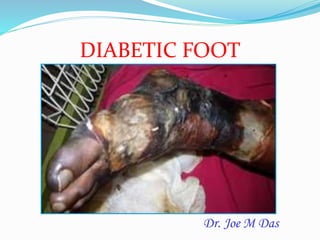
  • 1. Dr. Joe M Das DIABETIC FOOT
  • 2. - Bahara, Mills et al. International Wound Journal 2009.
  • 3.
  • 4. The theme – Diabetes education and prevention (2009 – 2013)
  • 5. Statistics  60-70% of those with diabetes will develop peripheral neuropathy, or lose sensation in their feet.  Up to 25% of those with diabetes will develop a foot ulcer.  More than half of all foot ulcers (wounds) will become infected, requiring hospitalization and 1 in 5 will require an amputation.  After a major amputation, 50% of patient will have their other limb amputated within 2 years.
  • 6. The History of Diabetes  1552 BC: the first known mention of diabetes – found on the Ebers Papyrus  Egyptian physician Hesy-Ra of the 3rd Dynasty makes  Lists remedies to combat the ‘passing of too much urine’ Wood Carving of Hesy-Ra
  • 7. The History.. Contd…  250 BC: Diabetes described as ‘the melting down of flesh and limbs into urine’  By Greek physician Aretaeus of Cappodocia who gives the first complete medical description of diabetes  1425 AD: Diabetes first appears in the English language as the Middle English word ‘diabete’.
  • 8.  16th Century: Swiss physician Phillipus Aureolus considered the ‘Martin Luther of Medicine’ – identifies diabetes as a serious general disorder. 1776: English physician Mathew Dobson of Liverpool  evaporates two quarts of urine from a patient with diabetes.  The resulting residue is granulated and smells and tastes like sugar, conclusively establishing the presence of ‘saccharine materials’ as a diagnosis of diabetes. The History… Contd… Phillipus Aureolus
  • 9.  1869  German medical student  Paul Langerhans  The islet cells of the pancreas but is unable to explain their function.  The find is dubbed the ‘islets of Langerhans.” The History Continues..
  • 10.  1871: French physician Apollinaire Bouchardat notices the disappearance of glycosuria in his diabetes patients during food rationing of food under the Siege of Paris in the Franco- Prussian War, and formulates individualized diets to treat the condition.  1889: Scientists Oskar Minkowski and Joseph von Mering of the University of Strasbourg, France demonstrate how removing a dog’s pancreas produces diabetes. The History Continues.. Apollinaire Bouchardat Oskar Minkowski
  • 11.  1901: American pathologist Eugene Opie of John Hopkins University in Baltimore establishes a connection between the failure of the islets of Langerhans in the pancreas and the occurrence of diabetes.  1913: Prof. John J.R. Macleod writes a monograph on diabetes entitled ‘Diabetes: Its Pathological Physiology.’ The History Continues..
  • 12.  Dec. 1916: Boston pathologist Elliott Joslin  compiles 1,000 of his own cases and creates the textbook The Treatment of Diabetes Mellitus.  In it he reports that ‘the mortality of patients was approximately 20 per cent lower than for the previous year’, due to ‘the introduction of fasting and the emphasis on regular exercise.’  1919: Dr. Frederick Allen of the Rockefeller Institute in New York publishes  “Total Dietary Regulations in the Treatment of Diabetes” that introduces a therapy of strict dieting – dubbed the ‘starvation treatment’ –- as a way to manage diabetes The History Continues.. Dr. Frederick Allen Elliott Joslin
  • 13. Oct. 31, 1920 Sir Frederick Grant Banting  conceives of the idea of insulin after reading an article in The Journal Surgery, Gynecology and Obstetrics by Moses Barron, by an American pathologist, titled ‘The Relation of Islets of Langerhans to Diabetes with Special Reference to Cases of Pancreatic Lithiasis.’  With the support of Prof.. Macleod of the University of Toronto, and the assistance of  Best, a medical student he starts his research using a variety of different extracts on depancreatized dogs. The History Continues..
  • 14.  Summer 1921: Banting’s work leads to the discovery of insulin. On July 30, Dog 410 is the first to receive the extract. On August 4 the extract is called ‘Isletin’ for the first time.  Nov. 14, 1921 Dr. Banting and Charles Best deliver a preliminary report of their research to the Journal Club of the University of Toronto, Department of Physiology.  Nov. 17, 1921: Banting and Best discover that extract from cattle foetal pancreas lowers blood sugar levels of depancreatized dogs, leading them toward plentiful, cheap sources for insulin. Experiments begin to test the long-term effectiveness of insulin treatment.  Dec. 30, 1921 :Banting, Macleod, Best and Dr. James Bertram Collip, a biochemist present the results of their research at a session of the American Physiological Society at Yale University. The paper initially generates little interest.  The paper – ‘The Internal Secretion of the Pancreas’ – is published two months later in the prestigious Journal of Laboratory and Clinical Medicine More Of Banting & Best…
  • 15. Oct. 25, 1923  Banting and Macleod  The Nobel Prize in Physiology or Medicine.  Banting shares his award with Best  Macleod shares his with Collip. The Nobel Prize Goes To…. Banting & Macleod Banting's and Best's laboratory, where insulin was discovered.
  • 16.  11th January 1922  Leonard Thompson, 14,  A ‘charity patient’ at the Toronto General Hospital,  First person to receive and injection of insulin to treat Type 1 diabetes.  Thompson lives another 13 years before dying of pneumonia at age 27. The History Continues..
  • 17.  May 3, 1922: The word ‘insulin’ is used in public for the first time when Macleod presents the paper ‘The Effect Produced on Diabetes by the Extracts of Pancreas’ to the  May 30, 1922: Pharmaceutical manufacturer Eli Lilly & Co. of Indianapolis and the University of Toronto enter a deal for the mass production of insulin. The History Continues..
  • 18.  1966  First pancreas transplant  University of Manitoba The History Continues..
  • 19.  July 7, 1989: Her Majesty Queen Elizabeth The Queen Mother kindles the  Flame of Hope at Banting House National Historic Site – ‘The Birthplace of Insulin’ – in London, Ontario.  As a symbol of hope, the flame will burn until a cure for diabetes is found.
  • 20.  March 1999  Scientists conduct the first successful islet transplant at the University of Alberta Hospital.  The Edmonton Protocol. isolating islets from a cadaveric donor pancreas using a mixture of enzymes called Liberase The History Continues..
  • 21.  Dec. 20, 2006: The United Nations recognizes diabetes as a global threat and designates World Diabetes Day, November 14 – in honour of Frederick Banting’s birthday – as a UN Day to be observed every year starting in 2007.
  • 22. Pathogenesis  Chronic hyperglycaemia  Nerve microvascular dysfunction  ↑ free radical formation (due to ↓ NADPH)  Polyol pathway hyperactivity (Glucose → ↑sorbitol → ↓ myoinositol → ↓ Na-K ATPase)  PKC hyperactivity  Nonenzymatic  Abnormalities of nerve growth
  • 23.  Pathology:  Axonal loss distally, with a “dying back” phenomenon  A reduction in myelinated fiber density and  Focal areas of demyelination on teased fiber preparations.
  • 24.
  • 25.
  • 26. Neuropathy Motor Sensory Autonomic ↓ nociception ↓ Proprioception, Unawareness of foot position A-V Shunt* open Permanent Increase foot Blood flow Bulging foot veins, Warm foot Reduced sweating Dry skin Fissures and cracks Muscle wasting Foot weakness Postural deviation Deformities, stress and shear pressures Trauma Stress on bones & joints Plantar pressure Callus formation InfectionUlcer
  • 27. Symmetrical neuropathies Asymmetrical neuropathies Distal sensory and sensory- motor neuropathy Mononeuropathy Large-fiber type Mononeuropathy multiplex Small-fiber type Radiculopathies Distal small-fiber neuropathy Lumbar plexopathy or radiculoplexopathy “Insulin neuropathy” CIDP CIDP
  • 28.  Distal Symmetrical Neuropathy  The commonest neuropathic syndrome  “Diabetic neuropathy”  A “length-related” pattern of sensory loss  Toes → Feet → Leg (stocking pattern)  Later motor  Autonomic neuropathy is less common
  • 29.  Autonomic neuropathy:  ↓ sweating → dry skin that is likely to crack easily → risk of infection.  Warm because of the arterio-venous shunting → Distension of foot veins that fail to collapse even when the foot is elevated  A gangrenous toe in a foot that has bounding arterial pulses may be seen as there is impairment of the nutritive capillary circulation because of arteriovenous shunting.  The oxygen tension of the blood in these veins is typically raised.  Neuropathic edema, which is resistant to treatment with diuretics but may respond to treatment with ephedrine
  • 30.  High blood sugar expedites artherosclerosis giving peripheral vascular disease (reduction of blood supply to the foot).  The delivery of essential nutrients and oxygen to the foot is compromised leading to anaerobic infections and tissue necrosis. Peripheral arterial disease Artherosclerosis narrows or blocks the arterial lumen Foot ischaemia Foot ulcer Necrosis/ Gangrene Infection Artheroma plaque narrowing the arterial lumen Ischaemic toes due to artherosclerosis Peripheral Arterial Disease
  • 31. Clinical features (Contd..)  Ulcers  ↓ vibration sense  Glove and stocking distribution of sensory loss  +ve Romberg’s sign; ↓ ankle tendon reflex  Wasting of small muscles of feet → Unopposed pulling of long extensors and flexors → Clawing of toes → Elevated plantar pressure points at the metatarsal heads→ Callus formation & foot ulceration
  • 32. Charcot arthropathy  A progressive condition of the musculoskeletal system that is characterized by joint dislocations, pathologic fractures, and debilitating deformities.
  • 33. Charcot Foot  More dramatic – less common 1%  Severe non-infective bony collapse with secondary ulceration  Two theories  Neurotraumatic  Neurovascular
  • 34. Charcot Foot  Neurotraumatic  Decreased sensation + repetitive trauma = joint and bone collapse  Neurovascular  Increased blood flow → increased osteoclast activity → osteopenia → Bony collapse  Glycolization of ligaments → brittle and fail → Joint collapse
  • 35. Charcot’s Foot-- Classification Eichenholtz 1 – acute inflammatory process Often mistaken for infection 2 – coalescing phase 3 - consolidation Location Forefoot, midfoot (most common) , hindfoot Atrophic or hypertrophic •Radiographic finding •Little treatment implication
  • 36. Wagner’s classification Depth of penetration Extent of tissue necrosis Treatment given
  • 37. Grade 1  No open lesion, skin intact  Potential site for ulceration like prominent metatarsal heads, hammer toes, Charcot’s deformity  Treatment:  Education & prevention  Self foot care & proper shoes  Antibiotics for cellulitis
  • 38. Grade 2  Superficial ulcer – extension to subcutaneous adipose tissue  Treatment:  Wound care + antibiotics  Radiological evaluation  Modified weight bearing bone resection
  • 39. Grade 3  Ulcer penetrating joint, deep abscess, OM, tendon sheath infection  No digital gangrene  Treatment:  Hospitalisation  Debridement and IV antibiotics  Joint and bone resection
  • 40. Grade 4  Some part of the foot gangrenous  Most part salvagable  Treatment:  Similar to that for grade 3 + Local amputation
  • 41. Grade 5  Extensive gangrene  Limb not salvagable  Treatment:  Major amputation
  • 42. University of Texas Diabetic Wound Classification  Stages  Stage A: No infection or ischemia  Stage B: Infection present  Stage C: Ischemia present  Stage D: Infection and ischemia present  Grading  Grade 0: Epithelialized wound  Grade 1: Superficial wound  Grade 2: Wound penetrates to tendon or capsule  Grade 3: Wound penetrates to bone or joint
  • 43. Depth Classification Definition Treatment 0 At-risk foot, no ulceration Patient education, accommodative footwear, regular clinical examination 1 Superficial ulceration, not infected Offloading with total contact cast (TCC), walking brace, or special footwear 2 Deep ulceration exposing tendons or joints Surgical debridement, wound care, offloading, culture-specific antibiotics 3 Extensive ulceration or abscess Debridement or partial amputation, offloading, culture-specific antibiotics
  • 44. Ischemia Classification Definition Treatment A Not ischemic B Ischemia without gangrene Noninvasive vascular testing, vascular consultation if symptomatic C Partial (forefoot) gangrene Vascular consultation D Complete foot gangrene Major extremity amputation, vascular consultation
  • 45. International Working Group on Diabetic Foot Classification System  On the basis of the scientific literature and expert opinion, five categories were identified, which were considered the most relevant items for research projects in diabetic foot ulcers:  Perfusion  Extent/size  Depth/ tissue loss  Infection  Sensation
  • 47. History taking  Age  Mode of onset  Bare foot walking  Paraesthesia  Claudication pain  Fever  H/o DM – Duration, controlled/uncontrolled  Treatment history  H/o impotency
  • 48.  Diabetics present to the surgeon in a number of scenarios:  with any foot and ankle problem and diabetes as an incidental part of the past medical history - important to assess how much diabetes increases the risk of any proposed surgical or non-surgical treatment  with an acutely inflamed where the main differential diagnosis is between infection and Charcot arthropathy  with an ulcer which may or may not be complicated by deep infection or ischemia  with a deformity which has led to recurrent ulceration, or is felt to be a risk factor for ulceration - important to assess the relative risks of corrective surgery and continuing protective care
  • 49. Whatever the initial presentation, it is important to ask about  what type of diabetes does the patient have (type 1 or type 2)  how is it managed - diet, oral hypoglycaemics, insulin - and what agents and regime  who normally looks after the patient's diabetes - GP/practice nurse/hospital diabetic clinic and therefore who is likely to have the patient's diabetic records  how well controlled is the diabetes - review the patient's glucose monitoring record and ask when the last HbA1c test was done and what was the result and the previous trend  does the patient have any of the major complications of diabetes  cardiovascular disease  renal disease  eye disease  has the patient had an ulcer or Charcot arthropathy before - the single biggest risk factor for either of these conditions is to have had them before
  • 50. Clinical Examination  Nourishment, pallor  A) Limb affected B) Type of lesion:  Ulcer  Gangrene  Cellulitis / abscess
  • 51. Ulcer: “Malperforant ulcer”  Site  Size, shape, margin  Floor: Slough ±  Edge: Punched out  Surrounding skin  Base: May be bone  Infected – foul smell  Local anesthesia of skin
  • 52. Gangrene:  Usually wet  Forefoot → Leg  Edema  Foul smell  Skip lesion  No line of demarcation  Bone may be exposed
  • 53. Cellulitis / abscess:  Diffuse tender  Swelling ±
  • 54. Regional examination: Deformity of foot High arching Local anaesthesia Regional pulsations: Posterior tibial & Dorsalis pedis Lymphadenopathy Local thrombophlebitis Local fracture
  • 55. Nylon monofilament test  For identifying loss of protective sensation  Failure to feel the filament at four of 10 sites is 97 percent sensitive and 83 percent specific
  • 56. Procedure of NMT  10 gram Semmes-Weinstein nylon monofilament  The patient must not watch  Test the monofilament on the patients hand so that he/she knows what to anticipate.  Apply the monofilament perpendicular to the  Apply sufficient force to cause the filament to bend or buckle, using a smooth, not a jabbing motion.  Apply the filament along the perimeter and NOT ON an ulcer site, callus, scar or necrotic tissue
  • 57. • Apply a vibrating 128 Hz tuning fork to the bony prominence of the big toe • If the patient cannot feel the vibration, gradually move the fork upwards • The sensitivity of this test for demonstrating a deficit is ~53% • A biothesiometer is a portable device that measures the vibration perception threshold. A vibration threshold of more than 25V has a sensitivity of 83%. Tuning fork test Either an abnormal 10g monofilament test or a vibration threshold of more than 25V predicts foot ulceration with a sensitivity of 100% , hence the rationale for combining these two tests in clinical practice. Tuning Fork (vibration)
  • 58. Biothesiometer  To measure simply and accurately the threshold of appreciation of vibration in human subjects.  An "electrical tuning fork" whose amplitude may be set to any predetermined level or whose amplitude may be gradually increased until the threshold of vibratory sensation is reached.  Conversely, the amplitude may be lowered until the vibration is no longer discernible.  To detect neurological changes that are not disclosed with a tuning fork.
  • 59. Thermal Exam  Taking temperature readings on both the plantar and dorsal surfaces of the foot  may be a good indicator of foot condition. A wide difference of temperatures between left and  right feet can indicate possible infection, injury or circulatory problems. Done with gloves and  the patient sitting comfortably, shoes and socks off.  a. Dorsal Temp: Take a reading on the dorsum of the foot near the dorsalis pedis artery.  Plantar Temp: Take a reading on the plantar aspect of the foot in the medial arch area. Skin & Nail Exam:  Done with gloves and the patient sitting comfortably, shoes and socks off.  a. Corns & Calluses: Metatarsal heads, Dorsal aspect of 5th toe, Lateral aspect of 1st toe, Heel, Plantar surface  b. Skin appearance: Shiny, thin, fragile  c. Hair: Normal hair growth / no hair growth  d. Skin Features: Bruises, Cracks/breaks in skin, Soggy skin, Dry skin, Blisters,  Discoloration, Ulcerations, Previous surgeries, Tinea Pedis (Fungus)  e. Nails: Ingrown, Improperly cut, Sharp, Long, Absent
  • 60. Footwear assessment  Examine the inside of the shoes  Look at the type of footwear  Look at the fit of the footwear  Are there any areas of increased pressure?  Are there any accommodations made for any deformities if already present?  Is the footwear appropriate in size & shape for the patients feet?  Would the patient benefit from corrective footwear?  From inserts?
  • 61. Evaluation of Diabetic foot  Urine – sugar, acetone  FBS, PPBS  Local –  Radiology – deformity, osteomyelitis, #, infection  Doppler & Duplex  Transcutaneous oximetry  Mapping of pressure areas by foot prints  C&S  MRI, MRA  Angiography
  • 62.
  • 63.
  • 64. Transcutaneous oximetry  Application of heat to skin → Localised hyperemia → If O2 present in excess of skin requirement (depends on oxygen delivery & blood flow) → O2 diffuses across skin → Measured by electode  TcPO2 - 40 – 70 mm Hg (normal)  TcPO2 <20 mm Hg – Limb threatening ischaemia
  • 65. MRI foot  Most specific  Differentiates between bony destruction from OM and that from Charcot’s arthropathy  Iridium – labelled white cell studies
  • 68. Interventions in diabetic ulcer Offloading Debridement Wound dressing Antibiotics Revascularisation Limited amputation
  • 70. Offloading Devices  Offloading devices are devices used to decrease the pressure over a wound and protect wounds, thereby giving the wound a good chance of healing.  Although many offloading modalities are currently utilized, only small numbers of case series exist describing the frequency and rate of wound healing associated with these devices.
  • 71.  Reduction of pressure and shear forces on the foot may be the single most important-and most neglected-aspect of treating neuropathic ulceration.  Off-loading therapy is a key part of the treatment plan for diabetic foot ulcers.  The goal is to off-load (reduce) the pressure at the ulcer while keeping the patient ambulatory. Offloading Devices Contd
  • 72. Total Contact Casts (TCCs):  Most common modality for offloading used by diabetic foot specialists. This type of offloading was first described by Milroy Paul in treating cases of neuropathic foot wounds and became more popular by Dr.Paul Brand at the Hansen's disease Center in Carville, Louisiana  This modality of offloading is known as total contact cast (TCC) due to the technique used to make it. It employs a well-moulded, minimally padded cast that maintains contact with the entire planter aspect of the foot and lower leg.  TCCs have been shown to reduce pressure at the site of ulceration by 84 - 92%(7). It is also effective in treating a majority of non-infected, non-ischemic plantar diabetic foot wounds, with healing rates ranging from 72% - 100% over a course of 5 - 7% weeks(8)
  • 73. TCC Gait modification  Total contact casts (TCCs) – gold standard
  • 74. Advantages of TCCs  Offloading the foot(mainly the anterior aspect)  Protect the foot from infection  Help in reduce or control edema
  • 75. Disadvantages of TCCs  Adds pressure on posterior aspect of foot hence not useful in case of heal ulcers  Technically difficult to apply, need special skills.  Consuming time on its application & Improper cast application can cause skin irritation.  Improper cast application can cause frank ulceration  Loss of flexibility of daily wound assessment.  Difficulty in daily life activity such as bathing without wetting the cast  Users may have difficulty in sleeping comfortably.  Some designs of TCCs may affect gait stability
  • 76. Contraindications for use of TCCs  For wounds with ischemia  For infected wounds  For wounds with osteomylitis
  • 78. Advantages of the Removable Cast Walkers  Easily removed for self inspection of the wound  Easily removed for local application of topical therapies  Easier to do activities such as bathing, sleeping comfortably.  Can be used for infected wounds  Can be used for superficial ulcers.
  • 79.  The amount of pressure reduction for certain RCW is equivalent to TCCs  Although some RCWs reduce the pressure as well as TCCs other data showed that healing with TCCs is more readily achieved than healing with other modalities.  Because patients can remove RCWs easily, the best feature of this device is potentially the most hazardous point.  In a recently conducted study evaluated the activity of patients with diabetic foot ulcers and their adherence to their offloading regime. This study suggests that it is less than 30% of their total daily activity  Due to the disadvantages of the TCCs it has been suggested to make an alternative which might make the RCWs difficult to remove.This alternative called instant total contact cast (iTCC). Wrapping the RCW with either a layer of cohesive bandage or plaster / fiberglass, making it more difficult for patients to remove. The great advantage of iTCC is that it binds the benefit of offloading plus the benefit of forced compliance.
  • 80.
  • 81. Scotch Cast Boot  Finishing below ankle  It was developed when fiberglass materials were introduced. Its development was to alternate with the plaster of Paris casts.  The boot is made to fit each individual foot and a window cut at the site of ulceration
  • 82. Advantages of Scotch Cast boot  Light in weight  Removable and allowing regular inspection of the wound and facilitates the redressing of the wound.  Reduces pressure on the lesion  Maintains patient mobility  Protects the wound and remaining foot
  • 83. Some other offloading devices  The Neuropathic CROW (Charcot Restraint Orthotic Walker)  The off-loading of foot ulcers, unstable Charcot foot/ankle  This model provides a total contact interface, pretibial shell, and rocker bottom
  • 85. HALF SHOES  To relieve pressure on forefoot post operatively  ADVANTAGES Easy to apply Inexpensive Removable  DISADVANTAGES Efficacy decreased Unstable gait
  • 86.  Double H boot  Pressure relieving ankle foot orthosis (PRAFO)
  • 87. Healing Sandals Therapeutic Footwear Silicon Insoles Sandal with a rigid rocker limiting dorsi flexion of MCP joint
  • 88. CRUTCHES, WALKERS, WHEELCHAIRS The best!!...but  Requires upper body strength  Endurance  Pressure on opposite foot increased
  • 89. Hyperbaric Oxygen therapy  Adjunctive therapy in  Persistent critical limb ischemia, soft tissue infection, and impaired wound healing from osteomyelitis  If TcPO2 > 200 mmHg close to the trophic lesion in 2.5 ATA hyperbaric oxygen.  Peri-wound TcPO2 > 400 mmHg in 2.5 ATA hyperbaric oxygen or > 50 mmHg in normobaric pure oxygen ═> Healing chance very high
  • 90. MOA of hyperbaric oxygen  ↑ angiogenesis  ↓ infection by killing bacteria  ↓ pain and inflammation  Releases stem cells from bone marrow  ↓ risk of thrombosis  Enhances nerve regeneration  Improves bone density  Eliminates toxins from blood
  • 91.
  • 92. Advice to patients  Inspect your feet daily  Wash your feet in lukewarm water  Be gentle when bathing your feet  Moisturize your feet—but not between your toes  Cut nails carefully—and straight across  Never trim corns and calluses  Wear clean, dry socks  Avoid open-toe / open-heel shoes
  • 93. Advice to patients Contd Wear socks to bed Shake out shoes and inspect the inside before wearing Keep your feet warm and dry Never walk barefoot Take care of your diabetes Don't smoke Get periodic foot exams
  • 94. Annual foot examination  Assess  Blood flow  Sensation (monofilament testing, pin prick, tuning fork)  Ankle reflex  Nail care  Look for foot deformities  Identify sites of potential ulceration
  • 95. Lower Extremity Amputation Prevention (LEAP) Program  The L.E.A.P. Program consists of five relatively simple activities:  Annual foot screening  Patient education  Daily self inspection of the foot  Appropriate footwear selection  Management of simple foot problems
  • 96.  Check his/her feet daily for early lesion
  • 97. New developments  Bioengineered skin (Apligraf)  Human dermis (Dermagraft)  are new types of biologically active implants for ulcers that are derived from fibroblasts of neonatal foreskins  PDGF  Hyaluronan  Epalrestat (Aldose reductase inhibitor)  Lipoic acid, Thioptic acid  VAC  Hydrocolloid dressing
  • 98. Apligraf  Apligraf® is a unique, advanced treatment for healing. It is created from cells found in healthy human skin. Which explains why it looks like a thin, piece of real skin.  It is used to heal ulcers such as diabetic foot and venous leg ulcers that are not healing after 3-4 weeks, despite treatment with conventional therapies.  When healthy skin gets wounded, the proteins and growth factors in the skin stimulate the body to regenerate new skin. This is the normal wound healing process.  However with diseases like diabetes the skin is missing these biological substances, and the healing cycle is broken. This leads to the development of non-healing ulcers and wounds.
  • 99. Apligraf  Apligraf® contains two types of cells – an outer layer of protective skin cells, and an inner layer of cells contained within collagen. Both types of cells contain substances similiar to those found in human skin.  Apligraf® plays an active role in healing by providing to the wound living cells, proteins produced by the cells, and collagen, which are important for healing.
  • 100. Apligraf •Apligraf® is placed directly on the wound. •The wound is then covered with a non-adhesive dressing to keep Apligraf® in place. •The area is then wrapped with other dressings that are changed weekly by the doctor •The healing process now begins, and improvement of the wound can usually be seen within weeks.
  • 101.
  • 102. Apligraf: How is it Made?  Human keratinocytes and fibroblasts are derived from neonatal foreskins obtained for use under informed- consent guidelines  The foreskin is decontaminated with antibiotics, antifungals, and an ethyl alcohol rinse.  Production of cell stocks involves enzymatic digestion of the foreskin tissue and fibroblast/keratinocyte isolation1
  • 103. Dermagraft  Advanced BioHealing is a The Company that currently manufactures and markets Dermagraft®, a bio-engineered skin substitute that assists in restoring damaged tissue and supports the body’s natural healing process.  Dermagraft is approved by the FDA for the treatment of diabetic foot ulcers.  It is a Cryopreserved human fibroblast-derived dermal substitute, approved by the FDA for the treatment of diabetic foot ulcers.  A combination of living fibroblasts, matrix proteins, and bound factors that protect the wound and stimulate dynamic events that promote regeneration and repair.
  • 104. Dermagraft To treat Diabetic foot ulcers that have been present for more than 6 weeks It is used together with standard methods of treating foot ulcers, including cleaning and preparing the wound, applying cover dressings to hold it in place, and offloading devices.
  • 105.
  • 106.  Dermagraft is indicated for use in the treatment of full- thickness diabetic foot ulcers greater than 6 weeks duration, which extend through the dermis, but without tendon, muscle, joint capsule, or bone exposure..  In a trial, a 64% relative increase in complete wound closure was seen in the Dermagraft-treated patients; 30.0% of Dermagraft and 18.3% of conventional therapy patients achieved complete wound closure at 12 weeks.  The most frequently reported adverse events experienced by patients in the Dermagraft group (terms ≥ 5%) included infection, accidental injury, skin dysfunction/blister, flu syndrome, osteomyelitis, surgeries involving study ulcer, wound enlargement/skin ulcer, cellulitis and peripheral edema/localized swelling. These adverse events were similar to those seen in the control group.
  • 107. PDGF  Platelet-derived growth factor is mainly secreted by the platelets’ α-granule, but it is also produced by other cells involved in early wound healing, ie, macrophages, endothelial cells, fibroblasts, and keratinocytes  PDGF is a powerful chemoattractant and mitogen, exerting its action on fibroblasts, smooth muscle cells, and endothelial cells  It also induces production of fibronectin and hyaluronic acid.  There is a synergistic effect between PDGF and EGF, as well as TGF-β, and so PDGF has a pivotal role at all stages of wound healing
  • 108. PDGF  At present, recombinant PDGF  by DNA technology via incorporation of the gene for the β-chain of human PDGF into the yeast Saccharomyces cerevisiae.  The resultant homodimeric protein, Becaplermin, has a biological activity similar to the endogenous PDGF- BB  Approved by the US Food and Drug Administration for the treatment of diabetic neuropathic ulcers with adequate peripheral circulation
  • 109. Hydrocolloid Dressing  Duoderm[1], Granuflex[2], and 3M Tegaderm Hydrocolloid)  An opaque dressing to provide a moist wound-healing environment, while protecting from contamination.  Hydrocolloids were initially utilized in medicine as a reliable, skin-friendly adhesive, useful for securing colostomy appliances to the patient's abdomen  Clinicians observed that acute abdominal wounds from colostomy operations healed more rapidly when a hydrocolloid was used.  It is biodegradeable, nonbreathable and adheres to the skin so no separate taping is needed.
  • 110. Hydrocolloid Dressing Contd  The active surface of the dressing is coated with a cross-linked adhesive mass containing a dispersion of gelatin, pectin and carboxy-methylcellulose together with other polymers and adhesives forming a flexible wafer.  In contact with wound exudate, the polysaccharides and other polymers absorb water and swell, forming a gel which is held within the structure of the adhesive matrix.
  • 111. Hydrocolloid Dressing Contd  The moist conditions produced under the dressing promote fibrinolysis, angiogenesis and wound healing, without causing softening and breaking down of tissue.  The gel which is formed as a result of the absorption of wound exudate is not mobile and free running but held within the structure of the adhesive matrix.  Most hydrocolloid dressings are waterproof, allowing normal washing and bathing. [2]  Dressings may be used even if aerobic infection is present
  • 112. Colloidal Silver  Silver -- colloidal or ionic silver, silver in solution -- is a potent antibacterial product. It has been demonstrated as such for well over 100 years.  Silver is incorporated into the wound pad of the new bandages which come in a variety of sizes  Eg: Hydroheal AM Gel & Dressings
  • 113. Colloidal Silver  Silver interferes with the bacteria in at least three ways:  by interacting with the cell membrane,  binding to the DNA of cells, and  blocking the metabolism of the bacteria.  It reduces the growth of hundreds of different types of bacteria, including some that do not normally react to pharmaceutical antibacterial agents.  Because silver blocks the growth and spread of germs through multiple mechanisms, it is hard for bacteria to build up resistance