
Recomendados
Más contenido relacionado
La actualidad más candente
La actualidad más candente (20)
Similar a Cardiac anatomy and physiology
Similar a Cardiac anatomy and physiology (20)
Último
Último (20)
Cardiac anatomy and physiology
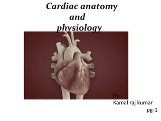
- 1. Cardiac anatomy and physiology by Kamal raj kumar pg-1
- 2. Normally located in the middle and slightly to the left side of the thoracic cavity on the diaphragm between 3rd and 5th ribs. Weighs about 325 gm is males and about 275 gm in females.
- 4. Heart has …. Base Apex Anterior Posterior
- 5. Right Atrium Receives venous blood from whole of the body via the superior vena cava (SVC) at its upper end and inferior vena cava (IVC) at its lower end. It pumps into Right ventricle (RV) through the tricuspid valve during the ventricular diastole. Right atrial appendage – arises from the antero superior part of the right atrium. It is to the left of the ascending aorta. Sulcus terminalis – shallow vertical groove along the right heart border between SVC and IVC. RA and SVC junction – Sino atrial node.
- 6. Right ventricle Triangular shaped or crescent shaped Opens into pulmonaryartery through pulmonaryvalve Most anteriorchamber RV wall measures 4-5 mmin thickness. Three portions – inflow , outflow and apical trabecular portion or body of RV.
- 7. Left atrium Posterior most chamber Receives oxygenated blood from pulmonary veins Pulmonary veins open into LA from the posteriorwall Left atrial appendage is anterior.
- 8. Left ventricle Bullet shaped Blunt tip forms the apex of the heart LVwall is 8-15 mm in thickness.
- 9. Layers of Heart Wall Pericardium - membrane (sac) that surrounds and protects theheart, it has two layers: ( a ) Fibrous pericardium - superficial layer, tough,inelastic, prevents overstretching, provides protection, and anchors the heart in place ( b ) Serous pericardium - deeper layer,thin; (i) parietal layer - fused to the fibrous pericardium, and (ii) visceral layer (or epicardium) adheres to the heart itself Pericardial cavity (between the two layers) is filled with pericardial fluid which reduces friction.
- 11. FIBROUS:THIN INELASTIC, DENSE IRREGULAR CONNECTIVE TISSUE ---HELPS IN PROTECTION, ANCHORS HEART TO MEDIASTINUM SEROUS: THINNER, MORE DELICATE DIVIDED INTOPARIETAL AND VISCERAL 13
- 12. MYOCARDIUM:RESPONSIBLE FOR PUMPING ENDOCARDIUM: THIN LAYER OF ENDOTHELIUM WHICH IS CONTINOUS WITH THE LINING OF THE LARGE BLOOD VESSELS ATTACHED TO THE HEART. 1 EPICARDIUM: COMPOSED OF MESOTHELIUM AND DELICATE CONNECTIVE TISSUE (IMPARTS A SLIPPERY TEXTURE TO THE OUTER SURFACE OF THE HEART).
- 13. Coronary Circulation • Def: Blood flow to heart through coronary arteries is coronary circulation • Normal value: 250 mL/min at rest or 70 ml / 100 gm / min • This may increase upto to 2000 ml / min
- 14. Coronary circulation • Consist of 1) Arterial supply 2) Venous drainage 3) Lymphatic drainage
- 15. Arterial supply • The cardiac muscle is supplied by two coronary arteries the right and left coronary arteries. • Both arteries arises from the sinuses behind the cusps of the aortic valves at the root of the aorta.
- 17. Rt. Coronary artery • Smaller than left coronary artery • Arises from anterior coronary sinus • Emerges from the surface of heart between pulmonary trunk and right auricle. • Winds round the inferior border to diaphragmatic surface to reach the posterior inter-ventricular groove. • Terminates by anastomising with left coronary artery EKG: lead II, III & AVF
- 18. Branches • Large Branches – Acute marginal – Post-interventricular • Small branches: – Right atrial – Infundibular – Nodal – in 60% cases – Terminal
- 19. Anterior schematic diagram of heart shows course of dominant right coronary artery and its tributaries. AV = atrioventricular, PDA = posterior descending artery, RCA = right coronary artery, RV = right ventricular, SA = sinoatrial
- 20. AREAS OF DISTRIBUTION • Right atrium • Ventricles – Greater part of right ventricle, except the area adjoining the anterior inter-ventricular groove. – A small part of the left ventricle adjoining the posterior interventricular groove. • Posterior part or the inter-ventricular septum • Whole of the conducting system of the heart except a part of the left branch of AV bundle. The SA node is supplied by left coronary artery in 40% cases
- 21. LEFT CORONARY ARTERY • Arises from left posterior aortic sinus • Runs forward and to the left and emerges between the pulmonary trunk and the left auricle. • Here the anterior inter-ventricular branch is given • After giving off the anterior interventricular branch it runs into the left anterior coronary sulcus as the circumflex artery. • It winds around the left border and near posterior interventriular groove it terminates by anastomosing with the right coronary artery. EKG: lead V3 &V5
- 22. BRANCHES: • Large Branches: – Left anterior desending arteryEKG: Lead V5 – Left circumflex branch EKG: lead 1& AVL • Small Branches: ― Left atrial ― Pulmonary ― Terminal
- 23. Dominant left coronary artery anatomy. Left anterior oblique schematic diagram of dominant left coronary artery anatomy, including left anterior descending artery and left circumflex artery tributaries, is shown. AVGA = atrioventricular groove artery, PDA = posterior descending artery.
- 24. Areas of distribution • Left atrium • Ventricles: Greater part of left ventricle, except the area adjoing the posterior interventricular groove. A small part of right ventricle adjoining the anterior interventricular groove. • Anterior part of interventricular septum. • Part of left branch of AV bundle
- 25. Collateral circulation • These channels open up in the emergencies when the coronary arteries are blocked. • Cardiac anatomosis: The two coronary arteries anastomose in the myocardium • Extra cardiac anastomosis: The coronary arteries anastomose with the • Vasa vasorum of the aorta, • Vasa vasorum of pulmonary arteries, • Internal thoracic arteries • The bronchial arteries • Phrenic arteries.
- 26. CORONARY ARTERY DOMINANCE • Artery that gives the posterior interventricular artery determines the coronary dominance • If Posterior interventricular artery is supplied by right coronary artery (RCA), “right-dominant”(70%) • Posterior interventricular artery is supplied by the circumflex artery (CX), a branch of the left artery, “left-dominant”(10%) • Posterior interventricular artery is supplied by both the right coronary artery (RCA) and the circumflex artery, then “co- dominant”(20%)
- 27. VENOUS DRAINAGE OF THE HEART • The venous drainage of the heart is by three means: – Coronary sinus – Anterior cardiac veins – Venae Cordis minimae
- 28. CORONARY SINUS • This is the largest of vein of heart situated in the left posterior coronary sulcus. It is about 3 cm long and ends by opening into the posterior wall of the right atrium • Its tributaries are: − Great cardiac vein: It accompanies LAD enters the left end of the coronary sinus − Middle cardiac vein: It accompanies the posterior interventricular artery and joins the right end of the coronary sinus − Small cardiac vein: It accompanies the right coronary artery and joins the right end of the coronary sinus
- 29. − Posterior vein of left ventricle: It runs on the diaphragmatic surface of the left ventricle and ends in the middle of the coronary sinus. − Oblique vein of left atrium ( of Marshall): It runs on the posterior surface of the left atrium, joins the left end of coronary sinus and develops from the left common cardinal vein. − The right marginal vein: It accompanies the marginal branch of the right coronary artery.
- 30. ANTERIOR CARDIAC VEIN 3 to 4 small veins run on the anterior wall of the right ventricle, open directly into the right atrium. VENAE CORDIS MINIMAE (also called smallest cardiac veins, venae cardiacae minimae, or Thebesian veins) • Numerous small veins present in all 4 chambers of heart which open directly into the cavities. • The Thebesian venous network is considered an alternative (secondary) pathway of venous drainage of the myocardium.
- 31. Lymphatics of heart • Lymphatics of the heart accompany the coronary arteries and form 2 trunks. • Right trunk ends in brachiocephalic nodes and the left trunk into the tracheobronchial lymph nodes at the bifurcation of the trachea.
- 32. PECULIARITIES OF COR.CIRCULATION • BF during diastole • End arteries • Anatomical anastomosis • High capillary density • High 02 extraction • Regulation is mainly by metabolites • The coronary vessels are susceptible to degeneration and atherosclerosis. • There is evident regional distribution: The subendocardial layer in the left ventricle receives less blood, due to more myocardial compression (but this is normally compensated during diastoles by V.D). However, this renders this area more liable to ischemia and infarction
- 33. Myocardial Oxygen Supply Determined by: Coronary Blood Flow & O2 Carrying Capacity Oxygen saturation of the blood Hemoglobin content of the blood (CorBF = CorPP /CorVasRes) Coronary perfusion pressure (pressure gradient across the myocardium) Coronary vascular resistance
- 34. CORONARY BLOOD FLOW (CBF): • The resting coronary blood flow is about 250 ml/min., which is about 0.7 – 0.8 ml/gm of heart muscle, or 4- 5 % of the total cardiac output • In severe muscular exercise, the work of the heart increased and the CBF may be increased up to 2 liters/ minute
- 35. Physiology of the coronary flow • C.B.F. occurs mainly during diastole due to compression of coronary blood vessels in systole by the contracted muscle fibers . • Coronary perfusion pressure : • Driving force for blood flow in coronary arteries • Pressure gradient across the myocardium (diff between aortic pressure and intraventricular pressure) • It varies throughout the cardiac cycle being greater during diastole than in systole (phasic). Dependent on: • Arterial pressure • Intraventricular pressure • Coronary sinus / RAP • CorPP changes during sysole and diastole Left Ventricle Right Ventricle Systole CorPP = aortic systolic P – LV systolic P =120 – 120 = 0mmHg = aortic sys P – RV sys P = 120 – 25 = 75mmHg Diastole CorPP aortic dias P – LV dias P = 80 – 5 = 75mmHg aortic dias P – RV dias P = 80 – 5 = 75mmHg
- 36. Perfusion time (diastolic time) • Most of coronary flow occurs during diastole • Flow is rate dependent → i.e. ↑HR → ↓diastolic time → ↓CorBF Coronary Vascular resistance External compression Calibre of vessels ↓with ↑intraventricular pressure which compresses the vessels overlying it → interrupt flow Intrinsic regulation Local metabolites Endothelial factors Neural factors (esp. sympathetic nervous system)
- 37. Coronary autoregulation • Ability of the organ to regulate their own blood supply as per metabolic need by constricting or dilatation of blood vessels using vasoactive substance • If there is sudden change in aortic pressure, coronary vascular resistance will adjust itself proportionally within few seconds; so that a constant blood flow is maintained. • Range of autoregulation: 60 – 140 mmHg of MAP. • The myocardial oxygen tension and presence of vasoconstrictors or vasodilators influence the range of coronary autoregulation
- 38. Coronary Autoregulation • Myocardial muscle cell - produces byproducts of aerobic metabolism (lactate,adenosine, etc) • Vascular endothelial cell (arteriole) - reacts to metabolic byproducts • Vascular smooth muscle cell (arteriole) - signaled by endothelial cell to contract (vessel constriction) or relax (vessel dilation) Involves 3 different cells
- 39. Mechanism: Myogenic response: an increase in passive stretch, caused by increased perfusion pressure, causes active smooth muscle contraction. Chemical theory: • ↑metabolite production (↑pCO2, ↑K+, ↑H+, ↓pH, ↓pO2, adenosine ) → vasodilation of coronary vessels via release of vasoactive substances (1° NO, prostacyclin) o This is most important controlling factor of vessel size
- 40. Endothelium derived relaxation factor (EDRF): • Hypoxia, ADP, VIP, muscular exercise (increase distention force), stimulate vascular endothelium to secrete EDRF, which is a potent vasodilator, that causes coronary dilatation and increase CBF.
- 41. Autoregulators • Other endothelial- derived factors contribute to autoregulation Dilators include: EDRF (NO) Prostacyclin Bradykinin Constrictors include: Endothelin-1 thromboxane A2
- 42. Other factors influencing the vascular resistance Myocardial metabolism • Vasomotor tone is almost exclusively determined by local metabolic oxygen demand. • Pre-capillary sphincters are relaxed and more capillaries recruited. Hypoxia Release adenosine Coronary vasodilatation opens ATP- sensitive potassium channels
- 43. Neural control: Direct effect: • Parasympathetic: vagus has very slight distribution to coronary, so its stimulation has slight dilator effect. • Sympathetic: Both alpha and Beta receptors exist in the coronary vessels. Sympathetic stimulation causes slight direct coronary constriction. Indirect effect: • Plays a far more important role in normal control of coronary blood flow than the direct. Sympathetic stimulation increase both heart rate and myocardial contractility, as well as its rate of metabolism leading to dilatation of coronary blood vessels. The blood flow increase proportional to the metabolic need of heart muscle
- 44. Humoral control • Peptide hormones like antidiuretic hormone, atrial natriuretic peptide, vasoactive intestinal peptide, and calcitonin gene- related peptide require an intact vascular endothelium. • Antidiuretic hormone in physiological concentration has little effect on the coronary circulation but causes vasoconstriction in stressed patients. • Other peptides cause endothelium-mediated vasodilatation. • Angiotensin II causes coronary vasoconstriction by releasing endothelin, the strongest vasoconstrictor peptide in humans • Angiotensin-converting enzyme inactivates bradykinin, a
- 45. Vascular endothelium • Final common pathway regulating vasomotor tone. • It modulates the contractile activity of the underlying smooth muscle through synthesis and secretion of vasoactive substances in response to blood flow, circulating hormones and chemical substances. • Vasorelaxants are endothelium-derived relaxing factor, nitric oxide, prostacyclin and bradykinin. • Vasoconstrictors include endothelin and thromboxane A2. The net response depends on the balance between the two opposing groups
- 46. REFLEX CONTROL • Anrep’s reflex: Increased venous return causes increased pressure in right atrium, leading to reflex increase in CBF e.g. during muscular exercise. • Gastro-coronary reflex: Distention of the stomach with heavy meal causes reflex vasoconstriction of coronary blood vessels decreasing CBF.
- 47. Intrinsic Cardiac Conduction System Approximately 1% of cardiac muscle cells are autorhythmic rather than contractile 70-80/min 40-60/min 20-40/min
- 48. Electrical Signal Flow - Conduction Pathway • Cardiac impulse originates at SA node • Action potential spreads throughout right and left atria • Impulse passes from atria into ventricles through AV node (only point of electrical contact between chambers) • Action potential briefly delayed at AV node (ensures atrial contraction precedes ventricular contraction to allow complete ventricular filling) • Impulse travels rapidly down interventricular septum by means of bundle of His • Impulse rapidly disperses throughout myocardium by means of Purkinje fibers • Rest of ventricular cells activated by cell- to-cell spread of impulse through gap junctions
- 49. Electrical Conduction in Heart • THE CONDUCTING SYSTEM OF THE HEART AV node Purkinje fibers Bundle branches A-V bundle AV node SA node Internodal pathways SA node depolarizes. Depolarization spreads more slowly across atria. Conduction slows through AV node. Depolarization wave spreads upward from the apex. 4 Depolarization moves rapidly through ventricular conducting system to the apex of the heart. 5 3 2 Electrical activity goes rapidly to AV node via internodal pathways. 1 4 5 3 2 Atria contract as single unit followed after brief delay by a synchronized ventricular contraction 1 SA node Purple shading in steps 2–5 represents depolarization.
- 50. Electrical Conduction • SA node - 75 bpm – Sets the pace of the heartbeat • AV node - 50 bpm – Delays the transmission of action potentials • Purkinje fibers - 30 bpm – Can act as pacemakers under some conditions
- 51. Intrinsic Conduction System • Autorhythmiccells: – – – – Initiate action potentials Have “drifting” resting potentials called pacemaker potentials Pacemaker potential - membrane slowly depolarizes “drifts” to threshold, initiates action potential, membrane repolarizes to -60 mV. Use calcium influx (rather than sodium) for rising phase of the action potential
- 52. Pacemaker Potential • • • • • • Decreased efflux of K+, membrane permeability decreases between APs, they slowly close at negative potentials Constant influx of Na+, no voltage-gated Na + channels Gradual depolarization because K+ builds up and Na+ flows inward As depolarization proceeds Ca++ channels (Ca2+ T) open influx of Ca++ further depolarizes to threshold (-40mV) At threshold sharp depolarization due to activation of Ca2+ L channels allow large influx of Ca++ • Falling phase at about +20 mV the Ca-L channels close, voltage-gated K channels open, repolarization due to normal K+ efflux At -60mV K+ channels close
- 53. AP of Contractile Cardiac cells – Rapid depolarization – Rapid, partial early repolarization, prolonged period of slow repolarization which is plateau phase – Rapid final repolarization phase PX = Permeability to ionX +20 -20 -40 -60 -80 -100 Membranepotential (mV) 0 0 100 200 300 Time (msec) PK and PCa PNa PK and PCa PNa Membrane channels Na+ channels open Na+ channelsclose Ca2+ channels open; fast K+ channelsclose Ca2+ channels close; slow K+ channels open Resting potential 1 2 30 4 4 Phase 0 1 2 3 4
- 54. AP of Contractile Cardiac cells • Action potentials of cardiac contractile cells exhibit prolonged positive phase (plateau) accompanied by prolonged period of contraction – Ensuresadequate ejection time – Plateau primarily due to activation of slow L-type Ca2+channels
- 55. Why A Longer AP In Cardiac Contractile Fibers? • • • We don’t want Summation and tetanus in our myocardium. Because long refractory period occurs in conjunction with prolonged plateau phase, summation and tetanus of cardiac muscle is impossible Ensures alternate periods of contraction and relaxation which are essential for pumping blood
- 57. Membrane Potentials in SA Node and Ventricle
- 59. Excitation-Contraction Coupling in Cardiac Contractile Cells • Ca2+entry through L-type channels in T tubules triggers larger release of Ca2+from sarcoplasmic reticulum – Ca2+induced Ca2+release leads to cross-bridge cycling and contraction
- 60. Cardiac cycle
- 62. Introduction Principal function of cardiovascular system is to deliver oxygen and nutrients to and remove carbon dioxide and wastes from metabolizing tissues. It is by two specialized circulations in series 1.a low resistance pulmonary driven by right heart 2.a high resistance systemic driven by left heart Systolic pressure in the vascular system refers to the peak pressure reached during the systole,not the mean pressure. The diastolic pressure refers to the lowest pressure during diastole.
- 64. Normal values PRESSURES (mm Hg) Right atrium mean 0-5 a wave 1-7 v wave 1-7 Right ventricle peak systolic/end diastolic 17-32/1-7 Pulmonary artery peak systolic/end diastolic 17-32/1-7 mean 9-19 PCWP 4-12 LA mean 4-12 a wave 4-15 v wave 4-15 LV peak systolic/end diastolic 90-140/5-12 Aorta peak systolic/end diastolic 90-140 /60-90 mean 70-105 Resistance (dynes/cm2) SVR 900-1400 PVR 40-120 Oxygen consumption index (L-min/m2) 115-140 Cardiac index (L-min/m2) 2.8-4.2
- 65. Wiggers diagram The X axis is used to plot time, The Y axis contains all of the following on a single grid: Blood pressure Aortic pressure Ventricular pressure Atrial pressure Ventricular volume Electrocardiogram Arterial flow (optional) Heart sounds (optional) JVP Illustration of the coordinated events makes it easier to correlate the changes during the cardiac cycle.
- 70. Cardiaccycle • The cardiac cycle describes pressure,volume and flow phenomena in the ventricles as a function of time. • Similar for both LV and RV except for the timing,levels of pressure.
- 72. Mechanical events in Cardiac cycle • Systole • Isovolumic contraction • Maximal ejection • Reduced ejection • Diastole • Isovolumic relaxation • Rapid filling phase • Slow filling phase /diastasis • Atrial systole
- 73. • The letters are arbitrarily allocated so that atrial systole(a) coinicides with the a wave and (c ) with the c wave of JVP. • Isovolumic contraction(b) • Maximal Ejection( c) LV CONTRACTION • Isovolumic relaxation(e) • Start of relaxation and reduced ejection (d) LV RELAXATION • Rapid phase(f) • Slow filling (diastasis)(g) • Atrial systole or booster(a) LVFILLING
- 74. LVcontraction • Actin myosin contraction triggered by arrival of Calcium ions at contractile proteins. • ECG – peak of R wave • LVpressure >LA pressure (10-15mm Hg) • Followed approx. 50msec by M1. • Delay of M1 – inertia of the blood flow – valve is kept open. • Isovolumic contraction as volume is fixed. • Increased pressure as more fibers enter contractile state. • LVpressure >Aorta – Aortic valve opens – silentevent clinically.
- 76. • Phase of rapid ejection: • Pressure gradient across the aortic valve • Elastic properties of aorta,arterial tree (systolic expansion) • LVpressure rises to peak and then falls.
- 78. LVrelaxation • Activated phospholamban causes calcium transfer into SR. • Decreases contractile apparatus function – phase of reduced ejection – blood flow from LVto aorta decreases but maintained by aortic recoil – WINDKESSEL EFFECT*. • Aortic valve closes as LVpressure drops • Isovolumic relaxation • Mitral valve opens – clinically silent event. † Giovanni Borelli,Stephen Hales,Fick(mathematical foundation). †WINDKESSEL in german AIRCHAMBER or elastic resorvoir.
- 79. LVfilling • Rapid filling phase – active diastolic relaxation of LV. • LA- LV pressures equalize - diastasis. • LA booster atrial systole – important in exercise and LVH. • First phase of diastole –isovolumic phase – no ventricular filling. • Most of ventricular filling –rapid filling phase. • Diastasis -5% • Final atrial booster phase -15%. • The sucking effect of ventricle – myosin is pulled into the space between the two anchoring segments of titin. • Dominant backward pressure wave –diastolic coronary filling.
- 82. Physiologicsystole,diastole • It is related to the events occuring at the cellular level ,by change of pressure and the electrical events. • Physiological systole –start of isovolumic contraction to the peak of ejection phase. • Physiological diastole – commences as pressure falls. • Fits well in pressure volume curve .
- 83. Cardiological systole,diastole • It is related to the valve closures. • Systole – M1-A2 • Thereby starts later than physiological systole and ends later. • Diastole –A2-M1
- 85. Protodiastole • Proto –means original,first • The period of start of ventricular relaxation. • Lasts until the semilunar valves are closed. • It is 0.04 sec.
- 88. Pressure volumeloop • Best of the current approaches to the assessment of the contractile behaviour of the intact heart. • Es,the pressure – volume relationship . • Changes in the slope of this line joining the different Es points are generally good load independent index of the contractile performance of the heart. • Enhanced inotropic effect, Es shifted upward and to the left. • Lusitropic effect shifted Es downward and to right. • The P-V relationship is linear in smooth muscle,curvilinear in cardiac muscle(exponential).
- 90. Determinants of Ventricular Performance • Cardiac output (CO) is the product of heart rate (HR) and stroke volume (SV): CO = HR x SV • Stroke volume is determined by three main factors: preload, afterload and contractility. • Preload is the ventricular volume at the end of diastole.
- 91. Starling's law of the heart. • The relationship between ventricular end-diastolic volume and stroke volume is known as Starling's law of the heart. • An increase in preload (end-diastolic volume) increases stroke volume.
- 92. Determinants of Ventricular Performance • Afterload is the resistance to ventricular ejection. This is caused by the resistance to flow in the systemic circulation and is the systemic vascular resistance. – Is affected mainly by: • ventricular volume (size) • arterial vasomotor tone (arterial resistance) • ventricular wall thickness – Afterload is increased by: • increase in ventricular volume • increase in arterial vasomotor tone • decrease in ventricular wall thickness – Afterload is decreased by the opposite changes
- 93. Determinants of Ventricular Performance • Contractility describes the ability of the myocardium to contract in the absence of any changes in preload or afterload. • In the sympathetic nervous system, Beta-adrenergic receptors are stimulated by noradrenaline released from nerve endings, and contractility increases. • A similar effect is seen with circulating adrenaline and drugs such as ephedrine, digoxin and calcium. • Contractility is reduced by acidosis, myocardial ischemia, and the use of beta-blocking and anti-arrhythmic agents.