STEREOTAXY EXPERIENCE- SRS.SBRT
•Descargar como PPTX, PDF•
7 recomendaciones•809 vistas

STEREOTAXY EXPERIENCE- SRS.SBRT

Recomendados
Más contenido relacionado
La actualidad más candente
La actualidad más candente (20)
Similar a STEREOTAXY EXPERIENCE- SRS.SBRT
Similar a STEREOTAXY EXPERIENCE- SRS.SBRT (20)
Más de Kanhu Charan
Más de Kanhu Charan (20)
Último
Último (20)
STEREOTAXY EXPERIENCE- SRS.SBRT
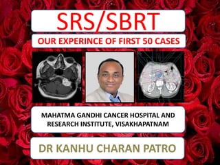
- 1. SRS/SBRT MAHATMA GANDHI CANCER HOSPITAL AND RESEARCH INSTITUTE, VISAKHAPATNAM OUR EXPERINCE OF FIRST 50 CASES DR KANHU CHARAN PATRO 1
- 2. WHAT IS SRS/SBRT? • Stereotactic radiosurgery (SRS) uses many precisely focused radiation beams to treat tumors and other problems in the brain, neck, lungs, liver, spine and other parts of the body. • It is not surgery in the traditional sense because there's no incision. • SRS is for cranial • SBRT for extracranial 2
- 3. THE DIFFERNCE CONVENTIONAL VS STEREOTAXY E. H. Balagamwala/Technology in Cancer Research and Treatment/2012 3
- 4. The duo • High dose • Strict immobilization 4
- 5. 5
- 6. • Massive vascular damage causes indirect tumor death-it is endothelial cell inflammation and apoptosis via the sphingomyelin pathway causing subsequent microvascular dysfunction that are the triggers for tumor cell death • 4 r of radiobiology in different manner • No Repair after ablative dose • Treatment is for short period no chance of Repopulation • No Reoxygenation of hypoxic cells due to massive vascular destruction by SRS/SBRT • Redistribution dose not happen as more cells die because of massive cell death • Massive immunogenic reaction • Abscopal effect RADIOBIOLOGY BEHIND STEROTAXY 6
- 7. The spectrum • SRS – Smaller lesion usually less than 3 cm. – Single fraction • FRACTIONATED SRS – Relatively larger tumor – 1 to 5 fractions • SRT – Larger tumor usually more than 3 cm – Close to vital structures 7
- 8. The spectrum • Malignant – Metastasis – Recurrent Gliomas • Benign – Arteriovenous Malformation – Vestibular Schwannoma – Pituitary – Cavernomas • Functional – Trigeminal Neuralgia – Tremor – Epileptic Focus 8
- 9. The wide spectrum Cranial – Metastasis • De novo • After WBRT – Arteriovenous malformation – Vestibular schwannoma – Reirradiation glioma – Glomus jugularae – Hamartoma – Cavernoma – Meningioma – Trigeminal neuralgia – Tremor – Epilepsy Extracranial – Bone metastasis – Prostate – Lung primary/ metastasis – Pancreas – Adrenal metastasis – Liver metastasis/HCC – Spine metastasis – Nodal recurrence – Head and neck reirradiation 9
- 10. • Malignant cases -- Weeks to months • Benign cases -- Months to years • Functional cases -- Days • Different criteria for different tumors e.g – RECIST – PERCIST – RANO – And many more RESPONSE EVALUATION 10
- 11. WHAT are the requirements? • Micro MLC/cone • Planning system • Imaging • Immobilization • Respiratory Motion management system • QA accessories • CBCT • Protocols 11
- 12. WHAT we have – machine 12
- 13. WHAT we have – micro MLC 13
- 14. WHAT we have – CONE 14
- 15. WHAT we have – immobilization 15 FRAXION
- 16. WHAT we have – immobilization 16
- 17. WHAT we have – motion management 17 SYMMETRY
- 18. WHAT we have – motion management 18 Abdominal compression
- 19. We have – ABC 19 ABC- ACTIVE BREATH COORDINATOR
- 20. • CT • PETCT – DOTA – PSMA – FDG • MRI IMAGING WE HAVE 20
- 21. WHAT we have – planning system 21
- 22. WHAT we have? – Ray Search planning system 22 FIRST IN INDIA-PHOTON
- 23. WHAT we have – verification system 23
- 24. • CBCT CORRECTIONS Set-up verification-CBCT 24
- 25. WHAT we have – Hexapod 25
- 26. • HEXAPOD CORRECTIONS Set-up verification- HEXAPOD 26
- 27. • MECHANICAL ISOCENTER CHECK – WINSTON LUTZ TEST • POINT DOSE VERIFICATION • TOLERANCE-1MM Travis R. Denton/JOURNAL OF APPLIED CLINICAL MEDICAL PHYSICS/2015 QA part 27
- 29. 29
- 30. 30
- 31. 31
- 32. 32 Approval
- 33. Started on July 2019 33
- 34. Cases completed till today 34
- 35. Cases completed till today 35 Brain metastasis 16 Brain metastasis after whole brain RT 8 Brain metastasis cavity SRS 3 Vestibular schwannoma 5 Arteriovenous malformation 1 Meningioma 5 Pituitary 1 Spine metastasis 3 Bone metastasis 1 Cranial cavernoma 2 Liver metastasis 3 Hepatocellular carcinoma 1 Lung cancer 1 Nodal recurrence 1 TOTAL 51
- 38. • NAME • UMR • PRESENTATION • 70 YEAR FEMALE • TRIPLE NEGATIVE BREAST CANCER • POST MRM • POST RT/CHEMO • 6 MONTH FOLLOW UP • PRESENTED WITH HEADACHE AND GIDDINESS • MRI • 2.2cm x2.2 cm LESION • LT.OCCIPITAL LOBE • RING ENHANCEMENT • NO MASS EFFECT • NO MID LINE SHIFT • MINIMAL EDEMA • PET CT • MULTIPLE LUNG NODULES • BRAIN LESION INCREASED Uptake • SRS • SRS • 18Gy/1# Case details 38
- 39. Pre SRS 39
- 40. SRS PALN 40
- 41. POST SRS 3 MONTHS 41
- 42. BRAIN METASTASIS CAVITY SRS CASE-2 42
- 43. • NAME • UMR • PRESENTATION • SEIZURES, UNCONSCIOUS • MRI • 3.2X3.2CM, LEFT OCCIPITAL LOBE,EDEMA,CONTRAST ENHANCEMNET • SURGERY • TOTAL EXCISON • BIOPSY • METASTATIC PAPILLARY ADENOCARCINOMA • PET CT • RT LUNG LESION • MULTIPLE NODULE BOTH LUNG • LESION IN BRAIN • MEDIASTINAL NODE • SRS • CAVITY SRS 30Gy/5# • IHC • EGFR+VE • EXON21 MUTATION • ALK NEG • TTF1 +VE • CHEMO • PEMETRXED AND CARBOPLATIN • OMERTINIB 80MG Case details 43
- 44. Pre op 44
- 45. Post op 1 month 45
- 46. 46 FRACTIONATED SRS • ADJACENT DURA and SURGICAL TRACT • BONE FLAP INNER PART • CAVITY PROPER • DURAL SINUS • ENHANCING COMPONENT
- 47. Post RT 3 months 47
- 49. • NAME • UMR • PRESENTATION • 59 year male • Diagnosed case of vestibular schwannoma • Right side • P/w Slight decreasing in hearing loss-4 - 5 months • No facial numbness • MRI • Intracanalicular and extra canalicular component • Touching brainstem • No cystic component • Minimal enhancing • Impending 5th nerve • SRS • SRS • 25Gy/5# Case details 49
- 53. NODAL RECURRENCE SBRT CASE- 4 53
- 54. • NAME • UMR • Diagnosis • Cancer cervix with common iliac node • RADIATION • EBRT –VMAT SIB 50Gy/25# -56Gy/28# • REGULAR FOLLOW UP • Post RT 3month - CR • Presented with • DVT and left leg pain • PET • Nodal recurrence same area • Planned SBRT • 30Gy/5# • PET POST SBRT 3M • Decreased SUV value • Now • Follow/up Case details 54
- 55. 55 At diagnosis
- 56. 56 At 3 month post RT
- 57. 57 At 1 year post RT
- 58. 58 Planned SBRT
- 59. 59 At 3 month post SBRT
- 60. 60 At 6 month post SBRT
- 62. • NAME • UMR • PRESENTATION • 50 YEAR • MALE • COLON CANCER • FOUND LIVER MET DURING SURGERY • 2 LESIONS • PET • 2 LESIONS • SEGMENT VIII SUV-13 • SEGMENT V • FNAC • ADENO • CP SCORE • A • SBRT • 40Gy/5# WITH DIBH Case details 62
- 63. 63
- 64. 64
- 66. • NAME • UMR • Diagnosis • Ca Lung left lower lobe with D11 bone metastasis • Presented with • Cough with expectoration, Pain over left chest wall, Upper backache • PET • Soft tissue enhancing lesion 5.2cm in LLL abutting pleura s/o primary • Hypermetabolic lesion in D11 vertebra (SUV max- 8) – s/o metastasis • Planned SBRT • 25Gy/5# • PET POST SBRT 3M • Complete metabolic resolution of the D11 vertebral lesion s/o favourable response to treatment • COURTESY • DR VKR Case details 66
- 69. 69 Planned SBRT
- 70. 70 3 month follow up scan
- 72. • NAME • UMR • Diagnosis • Hepatocellular carcinoma • Presented with • Diagnosed during screening • PET • Small lesion in segment 7 • Planned SBRT • 45Gy/3# • PET POST SBRT 3M • Complete resolution • Now • f/up • COURTESY • DR VKR Case details 72
- 73. CT/MRI 73
- 74. TARGET 74
- 75. SBRT PLAN 75
- 76. 3 month follow up 76
- 78. • NAME • UMR • Diagnosis • Metastatic Carcinoma Breast • Presented with • Pain over left hip • PET • Increased tracer uptake is seen in left acetabulum along the posterior margin and the left ischium showing sclerotic changes (SUV max - 6) • Planned SBRT • 33Gy/3# • PET POST SBRT 3M • No definite focal hypermetabolic or abnormally enhancing lesion • Increased sclerotic changes in the lesions noted in left acetabulum, ischium and inferior pubic ramus – s/o complete metabolic response • COURTESY • DR PSB Case details 78
- 80. TARGET 80
- 81. 81 Planned SBRT
- 82. 82 At 6 month follow up
- 84. • NAME • UMR • PRESENTATION • 23 year female • ECOG-1 • Sudden onset headache • Weakness of left upper and lower limb • Evaluated outside • Images not available • MRI • Location-Right high posterior parietal vascular malformation • Malformation size 3.4cm x 2.9cm x3.4cm • Nidus size 1.6cm x 1.4cm • Arterial supply- Pericollasal and collasomarginal branches of right anterior cerebral artery • Venous drainage- cortical veins along the right posterior parietal region • Hemoglobin degradation products with gliosis and enchephalomalacia. • SBRT • 18Gy/1# 84
- 85. MR ANGIO after 3 months 85
- 86. T1/T2- after 3 months 86
- 87. DSA THE GOLD STANDARD 87
- 88. CT ANGIO 88
- 90. 6 month follow up 90
- 92. • NAME • UMR • PRESENTATION • HEADACHE LEFT SIDED, LEFT EYE PAIN • MRI • 2.2×1.9×2.3CM, INTENSELY ENHANCING EXTRA AXIAL LESION T2W HYPOINTENSE & ISOINTENSE T1W ON LEFT SIDE POSTERIOR TO CAVERNOUS SINUS AND INDENTING PONS. LATERALLY ENCASING LEFT TRIGE • SURGERY • NEAR TOTAL EXCISON • HPE • S/O TRANSITIONAL MENINGIOMA (WHO GRADE I) • DOTA PET CT • 1.2×0.6 CM LESION NOTED IN THE LEFT PETROCLIVAL REGION ,POSTERIOR TO THE CAVERNOUS SINUS WITH SUV MAX - 7 • PLAN • SRS – 15Gy IN 1# Case details 92
- 94. 94 Dota scan
- 95. 95 SRS plan
- 96. 6 month follow up 96
- 98. • NAME • UMR • PRESENTATION • Vomiting, Head reeling sensation, Involuntary movements of all limbs • MRI 1. 2.3 × 1.6 × 1.6 cm, Dumbbell shaped lesion in sellar region 2. Extending into Suprasellar location 3. Pituitary gland not separated from lesion 4. Optic chiasm – compressed & superiorly displaced 5. Doubtful B/L Parasellar extension (R>L) with encasement of cavernous segment B/L ICA (R>L) • SURGERY • Endoscopic Trans sphenoid Excision and Near total excision • BIOPSY • F/S/O Pituitary Macro adenoma • SRS • FSRT – 25Gy / 5# • IHC • Synaptophysin +VE , • Chromogranin +VE Case details 98
- 99. Target 99
- 100. SRS PLAN 100
- 101. 6 month follw up- awaited 101
- 103. • NAME • UMR • PRESENTATION • Headache, difficulty in swallowing, Hoarseness of voice, Tinnitus , reduced hearing, Nasal regurgitation × 6 months • MRI • 2.5 x2 cm, Brilliantly enhancing, extracranial lesion in Left jugular foramen • Hypo on T1 and Iso on T2 • Erosion of carotid canal and jugular foramen • SURGERY • Excision of Glomus jugulare done by FISCH type approach • BIOPSY • well defined nests separated by highly vascularized fibrous septae[zelle ballen pattern] • SRS • 14Gy/1# • IHC • Synaptophysin positive • S100 positive • COURTESY • DR PSB Case details 103
- 104. GTV WITH PTV 1MM 104
- 105. SRS PLAN 105
- 106. 6 month follow up- awaited 106
- 108. 108
- 109. 109
- 110. 110
- 111. 111
- 112. 112
- 113. 113
- 114. if you are thinking about me as legend 114
- 115. ACKNOWLEDGMENTS-CONSULTANTS 115 DR C R KUNDU DR P S BHATTACHARYYA DR V K REDDY DR M MRUTYUNJAYA
- 116. ACKNOWLEDGMENTS- PHYSICISTS 116 A C PRABU A ANIL KUMAR A SRINU P PRASAD
- 118. THE GUIDANCE 118
- 119. 119