

Recomendados
Más contenido relacionado
La actualidad más candente
La actualidad más candente (20)
Destacado
Destacado (20)
Similar a Ophthalmoscopy
Similar a Ophthalmoscopy (20)
Último
Último (20)
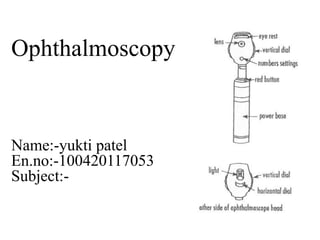
Ophthalmoscopy
- 2. Ophthalmoscopy • Ophthalmoscope is a test that allows a health professional to see inside the back of the eye (called the funds) and other structures using a magnifying instrument (ophthalmoscope) and a light source. It is done as part of an eye examination and may be done as part of a routine physical examination • An instrument used for assessment of ocular health • Posterior eye • Can also be used for the anterior eye
- 3. The modern ophthalmoscope:• Here light source from the batteries is reflected at 90o using a mirror placed in the head portion at 45o angle. The examiner looks through a hole in the mirror that is through the light.
- 4. Choosing the appropriate lens: The structures closer to the ophthalmoscope are best seen using positive lenses, which are labeled with black letters. The retina comes into focus at the 0 diopter, the cup at 2 red. 14 diopter 10 diopter 7 diopter 0 diopter -2 diopter
- 5. Type of ophthalmoscopy Direct ophthalmoscopy • During direct ophthalmoscopy, you may hear a clicking sound as the instrument is adjusted to focus on different structures in the eye. The light is sometimes very intense, and you may see spots for a short time following the examination. Some people report seeing light spots or branching images. These are actually the outlines of the blood vessels of the retina Indirect ophthalmoscopy • With indirect ophthalmoscopy, the light is much more intense and may be somewhat uncomfortable. Pressure applied to your eyeball with the blunt instrument also may be uncomfortable. After-images are common with this test. If the test is painful, let the health professional know
- 6. Risks • In some people, the dilating or anesthetic eyedrops can cause • Brief episodes of nausea, vomiting, dry mouth, flushing, and dizziness • An allergic reaction • A sudden increase in pressure inside the eyeball (closed-angle glaucoma) • Call your health professional immediately if you have severe and sudden eye pain, vision problems (halos may appear around light), or loss of vision after the examination
- 7. The head of the ophthalmoscope:• The head consists of a window for viewing the retina, and one for viewing the lens numbers and a wheel for changing them. Lens numbers are marked in black (positive) & red (negative) Type of portable Ophthalmoscope:- Specialist Ophthalmosco pe Professional Ophthalmoscope Pocket Ophthalmosc ope
- 8. STRUCTURE AND OPERATION diagram of a prior art binocular ophthalmoscope
- 9. Procedure • Ideally should be examined in a dark room. • Ask the patient to fix stare at an object. • Turn on scope and set dial at 0. • Right with right • Begin at arms length,should see red reflex. • Move close until optic disc is visible. • Turn dial until disc is in focus - +/-
- 10. Limitations of direct ophthalmoscopy • Direct ophthalmoscopy of the anterior eye is a screening technique – Instrument of choice is the slit lamp – We will cover this later in the year • Low magnification (2.5x for the anterior eye) • No stereopsis (3D vision) • Minimal lighting variability