SlideShare una empresa de Scribd logo
1 de 5
Descargar para leer sin conexión
CHAPTER 1

THE FUNDAMENTAL
UNIT OF LIFE
Objectives :
à Abc
à Abc
à Abc

O

ur planet Earth is inhabited by different types of living organisms, some that look similar while
some look very different from each other. Irrespective of the type of living organisms, each organism
is made up of a basic structural and functional unit called “Cell”. Let us learn in detail about the
discovery and the function of a cell in the subsequent sections below.

5.1 WHAT ARE LIVING ORGANISMS MADE UP OF?
5.1.1 Discovery of a “Cell”
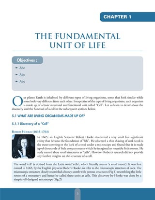
ROBERT HOOKE: (1635-1703)
In 1665, an English Scientist Robert Hooke discovered a very small but significant
entity that became the foundation of “life”. He observed a thin shaving of cork (cork is
the outer covering or the bark of a tree) under a microscope and found that it is made
up of thousands of little compartments which he imagined to resemble little rooms. He
aptly named these small structures as “cells”. However Robert’s research did not provide
any further insights on the structure of a cell.
The word ‘cell’ is derived from the Latin word ‘cella’, which literally means ‘a small room’). It was first
coined in 1665, by the English physicist Robert Hooke, to refer to the microscopic structure of cork. The
microscopic structure closely resembled a honey-comb with porous structures (Fig 1) resembling the little
rooms of a monastery and hence he called these units as cells. This discovery by Hooke was done by a
simple self-designed microscope (Fig 2)

3
Biology Education

Eyepiece

Barrel
Water Flask
Oil lamp
Focusing Screw
Objective
Specimen holder

Fig. 2: Robert Hooke’s microscope

Fig. 1: Cork cells as seen under a microscope

ANTON VON LEEUWENHOEK (1632-1723)
Through the simple microscope, Robert Hooke could only see the outer
thickened walls of the cork and could not provide any further insights. Anton Van
Leeuwenhoek, a Dutch microscopist made a technologically advanced microscope
(Fig 3). He observed the free floating ‘Algae Spirogyra’ swimming in pond water
and discovered that these little animals were actually single cells themselves. He is
considered the father of microscopy because of the advances he made in microscope
design and use. It was he who discovered the bacteria, yeast plants and much more.
Anton Von Leeuwenhoek is considered the father of microscope and his researches,
which were widely circulated, opened up an entire world of microscopic life to the
awareness of scientists.

Fig. 3: Anton Von
Leeuwenhoek’s microscope

MICROSCOPES: INSTRUMENTS FOR STUDYING CELLS
Cells are basically too small to be seen by the naked eye without any technological aid. They can only be
studied with the help of instruments called as ‘Microscopes’.
Microscope gets its name from the Greek word “small” and “to look”. It is essentially an instrument that is
used to view objects that are too small for the naked eye and the science of investigating these small objects
under a microscope is called ‘Microscopy’.
Types of Microscopes:
1. Light or Compound Microscope
The light microscope is so called because it employs visible light (generally sunlight) to illuminate small
objects. It is probably the most well-known and commonly used instrument in biology labs today. Optical
or light microscopy involves passing this visible light transmitted through a single or combination of
lenses to allow a magnified view of the sample. The resulting image can be detected directly by the eye or
imaged on a photographic plate

4
The Fundamental Unit of Life
Referring to figure 4 above, a good quality
microscope has a built-in reflector (mirror),
adjustable condenser below the stage, mechanical
stage, and binocular eyepiece tube. The sample or
specimen is mounted on a glass slide and kept on
the mechanical stage under the binocular eye piece
tube. The condenser is used to focus light on the
specimen through an opening in the stage. Sharp
image can be obtained by carefully focusing the side
knobs. The larger upper knob is meant for coarse
adjustments while the lower small knob is used to
for fine adjustments and getting a perfect image of
the specimen. After passing through the specimen,
the light is displayed to the eye with an apparent
field that is much larger than the area illuminated.
The magnification of the image can be obtained by
changing the objective lens magnification (usually
stamped on the lens body).

Eyepiece
Bodytube

Arm
Stage clip
Coarse
adjustment knob
Fine
adjustment knob

Revolving Nosepiece
Objectives
Stage
Diaphragm
Light source
Base

Fig 4 : A light microscope

2. Electron Microscope
Electron microscope is a significant alternative to
light microscopy which uses electrons rather than
light to generate the image. It was developed by
Ernst Ruska and later by Max Knoll of Germany
and uses a beam of electrons to illuminate the
specimen and produce a magnified image. Electron
microscopes have a greater resolution power than
a light-powered optical microscope as electrons
have wavelengths about 100,000 times shorter than
visible light (photons), and hence can achieve better
magnifications of up to about 500,000 times, as
compared to light microscopes which are limited by
magnifications below 2000x.Due to this property
Electron microscopes are helpful in observing sub
cellular structured which cannot be clearly seen
through a light microscope. Internal vacuum is
essential for the working and care must be taken to
ensure that the sample being used in an Electron
microscope is ultra thin and absolutely dry.

Fig. 5: An Electron Microscope

Fig. 6: Parts of an Electron Microscope

5
Biology Education

Table 1: Differences between light microscope and electron microscope

Light Microscope

Electron Microscope

It uses a beam of visible light to illuminate the
object

It uses a beam of electrons to illuminate the
object

It uses a single or combination of lenses

Uses electromagnets

Magnification obtained is usually 300 to
1500 times

Magnification obtained is 100,00 to 500,000
times

Internal vacuum is not required

Internal vacuum is essential

5.1.2: Cell Theory
As mentioned earlier, Robert Hooke had only seen the thickened walls of the cells and could not view the
inner contents of the cell. However, the first sightings of the internal action of the cell were made by a
Scottish botanist Robert Brown in 1831.He discovered and named the central substance of a plant cell as the
‘Nucleus’. Later in 1839, J.E. Purkinje, a Czech physiologist named the living fluid substance present inside
the cell as ‘protoplasm’.
Advancement in technology and continued research in this field helped Scientists develop a very important
foundation theories of Biology called – “The Cell Theory”
In 1838 Scientists Matthias Schleiden proposed that all plants comprise of cells. Later in 1839, Theodor
Schwann (1810-1882) a German Zoologist independently confirmed that all living things-both plants and
animals are made up of cells. Thus cell is the basic fundamental and structural unit of life. In 1855, another
German biologist Rudolf Virchow, further extended and refined the cell theory by proving that all cells arise
from pre-existing cells by dividing themselves i.e ‘Omnis cellulae a cellula’
The “Cell Theory” has three main aspects to it:
1. All living organisms are made up of single or many cells.
2. All cells arise from pre-existing cells by division. No cell can originate spontaneously on its own, instead
comes into existence only by division of an already existing cell.
3. The cell is the fundamental unit of structure in all living organisms. All metabolic reactions take place in
a cell.
Thus to conclude this discussion “What makes a Living Organism”; any self replicating living form comprises
of a basic fundamental structure called Cell.

6
The Fundamental Unit of Life

ACTIVITY 1
Objective: Observing Cells under a Microscope
Resources: Microscopes, slides, cover slips, source of
plant cells e.g. onion epidermis, leaf surface or petals
or even potato and tomato scrapings and appropriate
stains
Now that we have understood what is a “cell” let
us observe this under a microscope ourselves and
see if the shape and size or structure of cells within
the same organism is the same or different? For this
we can use any plant material like onion peel layer
(called epidermis) or surface of a leaf or petals. For
our experimental purpose we have considered the
thin onion epidermis.

Fig. 7: Cells of the onion peel

1. Take an onion peel layer and immerse it immediately in a watch glass containing water to avoid
dryness.
2. Prepare a microscope slide by gently placing a drop of water on it. Then with the help of a thin
paintbrush transfer the onion epidermis from the watch glass on this slide.
3. A drop of stain solution like safranine will colour these cells in the onion layer to make it more visible
to us under the microscope.
4. Place a cover slip on this mount taking care that no air bubbles emerge on the slide. The air bubbles
will cause hindrance while observing the mount under the microscope.
5. Now our slide is ready to be observed under a compound microscope.
6. Draw the structures as is on the sheet.
7. Repeat this experiment with different size of the onion peel taken from different parts of the same
onion.
What do you observe when you look through the lens of the microscope? Does it resemble the
figure 7 as shown above?
Interestingly, we observe that with different peels that may have been taken from either small or big onions
the basic structure of the cell does not change. These small structures (called cells) together form a big
structure called as the onion bulb. Thus, cell is the basic structure and the building block of all the onion
bulbs. This can be extrapolated to any living organism.

7

Más contenido relacionado

La actualidad más candente

La actualidad más candente (20)

Exudative Retinal Detachment- Etiopathogenesis
Exudative Retinal Detachment- EtiopathogenesisExudative Retinal Detachment- Etiopathogenesis
Exudative Retinal Detachment- Etiopathogenesis
 
Cure of leber congenital amaurosis
Cure of leber congenital amaurosisCure of leber congenital amaurosis
Cure of leber congenital amaurosis
 
Binocular vision
Binocular visionBinocular vision
Binocular vision
 
Ocular virology
Ocular virologyOcular virology
Ocular virology
 
Trabeculectomy.pptx
Trabeculectomy.pptxTrabeculectomy.pptx
Trabeculectomy.pptx
 
Atmospheric refraction
Atmospheric refractionAtmospheric refraction
Atmospheric refraction
 
Optic disc swelling
Optic disc swellingOptic disc swelling
Optic disc swelling
 
04 prism
04 prism 04 prism
04 prism
 
Abnormalities of pupillary reaction.pptx
Abnormalities of pupillary reaction.pptxAbnormalities of pupillary reaction.pptx
Abnormalities of pupillary reaction.pptx
 
Multiple evanescent white dot syndrome
Multiple evanescent white dot syndromeMultiple evanescent white dot syndrome
Multiple evanescent white dot syndrome
 
Subretinal hemorrhage
Subretinal hemorrhageSubretinal hemorrhage
Subretinal hemorrhage
 
Slit lump biomicroscope
Slit lump biomicroscopeSlit lump biomicroscope
Slit lump biomicroscope
 
Cardinal Points
Cardinal PointsCardinal Points
Cardinal Points
 
OCT IN DRY ARMD
OCT IN DRY ARMDOCT IN DRY ARMD
OCT IN DRY ARMD
 
02+semeiotica
02+semeiotica02+semeiotica
02+semeiotica
 
Management of dr
Management of drManagement of dr
Management of dr
 
Gonioscopy
GonioscopyGonioscopy
Gonioscopy
 
Pentacam
Pentacam Pentacam
Pentacam
 
Ocular manifestations in aids
Ocular manifestations in aidsOcular manifestations in aids
Ocular manifestations in aids
 
Retinal artery occlusion
Retinal artery occlusionRetinal artery occlusion
Retinal artery occlusion
 

Similar a Biology book layout, Layout Design, Book Layout, book inner page design

Unit 1 How to Use Microscope
Unit 1 How to Use MicroscopeUnit 1 How to Use Microscope
Unit 1 How to Use MicroscopeShally Rahmawaty
 
Unstained Check Cells
Unstained Check CellsUnstained Check Cells
Unstained Check CellsRachel Davis
 
CVA Biology I - B10vrv3071
CVA Biology I - B10vrv3071CVA Biology I - B10vrv3071
CVA Biology I - B10vrv3071ClayVirtual
 
Microbiology and its History.pptx
Microbiology and its History.pptxMicrobiology and its History.pptx
Microbiology and its History.pptxClydeDwynelhOGarcia
 
A. The development of the has allowed us to study cells and their pro.pdf
A. The development of the has allowed us to study cells and their pro.pdfA. The development of the has allowed us to study cells and their pro.pdf
A. The development of the has allowed us to study cells and their pro.pdfthangarajarivukadal
 
9 the fundamental unit of life
9   the fundamental unit of life9   the fundamental unit of life
9 the fundamental unit of lifethemassmaker
 
Lecture 1 The Cell.pdf
Lecture 1 The Cell.pdfLecture 1 The Cell.pdf
Lecture 1 The Cell.pdfAliSheikh116
 
Chapter 2 section 2 (2011): Viewing Cells
Chapter 2 section 2 (2011):  Viewing CellsChapter 2 section 2 (2011):  Viewing Cells
Chapter 2 section 2 (2011): Viewing CellsMr. Motuk
 
PP3_History_of_microscopy_1453876435.ppt
PP3_History_of_microscopy_1453876435.pptPP3_History_of_microscopy_1453876435.ppt
PP3_History_of_microscopy_1453876435.pptMuhammad Rawat
 
How to use microscope (basic biology) unm
How to use microscope (basic biology) unmHow to use microscope (basic biology) unm
How to use microscope (basic biology) unmJeny Hardiah
 
Bio A2 Cells
Bio A2 CellsBio A2 Cells
Bio A2 CellsR. Price
 
Microscopy_and_its_Application_in_Microbiology_and.pdf
Microscopy_and_its_Application_in_Microbiology_and.pdfMicroscopy_and_its_Application_in_Microbiology_and.pdf
Microscopy_and_its_Application_in_Microbiology_and.pdfImeneFl
 
Laporan Praktikum Biologi Dasar - How to use the microscope
Laporan Praktikum Biologi Dasar - How to use the microscopeLaporan Praktikum Biologi Dasar - How to use the microscope
Laporan Praktikum Biologi Dasar - How to use the microscopeWiwi Pratiwie
 
introduction to Celltheory
 introduction to Celltheory introduction to Celltheory
introduction to Celltheorysudarshanamigo
 
Celltheory and its types
Celltheory and its typesCelltheory and its types
Celltheory and its typessanthoshcsanthu
 

Similar a Biology book layout, Layout Design, Book Layout, book inner page design (20)

Unit 1 How to Use Microscope
Unit 1 How to Use MicroscopeUnit 1 How to Use Microscope
Unit 1 How to Use Microscope
 
Life Science
Life ScienceLife Science
Life Science
 
Unstained Check Cells
Unstained Check CellsUnstained Check Cells
Unstained Check Cells
 
CVA Biology I - B10vrv3071
CVA Biology I - B10vrv3071CVA Biology I - B10vrv3071
CVA Biology I - B10vrv3071
 
Microbiology and its History.pptx
Microbiology and its History.pptxMicrobiology and its History.pptx
Microbiology and its History.pptx
 
A. The development of the has allowed us to study cells and their pro.pdf
A. The development of the has allowed us to study cells and their pro.pdfA. The development of the has allowed us to study cells and their pro.pdf
A. The development of the has allowed us to study cells and their pro.pdf
 
9 the fundamental unit of life
9   the fundamental unit of life9   the fundamental unit of life
9 the fundamental unit of life
 
Lecture 1 The Cell.pdf
Lecture 1 The Cell.pdfLecture 1 The Cell.pdf
Lecture 1 The Cell.pdf
 
Cell theory
Cell theoryCell theory
Cell theory
 
Chapter 2 section 2 (2011): Viewing Cells
Chapter 2 section 2 (2011):  Viewing CellsChapter 2 section 2 (2011):  Viewing Cells
Chapter 2 section 2 (2011): Viewing Cells
 
PP3_History_of_microscopy_1453876435.ppt
PP3_History_of_microscopy_1453876435.pptPP3_History_of_microscopy_1453876435.ppt
PP3_History_of_microscopy_1453876435.ppt
 
How to use microscope (basic biology) unm
How to use microscope (basic biology) unmHow to use microscope (basic biology) unm
How to use microscope (basic biology) unm
 
Bio A2 Cells
Bio A2 CellsBio A2 Cells
Bio A2 Cells
 
Lab 1
Lab 1Lab 1
Lab 1
 
Microscopy_and_its_Application_in_Microbiology_and.pdf
Microscopy_and_its_Application_in_Microbiology_and.pdfMicroscopy_and_its_Application_in_Microbiology_and.pdf
Microscopy_and_its_Application_in_Microbiology_and.pdf
 
Laporan Praktikum Biologi Dasar - How to use the microscope
Laporan Praktikum Biologi Dasar - How to use the microscopeLaporan Praktikum Biologi Dasar - How to use the microscope
Laporan Praktikum Biologi Dasar - How to use the microscope
 
introduction to Celltheory
 introduction to Celltheory introduction to Celltheory
introduction to Celltheory
 
Celltheory and its types
Celltheory and its typesCelltheory and its types
Celltheory and its types
 
Cells
CellsCells
Cells
 
00introhistologytext
00introhistologytext00introhistologytext
00introhistologytext
 

Más de KS Designers

Desk Calender Design
Desk Calender DesignDesk Calender Design
Desk Calender DesignKS Designers
 
Traveloholic Design 1- Branding Design, Branding, Corporate Identify, Brand D...
Traveloholic Design 1- Branding Design, Branding, Corporate Identify, Brand D...Traveloholic Design 1- Branding Design, Branding, Corporate Identify, Brand D...
Traveloholic Design 1- Branding Design, Branding, Corporate Identify, Brand D...KS Designers
 
Atos Broucher - Magazine Design India, Journal Design, Layout Design, Magazin...
Atos Broucher - Magazine Design India, Journal Design, Layout Design, Magazin...Atos Broucher - Magazine Design India, Journal Design, Layout Design, Magazin...
Atos Broucher - Magazine Design India, Journal Design, Layout Design, Magazin...KS Designers
 
Directory Design and Layout
Directory Design and LayoutDirectory Design and Layout
Directory Design and LayoutKS Designers
 
Magazine Design & Layout
Magazine Design & LayoutMagazine Design & Layout
Magazine Design & LayoutKS Designers
 
Magazine Design and Layout
Magazine Design and LayoutMagazine Design and Layout
Magazine Design and LayoutKS Designers
 
Fantastic and modern magazine design layouts
Fantastic and modern magazine design layouts Fantastic and modern magazine design layouts
Fantastic and modern magazine design layouts KS Designers
 
Presto calender layout 2015
Presto calender layout 2015Presto calender layout 2015
Presto calender layout 2015KS Designers
 
Calender layout design by KS Designers
Calender layout design by KS DesignersCalender layout design by KS Designers
Calender layout design by KS DesignersKS Designers
 
Personal Magazine Design, Magazine Layout Design, Layout Design, Design Agenc...
Personal Magazine Design, Magazine Layout Design, Layout Design, Design Agenc...Personal Magazine Design, Magazine Layout Design, Layout Design, Design Agenc...
Personal Magazine Design, Magazine Layout Design, Layout Design, Design Agenc...KS Designers
 
advertising agency, advertising agencies, ad agency in delhi, advertising age...
advertising agency, advertising agencies, ad agency in delhi, advertising age...advertising agency, advertising agencies, ad agency in delhi, advertising age...
advertising agency, advertising agencies, ad agency in delhi, advertising age...KS Designers
 
Child Friendly News Magazine
Child Friendly News MagazineChild Friendly News Magazine
Child Friendly News MagazineKS Designers
 
Delhi public school magazine
Delhi public school magazineDelhi public school magazine
Delhi public school magazineKS Designers
 
Child Friendly News Jan 2nd issue
Child Friendly News Jan 2nd issueChild Friendly News Jan 2nd issue
Child Friendly News Jan 2nd issueKS Designers
 
Rockland flyer-sample
Rockland flyer-sampleRockland flyer-sample
Rockland flyer-sampleKS Designers
 
Table Calender design sample 1
Table Calender design sample 1Table Calender design sample 1
Table Calender design sample 1KS Designers
 
Table Calender design sample 3
Table Calender design sample 3Table Calender design sample 3
Table Calender design sample 3KS Designers
 
Table Calender design sample 4
Table Calender design sample 4Table Calender design sample 4
Table Calender design sample 4KS Designers
 
Table Calender design sample 5
Table Calender design sample 5Table Calender design sample 5
Table Calender design sample 5KS Designers
 

Más de KS Designers (20)

Broucher Design
Broucher DesignBroucher Design
Broucher Design
 
Desk Calender Design
Desk Calender DesignDesk Calender Design
Desk Calender Design
 
Traveloholic Design 1- Branding Design, Branding, Corporate Identify, Brand D...
Traveloholic Design 1- Branding Design, Branding, Corporate Identify, Brand D...Traveloholic Design 1- Branding Design, Branding, Corporate Identify, Brand D...
Traveloholic Design 1- Branding Design, Branding, Corporate Identify, Brand D...
 
Atos Broucher - Magazine Design India, Journal Design, Layout Design, Magazin...
Atos Broucher - Magazine Design India, Journal Design, Layout Design, Magazin...Atos Broucher - Magazine Design India, Journal Design, Layout Design, Magazin...
Atos Broucher - Magazine Design India, Journal Design, Layout Design, Magazin...
 
Directory Design and Layout
Directory Design and LayoutDirectory Design and Layout
Directory Design and Layout
 
Magazine Design & Layout
Magazine Design & LayoutMagazine Design & Layout
Magazine Design & Layout
 
Magazine Design and Layout
Magazine Design and LayoutMagazine Design and Layout
Magazine Design and Layout
 
Fantastic and modern magazine design layouts
Fantastic and modern magazine design layouts Fantastic and modern magazine design layouts
Fantastic and modern magazine design layouts
 
Presto calender layout 2015
Presto calender layout 2015Presto calender layout 2015
Presto calender layout 2015
 
Calender layout design by KS Designers
Calender layout design by KS DesignersCalender layout design by KS Designers
Calender layout design by KS Designers
 
Personal Magazine Design, Magazine Layout Design, Layout Design, Design Agenc...
Personal Magazine Design, Magazine Layout Design, Layout Design, Design Agenc...Personal Magazine Design, Magazine Layout Design, Layout Design, Design Agenc...
Personal Magazine Design, Magazine Layout Design, Layout Design, Design Agenc...
 
advertising agency, advertising agencies, ad agency in delhi, advertising age...
advertising agency, advertising agencies, ad agency in delhi, advertising age...advertising agency, advertising agencies, ad agency in delhi, advertising age...
advertising agency, advertising agencies, ad agency in delhi, advertising age...
 
Child Friendly News Magazine
Child Friendly News MagazineChild Friendly News Magazine
Child Friendly News Magazine
 
Delhi public school magazine
Delhi public school magazineDelhi public school magazine
Delhi public school magazine
 
Child Friendly News Jan 2nd issue
Child Friendly News Jan 2nd issueChild Friendly News Jan 2nd issue
Child Friendly News Jan 2nd issue
 
Rockland flyer-sample
Rockland flyer-sampleRockland flyer-sample
Rockland flyer-sample
 
Table Calender design sample 1
Table Calender design sample 1Table Calender design sample 1
Table Calender design sample 1
 
Table Calender design sample 3
Table Calender design sample 3Table Calender design sample 3
Table Calender design sample 3
 
Table Calender design sample 4
Table Calender design sample 4Table Calender design sample 4
Table Calender design sample 4
 
Table Calender design sample 5
Table Calender design sample 5Table Calender design sample 5
Table Calender design sample 5
 

Último

Jordan_Amanda_DMBS202404_PB1_2024-04.pdf
Jordan_Amanda_DMBS202404_PB1_2024-04.pdfJordan_Amanda_DMBS202404_PB1_2024-04.pdf
Jordan_Amanda_DMBS202404_PB1_2024-04.pdfamanda2495
 
Call Girls In Ratnagiri Escorts ☎️8617370543 🔝 💃 Enjoy 24/7 Escort Service En...
Call Girls In Ratnagiri Escorts ☎️8617370543 🔝 💃 Enjoy 24/7 Escort Service En...Call Girls In Ratnagiri Escorts ☎️8617370543 🔝 💃 Enjoy 24/7 Escort Service En...
Call Girls In Ratnagiri Escorts ☎️8617370543 🔝 💃 Enjoy 24/7 Escort Service En...Nitya salvi
 
Top profile Call Girls In Sonipat [ 7014168258 ] Call Me For Genuine Models W...
Top profile Call Girls In Sonipat [ 7014168258 ] Call Me For Genuine Models W...Top profile Call Girls In Sonipat [ 7014168258 ] Call Me For Genuine Models W...
Top profile Call Girls In Sonipat [ 7014168258 ] Call Me For Genuine Models W...nirzagarg
 
How to Build a Simple Shopify Website
How to Build a Simple Shopify WebsiteHow to Build a Simple Shopify Website
How to Build a Simple Shopify Websitemark11275
 
Just Call Vip call girls Kasganj Escorts ☎️8617370543 Two shot with one girl ...
Just Call Vip call girls Kasganj Escorts ☎️8617370543 Two shot with one girl ...Just Call Vip call girls Kasganj Escorts ☎️8617370543 Two shot with one girl ...
Just Call Vip call girls Kasganj Escorts ☎️8617370543 Two shot with one girl ...Nitya salvi
 
怎样办理伯明翰大学学院毕业证(Birmingham毕业证书)成绩单留信认证
怎样办理伯明翰大学学院毕业证(Birmingham毕业证书)成绩单留信认证怎样办理伯明翰大学学院毕业证(Birmingham毕业证书)成绩单留信认证
怎样办理伯明翰大学学院毕业证(Birmingham毕业证书)成绩单留信认证eeanqy
 
NO1 Top Pakistani Amil Baba Real Amil baba In Pakistan Najoomi Baba in Pakist...
NO1 Top Pakistani Amil Baba Real Amil baba In Pakistan Najoomi Baba in Pakist...NO1 Top Pakistani Amil Baba Real Amil baba In Pakistan Najoomi Baba in Pakist...
NO1 Top Pakistani Amil Baba Real Amil baba In Pakistan Najoomi Baba in Pakist...Amil baba
 
Q4-Trends-Networks-Module-3.pdfqquater days sheets123456789
Q4-Trends-Networks-Module-3.pdfqquater days sheets123456789Q4-Trends-Networks-Module-3.pdfqquater days sheets123456789
Q4-Trends-Networks-Module-3.pdfqquater days sheets123456789CristineGraceAcuyan
 
Top profile Call Girls In fatehgarh [ 7014168258 ] Call Me For Genuine Models...
Top profile Call Girls In fatehgarh [ 7014168258 ] Call Me For Genuine Models...Top profile Call Girls In fatehgarh [ 7014168258 ] Call Me For Genuine Models...
Top profile Call Girls In fatehgarh [ 7014168258 ] Call Me For Genuine Models...gajnagarg
 
Q4-W4-SCIENCE-5 power point presentation
Q4-W4-SCIENCE-5 power point presentationQ4-W4-SCIENCE-5 power point presentation
Q4-W4-SCIENCE-5 power point presentationZenSeloveres
 
Essential UI/UX Design Principles: A Comprehensive Guide
Essential UI/UX Design Principles: A Comprehensive GuideEssential UI/UX Design Principles: A Comprehensive Guide
Essential UI/UX Design Principles: A Comprehensive GuideDesign Studio UI UX
 
一比一定(购)滑铁卢大学毕业证(UW毕业证)成绩单学位证
一比一定(购)滑铁卢大学毕业证(UW毕业证)成绩单学位证一比一定(购)滑铁卢大学毕业证(UW毕业证)成绩单学位证
一比一定(购)滑铁卢大学毕业证(UW毕业证)成绩单学位证wpkuukw
 
怎样办理巴斯大学毕业证(Bath毕业证书)成绩单留信认证
怎样办理巴斯大学毕业证(Bath毕业证书)成绩单留信认证怎样办理巴斯大学毕业证(Bath毕业证书)成绩单留信认证
怎样办理巴斯大学毕业证(Bath毕业证书)成绩单留信认证eeanqy
 
High Profile Escorts Nerul WhatsApp +91-9930687706, Best Service
High Profile Escorts Nerul WhatsApp +91-9930687706, Best ServiceHigh Profile Escorts Nerul WhatsApp +91-9930687706, Best Service
High Profile Escorts Nerul WhatsApp +91-9930687706, Best Servicemeghakumariji156
 
一比一定(购)西悉尼大学毕业证(WSU毕业证)成绩单学位证
一比一定(购)西悉尼大学毕业证(WSU毕业证)成绩单学位证一比一定(购)西悉尼大学毕业证(WSU毕业证)成绩单学位证
一比一定(购)西悉尼大学毕业证(WSU毕业证)成绩单学位证eqaqen
 
Eye-Catching Web Design Crafting User Interfaces .docx
Eye-Catching Web Design Crafting User Interfaces .docxEye-Catching Web Design Crafting User Interfaces .docx
Eye-Catching Web Design Crafting User Interfaces .docxMdBokhtiyarHossainNi
 
LANDSCAPE ARCHITECTURE PORTFOLIO - MAREK MITACEK
LANDSCAPE ARCHITECTURE PORTFOLIO - MAREK MITACEKLANDSCAPE ARCHITECTURE PORTFOLIO - MAREK MITACEK
LANDSCAPE ARCHITECTURE PORTFOLIO - MAREK MITACEKMarekMitek1
 
Mohanlalganj ! Call Girls in Lucknow - 450+ Call Girl Cash Payment 9548273370...
Mohanlalganj ! Call Girls in Lucknow - 450+ Call Girl Cash Payment 9548273370...Mohanlalganj ! Call Girls in Lucknow - 450+ Call Girl Cash Payment 9548273370...
Mohanlalganj ! Call Girls in Lucknow - 450+ Call Girl Cash Payment 9548273370...gargpaaro
 

Último (20)

Jordan_Amanda_DMBS202404_PB1_2024-04.pdf
Jordan_Amanda_DMBS202404_PB1_2024-04.pdfJordan_Amanda_DMBS202404_PB1_2024-04.pdf
Jordan_Amanda_DMBS202404_PB1_2024-04.pdf
 
Call Girls In Ratnagiri Escorts ☎️8617370543 🔝 💃 Enjoy 24/7 Escort Service En...
Call Girls In Ratnagiri Escorts ☎️8617370543 🔝 💃 Enjoy 24/7 Escort Service En...Call Girls In Ratnagiri Escorts ☎️8617370543 🔝 💃 Enjoy 24/7 Escort Service En...
Call Girls In Ratnagiri Escorts ☎️8617370543 🔝 💃 Enjoy 24/7 Escort Service En...
 
Top profile Call Girls In Sonipat [ 7014168258 ] Call Me For Genuine Models W...
Top profile Call Girls In Sonipat [ 7014168258 ] Call Me For Genuine Models W...Top profile Call Girls In Sonipat [ 7014168258 ] Call Me For Genuine Models W...
Top profile Call Girls In Sonipat [ 7014168258 ] Call Me For Genuine Models W...
 
How to Build a Simple Shopify Website
How to Build a Simple Shopify WebsiteHow to Build a Simple Shopify Website
How to Build a Simple Shopify Website
 
Just Call Vip call girls Kasganj Escorts ☎️8617370543 Two shot with one girl ...
Just Call Vip call girls Kasganj Escorts ☎️8617370543 Two shot with one girl ...Just Call Vip call girls Kasganj Escorts ☎️8617370543 Two shot with one girl ...
Just Call Vip call girls Kasganj Escorts ☎️8617370543 Two shot with one girl ...
 
怎样办理伯明翰大学学院毕业证(Birmingham毕业证书)成绩单留信认证
怎样办理伯明翰大学学院毕业证(Birmingham毕业证书)成绩单留信认证怎样办理伯明翰大学学院毕业证(Birmingham毕业证书)成绩单留信认证
怎样办理伯明翰大学学院毕业证(Birmingham毕业证书)成绩单留信认证
 
NO1 Top Pakistani Amil Baba Real Amil baba In Pakistan Najoomi Baba in Pakist...
NO1 Top Pakistani Amil Baba Real Amil baba In Pakistan Najoomi Baba in Pakist...NO1 Top Pakistani Amil Baba Real Amil baba In Pakistan Najoomi Baba in Pakist...
NO1 Top Pakistani Amil Baba Real Amil baba In Pakistan Najoomi Baba in Pakist...
 
Q4-Trends-Networks-Module-3.pdfqquater days sheets123456789
Q4-Trends-Networks-Module-3.pdfqquater days sheets123456789Q4-Trends-Networks-Module-3.pdfqquater days sheets123456789
Q4-Trends-Networks-Module-3.pdfqquater days sheets123456789
 
Abortion Pills in Oman (+918133066128) Cytotec clinic buy Oman Muscat
Abortion Pills in Oman (+918133066128) Cytotec clinic buy Oman MuscatAbortion Pills in Oman (+918133066128) Cytotec clinic buy Oman Muscat
Abortion Pills in Oman (+918133066128) Cytotec clinic buy Oman Muscat
 
Top profile Call Girls In fatehgarh [ 7014168258 ] Call Me For Genuine Models...
Top profile Call Girls In fatehgarh [ 7014168258 ] Call Me For Genuine Models...Top profile Call Girls In fatehgarh [ 7014168258 ] Call Me For Genuine Models...
Top profile Call Girls In fatehgarh [ 7014168258 ] Call Me For Genuine Models...
 
Q4-W4-SCIENCE-5 power point presentation
Q4-W4-SCIENCE-5 power point presentationQ4-W4-SCIENCE-5 power point presentation
Q4-W4-SCIENCE-5 power point presentation
 
Essential UI/UX Design Principles: A Comprehensive Guide
Essential UI/UX Design Principles: A Comprehensive GuideEssential UI/UX Design Principles: A Comprehensive Guide
Essential UI/UX Design Principles: A Comprehensive Guide
 
Hackathon evaluation template_latest_uploadpdf
Hackathon evaluation template_latest_uploadpdfHackathon evaluation template_latest_uploadpdf
Hackathon evaluation template_latest_uploadpdf
 
一比一定(购)滑铁卢大学毕业证(UW毕业证)成绩单学位证
一比一定(购)滑铁卢大学毕业证(UW毕业证)成绩单学位证一比一定(购)滑铁卢大学毕业证(UW毕业证)成绩单学位证
一比一定(购)滑铁卢大学毕业证(UW毕业证)成绩单学位证
 
怎样办理巴斯大学毕业证(Bath毕业证书)成绩单留信认证
怎样办理巴斯大学毕业证(Bath毕业证书)成绩单留信认证怎样办理巴斯大学毕业证(Bath毕业证书)成绩单留信认证
怎样办理巴斯大学毕业证(Bath毕业证书)成绩单留信认证
 
High Profile Escorts Nerul WhatsApp +91-9930687706, Best Service
High Profile Escorts Nerul WhatsApp +91-9930687706, Best ServiceHigh Profile Escorts Nerul WhatsApp +91-9930687706, Best Service
High Profile Escorts Nerul WhatsApp +91-9930687706, Best Service
 
一比一定(购)西悉尼大学毕业证(WSU毕业证)成绩单学位证
一比一定(购)西悉尼大学毕业证(WSU毕业证)成绩单学位证一比一定(购)西悉尼大学毕业证(WSU毕业证)成绩单学位证
一比一定(购)西悉尼大学毕业证(WSU毕业证)成绩单学位证
 
Eye-Catching Web Design Crafting User Interfaces .docx
Eye-Catching Web Design Crafting User Interfaces .docxEye-Catching Web Design Crafting User Interfaces .docx
Eye-Catching Web Design Crafting User Interfaces .docx
 
LANDSCAPE ARCHITECTURE PORTFOLIO - MAREK MITACEK
LANDSCAPE ARCHITECTURE PORTFOLIO - MAREK MITACEKLANDSCAPE ARCHITECTURE PORTFOLIO - MAREK MITACEK
LANDSCAPE ARCHITECTURE PORTFOLIO - MAREK MITACEK
 
Mohanlalganj ! Call Girls in Lucknow - 450+ Call Girl Cash Payment 9548273370...
Mohanlalganj ! Call Girls in Lucknow - 450+ Call Girl Cash Payment 9548273370...Mohanlalganj ! Call Girls in Lucknow - 450+ Call Girl Cash Payment 9548273370...
Mohanlalganj ! Call Girls in Lucknow - 450+ Call Girl Cash Payment 9548273370...
 

Biology book layout, Layout Design, Book Layout, book inner page design

  • 1. CHAPTER 1 THE FUNDAMENTAL UNIT OF LIFE Objectives : à Abc à Abc à Abc O ur planet Earth is inhabited by different types of living organisms, some that look similar while some look very different from each other. Irrespective of the type of living organisms, each organism is made up of a basic structural and functional unit called “Cell”. Let us learn in detail about the discovery and the function of a cell in the subsequent sections below. 5.1 WHAT ARE LIVING ORGANISMS MADE UP OF? 5.1.1 Discovery of a “Cell” ROBERT HOOKE: (1635-1703) In 1665, an English Scientist Robert Hooke discovered a very small but significant entity that became the foundation of “life”. He observed a thin shaving of cork (cork is the outer covering or the bark of a tree) under a microscope and found that it is made up of thousands of little compartments which he imagined to resemble little rooms. He aptly named these small structures as “cells”. However Robert’s research did not provide any further insights on the structure of a cell. The word ‘cell’ is derived from the Latin word ‘cella’, which literally means ‘a small room’). It was first coined in 1665, by the English physicist Robert Hooke, to refer to the microscopic structure of cork. The microscopic structure closely resembled a honey-comb with porous structures (Fig 1) resembling the little rooms of a monastery and hence he called these units as cells. This discovery by Hooke was done by a simple self-designed microscope (Fig 2) 3
  • 2. Biology Education Eyepiece Barrel Water Flask Oil lamp Focusing Screw Objective Specimen holder Fig. 2: Robert Hooke’s microscope Fig. 1: Cork cells as seen under a microscope ANTON VON LEEUWENHOEK (1632-1723) Through the simple microscope, Robert Hooke could only see the outer thickened walls of the cork and could not provide any further insights. Anton Van Leeuwenhoek, a Dutch microscopist made a technologically advanced microscope (Fig 3). He observed the free floating ‘Algae Spirogyra’ swimming in pond water and discovered that these little animals were actually single cells themselves. He is considered the father of microscopy because of the advances he made in microscope design and use. It was he who discovered the bacteria, yeast plants and much more. Anton Von Leeuwenhoek is considered the father of microscope and his researches, which were widely circulated, opened up an entire world of microscopic life to the awareness of scientists. Fig. 3: Anton Von Leeuwenhoek’s microscope MICROSCOPES: INSTRUMENTS FOR STUDYING CELLS Cells are basically too small to be seen by the naked eye without any technological aid. They can only be studied with the help of instruments called as ‘Microscopes’. Microscope gets its name from the Greek word “small” and “to look”. It is essentially an instrument that is used to view objects that are too small for the naked eye and the science of investigating these small objects under a microscope is called ‘Microscopy’. Types of Microscopes: 1. Light or Compound Microscope The light microscope is so called because it employs visible light (generally sunlight) to illuminate small objects. It is probably the most well-known and commonly used instrument in biology labs today. Optical or light microscopy involves passing this visible light transmitted through a single or combination of lenses to allow a magnified view of the sample. The resulting image can be detected directly by the eye or imaged on a photographic plate 4
  • 3. The Fundamental Unit of Life Referring to figure 4 above, a good quality microscope has a built-in reflector (mirror), adjustable condenser below the stage, mechanical stage, and binocular eyepiece tube. The sample or specimen is mounted on a glass slide and kept on the mechanical stage under the binocular eye piece tube. The condenser is used to focus light on the specimen through an opening in the stage. Sharp image can be obtained by carefully focusing the side knobs. The larger upper knob is meant for coarse adjustments while the lower small knob is used to for fine adjustments and getting a perfect image of the specimen. After passing through the specimen, the light is displayed to the eye with an apparent field that is much larger than the area illuminated. The magnification of the image can be obtained by changing the objective lens magnification (usually stamped on the lens body). Eyepiece Bodytube Arm Stage clip Coarse adjustment knob Fine adjustment knob Revolving Nosepiece Objectives Stage Diaphragm Light source Base Fig 4 : A light microscope 2. Electron Microscope Electron microscope is a significant alternative to light microscopy which uses electrons rather than light to generate the image. It was developed by Ernst Ruska and later by Max Knoll of Germany and uses a beam of electrons to illuminate the specimen and produce a magnified image. Electron microscopes have a greater resolution power than a light-powered optical microscope as electrons have wavelengths about 100,000 times shorter than visible light (photons), and hence can achieve better magnifications of up to about 500,000 times, as compared to light microscopes which are limited by magnifications below 2000x.Due to this property Electron microscopes are helpful in observing sub cellular structured which cannot be clearly seen through a light microscope. Internal vacuum is essential for the working and care must be taken to ensure that the sample being used in an Electron microscope is ultra thin and absolutely dry. Fig. 5: An Electron Microscope Fig. 6: Parts of an Electron Microscope 5
  • 4. Biology Education Table 1: Differences between light microscope and electron microscope Light Microscope Electron Microscope It uses a beam of visible light to illuminate the object It uses a beam of electrons to illuminate the object It uses a single or combination of lenses Uses electromagnets Magnification obtained is usually 300 to 1500 times Magnification obtained is 100,00 to 500,000 times Internal vacuum is not required Internal vacuum is essential 5.1.2: Cell Theory As mentioned earlier, Robert Hooke had only seen the thickened walls of the cells and could not view the inner contents of the cell. However, the first sightings of the internal action of the cell were made by a Scottish botanist Robert Brown in 1831.He discovered and named the central substance of a plant cell as the ‘Nucleus’. Later in 1839, J.E. Purkinje, a Czech physiologist named the living fluid substance present inside the cell as ‘protoplasm’. Advancement in technology and continued research in this field helped Scientists develop a very important foundation theories of Biology called – “The Cell Theory” In 1838 Scientists Matthias Schleiden proposed that all plants comprise of cells. Later in 1839, Theodor Schwann (1810-1882) a German Zoologist independently confirmed that all living things-both plants and animals are made up of cells. Thus cell is the basic fundamental and structural unit of life. In 1855, another German biologist Rudolf Virchow, further extended and refined the cell theory by proving that all cells arise from pre-existing cells by dividing themselves i.e ‘Omnis cellulae a cellula’ The “Cell Theory” has three main aspects to it: 1. All living organisms are made up of single or many cells. 2. All cells arise from pre-existing cells by division. No cell can originate spontaneously on its own, instead comes into existence only by division of an already existing cell. 3. The cell is the fundamental unit of structure in all living organisms. All metabolic reactions take place in a cell. Thus to conclude this discussion “What makes a Living Organism”; any self replicating living form comprises of a basic fundamental structure called Cell. 6
  • 5. The Fundamental Unit of Life ACTIVITY 1 Objective: Observing Cells under a Microscope Resources: Microscopes, slides, cover slips, source of plant cells e.g. onion epidermis, leaf surface or petals or even potato and tomato scrapings and appropriate stains Now that we have understood what is a “cell” let us observe this under a microscope ourselves and see if the shape and size or structure of cells within the same organism is the same or different? For this we can use any plant material like onion peel layer (called epidermis) or surface of a leaf or petals. For our experimental purpose we have considered the thin onion epidermis. Fig. 7: Cells of the onion peel 1. Take an onion peel layer and immerse it immediately in a watch glass containing water to avoid dryness. 2. Prepare a microscope slide by gently placing a drop of water on it. Then with the help of a thin paintbrush transfer the onion epidermis from the watch glass on this slide. 3. A drop of stain solution like safranine will colour these cells in the onion layer to make it more visible to us under the microscope. 4. Place a cover slip on this mount taking care that no air bubbles emerge on the slide. The air bubbles will cause hindrance while observing the mount under the microscope. 5. Now our slide is ready to be observed under a compound microscope. 6. Draw the structures as is on the sheet. 7. Repeat this experiment with different size of the onion peel taken from different parts of the same onion. What do you observe when you look through the lens of the microscope? Does it resemble the figure 7 as shown above? Interestingly, we observe that with different peels that may have been taken from either small or big onions the basic structure of the cell does not change. These small structures (called cells) together form a big structure called as the onion bulb. Thus, cell is the basic structure and the building block of all the onion bulbs. This can be extrapolated to any living organism. 7