Circulatory system
•Descargar como PPTX, PDF•
7 recomendaciones•1,498 vistas

The circulatory system transports blood, nutrients, gases, hormones, and waste products throughout the body. It consists of the heart, blood vessels, and blood. The heart pumps blood through two main circuits - the pulmonary circuit, which carries deoxygenated blood to the lungs and returns oxygenated blood, and the systemic circuit, which pumps oxygenated blood to the entire body and returns deoxygenated blood back to the heart. The cardiovascular system is further divided into four chambers, two atria that receive blood and two ventricles that pump blood out of the heart. Blood contains red blood cells, white blood cells, platelets, and plasma and comes in four main blood groups - A, B, AB, and

Recomendados
Más contenido relacionado
La actualidad más candente
La actualidad más candente (20)
Similar a Circulatory system
Similar a Circulatory system (20)
Último
Último (20)
Circulatory system
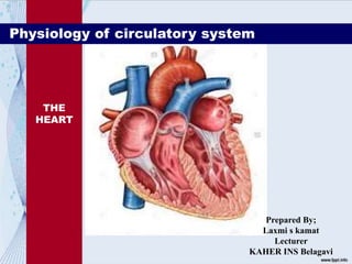
- 1. THE HEART Physiology of circulatory system Prepared By; Laxmi s kamat Lecturer KAHER INS Belagavi
- 2. INTRODUCTION The cardiovascular system is transport system of body It comprises blood, heart and blood vessels. The system supplies nutrients to and remove waste products from various tissue of body. The conveying media is liquid in form of blood which flows in close tubular system.
- 3. FUNCTION OF CARDIOVASCULAR SYSTEM
- 4. • Protection – protects against blood loss from injury and against pathogens, including foreign microbes and toxins introduced into the body. These substances can be categorized as follows: • Clotting– prevents blood loss when blood vessels are damaged • Immune function - protect against many disease- causing agents (pathogens). Performed by leukocytes (white blood cells).
- 5. Major Components of the Circulatory System Cardiovascular system consist of: • Heart • Blood • Blood vessels: form a tubular network that permits the flow of blood • Arteries, arterioles, veins, capillaries Lymphatic system consists of: • Lymphatic vessels • Lymphoid tissues • Found in the spleen, thymus, tonsil and lymph nodes • The circulatory system is divided into two major subdivisions: • The Cardiovascular system and the Lymphatic system
- 7. Composition of blood: 1.Solid component (Blood cells) 41% of total volume Red blood cells (erythrocytes), white blood cells (leukocytes), or platelets (thrombocytes) 2.Liquid Component (Blood plasma) • 55% of total blood volume • Composed of 91.5% water and 8.5% of nutrients, waste products, proteins, enzymes and hormones • Straw coloured or yellowy solution • Nutrients from the small intestine is absorbed into the plasma and transported around the body 3. Platelet
- 10. PLATELETS • Small colour-less bodies that usually appear as irregular spindles or discs that are much smaller than RBC and WBC • Produced in red bone marrow • Life cycle is approximately 5 to 9 days • Platelets are involved in the process of clotting and they help to repair slightly damaged blood vessels
- 11. WHITE BLOOD CELLS • Slightly larger than red blood cells • Classified according to the presence or absence of granules(tiny sacs) in their cytoplasm • Life cycle is from a few hours to a few days • Produced in bone marrow and lymph tissue • They move to areas of infection or disease to engulf invading bodies (puss is the accumulation of WBC)
- 13. Blood cell formation, • Also called hematopoiesis, or hemopoiesis, • Continuous process by which the cellular constituents of blood are replenished as needed. • Blood cells are divided into three groups: the red blood cells (erythrocytes), the white blood cells (leukocytes), and the blood platelets (thrombocytes). • The white blood cells are subdivided into three broad groups: granulocytes and agranulocytes.
- 15. • Blood cells do not originate in the bloodstream itself but in specific blood-forming organs, notably the marrow of certain bones. • In the human embryo, the first site of blood formation is the yolk sac. • Later in embryonic life, the liver becomes the most important red blood cell-forming organ, but it is soon succeeded by the bone marrow, which in adult life is the only source of both red blood cells and the granulocytes.
- 16. • In the human adult, the bone marrow produces all of the red blood cells, 60–70 percent of the white cells (i.e., the granulocytes), and all of the platelets. • The lymphatic tissues, particularly the thymus, the spleen, and the lymph nodes, produce the lymphocytes (comprising 20–30 percent of the white cells). • The reticuloendothelial tissues of the spleen, liver, lymph nodes, and other organs produce the monocytes (4–8 percent of the white cells). • The platelets, which are small cellular fragments rather than complete cells, are formed from bits of the cytoplasm of the giant cells (megakaryocytes) of the bone marrow.
- 17. • In a normal adult the red cells of about half a litre (almost one pint) of blood are produced by the bone marrow every week. • Almost 1 percent of the body’s red cells are generated each day, • The balance between red cell production and the removal of aging red cells from the circulation is precisely maintained. • The rate of blood cell formation varies depending on the individual, but a typical production might average 200,000,000,000 red cells per day, 10,000,000,000 white cells per day, and 400,000,000,000 platelets per day.
- 18. • A blood type (also called a blood group) is a classification of blood, based on the presence and absence of antibodies and inherited antigenic substances on the surface of red blood cells (RBCs). BLOOD GROUPING
- 19. History of Blood Groups and Blood Transfusions •Experiments with blood transfusions have been carried out for hundreds of years. Many patients have died and it was not until 1901, when the Austrian Karl Landsteiner discovered human blood groups, that blood transfusions became safer. •He found that mixing blood from two individuals can lead to blood clumping. The clumped RBCs can crack and cause toxic reactions. This can be fatal. http://nobelprize.org/medicine/educational/landsteiner/readmore.html
- 20. History of Blood Groups and Blood Transfusions (Cont.) •Karl Landsteiner discovered that blood clumping was an immunological reaction which occurs when the receiver of a blood transfusion has antibodies against the donor blood cells. •Karl Landsteiner's work made it possible to determine blood types and thus paved the way for blood transfusions to be carried out safely. For this discovery he was awarded the Nobel Prize in Physiology or Medicine in 1930.
- 21. Of What is Blood Made? An adult human has about 4–6 liters of blood circulating in the body. Blood consists of several types of cells floating around in a fluid called plasma. The red blood cells (RBCs) contain haemoglobin, a protein that binds oxygen. RBCs transport oxygen to, and remove carbon dioxide from the tissues. The white blood cells fight infection. The platelets help the blood to clot, if you get a wound for example. The plasma contains salts and various kinds of proteins.
- 22. •The differences in human blood are due to the presence or absence of certain protein molecules called antigens and antibodies. •The antigens are located on the surface of the RBCs and the antibodies are in the blood plasma. •Individuals have different types and combinations of these molecules. •The blood group you belong to depends on what you have inherited from your parents. What are the different blood groups?
- 23. •There are more than 20 genetically determined blood group systems known today •The AB0 and Rhesus (Rh) systems are the most important ones used for blood transfusions. •Not all blood groups are compatible with each other. Mixing incompatible blood groups leads to blood clumping or agglutination, which is dangerous for individuals. What are the different blood groups?
- 24. According to the ABO blood typing system there are four different kinds of blood types: A, B, AB or O (null).
- 27. Blood Types
- 28. There is an agglutination reaction between similar antigen and antibody (for example, antigen A agglutinates the antibody A and antigen B agglutinates the antibody B). Thus, transfusion can be considered safe as long as the serum of the recipient does not contain antibodies for the blood cell antigens of the donor. Note:
- 29. Blood group A If you belong to the blood group A, you have A antigens on the surface of your RBCs and B antibodies in your blood plasma. Blood group B If you belong to the blood group B, you have B antigens on the surface of your RBCs and A antibodies in your blood plasma. AB0 blood grouping system
- 30. Blood group AB If you belong to the blood group AB, you have both A and B antigens on the surface of your RBCs and no A or B antibodies at all in your blood plasma. Blood group O If you belong to the blood group O (null), you have neither A or B antigens on the surface of your RBCs but you have both A and B antibodies in your blood plasma.
- 31. Well, it gets more complicated here, because there's another antigen to be considered - the Rh antigen. Some of us have it, some of us don't. If it is present, the blood is RhD positive, if not it's RhD negative. So, for example, some people in group A will have it, and will therefore be classed as A+ (or A positive). While the ones that don't, are A- (or A negative). And so it goes for groups B, AB and O. The Rhesus (Rh) System
- 32. The Rhesus (Rh) System (Cont.) •Rh antigens are transmembrane proteins with loops exposed at the surface of red blood cells. •They appear to be used for the transport of carbon dioxide and/or ammonia across the plasma membrane. •They are named for the rhesus monkey in which they were first discovered. •RBCs that are "Rh positive" express the antigen designated D. •85% of the population is RhD positive, the other 15% of the population is running around with RhD negative blood.
- 33. According to above blood grouping systems, you can belong to either of following 8 blood groups: Do you know which blood group you belong to?
- 34. •A person with Rh- blood can develop Rh antibodies in the blood plasma if he or she receives blood from a person with Rh+ blood, whose Rh antigens can trigger the production of Rh antibodies. •A person with Rh+ blood can receive blood from a person with Rh- blood without any problems.
- 35. Several methods for testing the ABO group of an individual exist. The most common method is: Serology: This is a direct detection of the ABO antigens. It is the main method used in blood transfusion centres and hospital blood banks. This form of testing involves two components: a)Antibodies that are specific at detecting a particular ABO antigen on RBCs. b)Cells that are of a known ABO group that are agglutinated by the naturally occurring antibodies in the person's serum.
- 37. Blood transfusions – who can receive blood from whom? People with blood group O are called "universal donors" and people with blood group AB are called "universal receivers."
- 41. • Heart is a four chambered, hollow muscular organ approximately the size of your fist • Located in the thoracic cavity in the mediastinum, between the lungs and deep to the sternum • Contains four chamber • Its about the size of a fist, the hollow, cone-shaped • There is a layer of dense connective tissues b/t the atria and ventricle The Heart Structure
- 43. FUNCTIONS OF THE HEART • Generating blood pressure • Routing blood Heart separates pulmonary and systemic circulations • Ensuring one-way blood flow Heart valves ensure one-way flow • Regulating blood supply Changes in contraction rate and force match blood delivery to changing metabolic needs
- 44. Blood Circulation • Movement of blood through the vessels of the body that is induced by the pumping action of the heart and serves to distribute oxygen to and remove wasted products from all parts of the body • Two (2) types: • Pulmonary circulation • Systemic circulation
- 45. Blood Circulation Cont’d… • Pulmonary circuit carries deoxygenated blood away from the heart to the lungs and returns oxygenated blood to the heart • Systemic circuit carries oxygenated blood away from the heart to body system and returns deoxygenated blood to the heart • Pulmonary circuit begins in the right atrium and ends in the right ventricle • Systemic circuit beings in the left ventricle and ends in the right atrium
- 48. Valves of the Heart: Atrioventricular Valves • Found between the atria and ventricles • Constitutes; • Tricuspid valve • Bicuspid valve Tricuspid valve Bicuspid valve AV Valves • Bicuspid valves: left AV valve that prevents blood from flowing back into the left atrium when the left ventricle contract. • Tricuspid valve: right AV valve that prevents blood from flowing back into the right atrium when the right ventricle contract. It has three flaps of tissues
- 49. Valves of the Heart: Semilunar Valves • Shaped like half moons • Constitutes; • Pulmonary valve • Aortic valve • Aortic valve: beginning of the aorta. Prevents blood from flowing back into the left ventricle Aortic valve Pulmonary valve • Pulmonary valve: beginning of the pulmonary truck. Prevents blood from flowing back into the right ventricle
- 50. Pathway of Blood Flow Through the Heart
- 51. The Cardiac Cycle • Cardiac cycle refers to the repeating patterns of contraction and relaxation of the heart. The phase of contraction is called systole, and the phase of relaxation is called diastole • One heartbeat = one cardiac cycle Atria contract and relax Ventricles contract and relax • Right atrium contracts (1st Diastole) • Tricuspid valve opens • Blood fills right ventricle • Right ventricle contracts (1st Systole) • Tricuspid valve closes • Pulmonary semilunar valve opens • Blood flows into pulmonary artery • Left atrium contracts (2nd Diastole) • Bicuspid valve opens • Blood fills left ventricle • Left ventricle contracts (2nd Systole) • Bicuspid valve closes • Aortic semilunar valve opens • Blood pushed into aorta
- 52. Cardiac Output • Defined as the amount of blood each ventricle pumps out per minute. • Determined by: • Stroke volume – amount of blood that each ventricle pumps out per beat • Heart rate – number of times the heart beats in one minute • Cardiac Output = Heart rate X Stroke volume
- 53. • Normal resting stroke volume = 70 mL of blood • Normal resting heart rate = 70-72 beats per minute • When one factor changes, the body regulates the other factor to enhance the cardiac output. • Normal cardiac output = 4.9-5.4 L/min (based on the body size of an individual) • Normal physiological Resting Cardiac Output – 5 L/min • When the body begins to move, the cardiac output increases so as to enhance blood flow to the muscles.
- 54. Heart Sounds • There are 4 heart sounds, 3 normal, 2 of which are easily heard • The 4th heart sound may normally be heard in a young child, but is abnormal in adults • The 1st and 2nd heart sounds are associated with the closure of valves
- 55. 1st Heart Sound(Lubb) • When the ventricle contract, the tricuspid and bicuspid valves snap shut 2nd Heart Sound(Dubb) • When the atria contract and the pulmonary and aortic valves snap shut 3rd Heart Sound • Produced during diastole • Heard when the two inlet valves opens • Not usually audible, may be heard in young child 4th HeartSound • Caused by contraction of both atria • It’s heard when there is atrial hypertrophy • Thickening of the wall of the atria
- 56. The Heart: Cardiac Conduction System • Group of structures that send electrical impulses through the heart • Sinoatrial node (SA node) • Wall of right atrium • Generates impulse • Natural pacemaker • Sends impulse to AV node • Atrioventricular node (AV node) • Between atria just above ventricles • Atria contract • Sends impulse to the bundle of His • Bundle of His • Between ventricles • Two branches • Sends impulse to Purkinje fibers • Purkinje fibers • Lateral walls of ventricles • Ventricles contract
- 57. Cardiac Conduction System The conduction system of the heart. The conduction system consists of specialized myocardial cells that rapidly conduct the impulses from the atria into the ventricles.
- 58. Factors That Influence Blood Pressure Five factors influence blood pressure: • Cardiac output • Peripheral vascular resistance • Volume of circulating blood • Viscosity of blood • Elasticity of vessels walls • Blood pressure increases with increased cardiac output, peripheral vascular resistance, volume of blood, viscosity of blood and rigidity of vessel walls. • Blood pressure decreases with decreased cardiac output, peripheral vascular resistance, volume of blood, viscosity of blood and elasticity of vessel walls.
- 59. FACTORS AFFECTING BLOOD PRESSURE Short term factors include – Smoking – increases blood pressure, as the capillaries constrict or reduce in size when the nicotine is present which increases the resistance to blood flew. This effect lasts for about 20 minutes Exercise – increases BP and HR (heart rate) Fright, stress or anxiety – increases BP Body position – affects BP due to the pull of gravity. Standing increases BP while lying down decreases BP. Long term factors include: Diet – high intake of fat and salt can lead to a permanent increase in BP into the unhealthy range. Fatty deposits narrow the artery walls and lead to a loss of elasticity in artery walls Stress – can cause high BP due to an imbalance in hormone levels Exercise – regular exercise can lead to a decrease in blood pressure when at rest, if blood pressure has been high
- 60. HEART DISORDERS Abnormal heart rate: a regular heart rate lower than 60 and higher than 100 is abnormal. Heart block: SA node fails to send impulses which tell the heart to contract. Ventricles contract at their own rate slower than the atria. Arteriosclerosis: Hardening and narrowing of coronary arteries. If an artery blocks then myocardial infarction occurs (heart attack) Myocardial Infarction: blood flow is interrupted to heart muscle. This tissue dies. Complete rest is needed. If a large area is starved of oxygen, death will occur Heart muscle can become infected Valves of the heart can become ineffective Cardiomyopathy – virus of the heart causing heart to enlarge and harden making it ineffective. Death will result if the heart is not transplanted.