SlideShare una empresa de Scribd logo
1 de 48
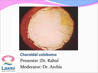
Choroidal coloboma
Presenter :Dr. Rahul
Moderator: Dr. Archis
Coloboma
Coloboma
(kolobomos :Greek for
mutilation)
Indicates “a condition
where a portion of the
structure of the eye is
lacking”
Epidemiology
Incidence is 5 per 1,00,000 newborns
Prevalence is 1 in 10,000
60% are bilateral
Molecular genetics
 deletion in gene which maps to chromosome 7q26.
 a mutation in the PAX6 g
 # Warburg M
J Med Genet. 1993 Aug;30(8):664-9
Types
According to site of
genesis
 Typical
 Atypical
As an isolated
anomaly with or
without
microophthalmia
Unilateral or Bilateral
Phenotypic classification
It may help the clinician to give a systematic
description of the anomalies
Two major classes
Total
Partial
Aetiological classification
• The aetiological classification consists of
three classes:
• Genetic
• Prenatally acquired
• Associations
• The aetiological classification can be
applied to other congenital birth defects
and improves counselling of families #
# Warburg M
J Med Genet. 1993 Aug;30(8):664-9
Classification of microphthalmos and coloboma
Embryonic Origin
The eye is derived
from the neural tube
(neuroectoderm),
from which arise the
retina proper
The neural crest cells
produce the
corneoscleral and
uveal tunics
 From the surface
ectoderm, lens.
The earliest stage of
eye development is the
formation of the
paired optic
vesicles on either side
of the forebrain.
These growing
diverticula expand
laterally into the
mesoderm of the head
and develop a stalk-
like connection to the
main portion of the
rudimentary central
nervous system
In humans, this process begins at about 22 days of
development
 The vesicles continue to grow, their connection to
the brain becomes progressively narrower and more
stalk-like.
This stalks will eventually become the rudiments of
the optic nerves.
There is a seam at the
bottom of each stalk,
where blood vessels
originally run.
This seam is known as
the optic fissure or
choroidal fissure or
embryonic fissure.
The closure starts roughly in the middle of the
developing eye, and runs in both directions.
This process is start at five week & finished by the
seventh week of gestation.
If, for some reason, the closure does not happen, a
uveal coloboma is formed
Depending on
where the closure
did not happen, the
baby can have
 an iris coloboma
(front of the fissure)
 a chorio-retinal
coloboma (back of
the fissure)
 any combination of
these.
Uveal coloboma can
affect one eye
(unilateral) or both
eyes (bilateral)
A uveal coloboma may go from front to back
continuous or have “skip lesions”.
The fact that the seam runs at the bottom of the stalk
is the reason why uveal coloboma is always located in
the lower inside corner of the eye.
Pathophysiology
Fusion or closure of
the fissure begins in its
central portion in the
11-mm embryo and
proceeds anteriorly
and posteriorly.
Closure of the fissure
is complete by the
sixth week (13-mm
stage).
Pathophysiology
In the normal eye, optic fissure closes 33 to 40
days after conception.
Failure of the embryonic fissure to close along the inferonasal
aspect of the optic cup and stalk
That causes Retinal neurosensory elements and RPE precursors
fail to become confluent.
Due to which underlying choroid fails to differentiate
That results in bridge of bare sclera or a cyst formation
Pathophysiology
Anteriorly
defects in the neuroectodermally derived iris pigment
epithelium produce iris coloboma
Posteriorly
The entire optic nerve head may be involved in a
colobomatous malformation
Typical/atypical
Colobomas are called typical if they are located
inferonasally in the region of the fissure
Typical colobomas can involve the iris, ciliary body,
choroid, or any of the three, and also may involve the
optic nerve
Atypical colobomas are not related to closure of the fissure
Multi-system involvement
A single-gene disorder
with multisystem
involvement
Aicardi's syndrome
Lenz microphthalmia
syndrome
Meckel's syndrome
Warburg's syndrome
Aicardi's syndrome
Absence of corpus
callousm
Consist of triad -
Agenesis of the corpus
callosum
 infantile spasms.
morning glory
syndrome
Multisystem disorder
without known genetic
cause
CHARGE association of
anomalies
 colobomatous
Microphthalmos
 heart defects
 choanal atresia
 retarded growth
 genital anomalies
 ear anomalies or deafness
(At least three of the
features are necessary
for the diagnosis)
Iris coloboma
Total
if they involve a whole sector
of the iris up to the ciliary
body“keyhole” defect
Partial
if they do not involve a
whole sector of the iris
may appear as a notch at the
pupillary margin
Histological
Complete coloboma
involves the whole thickness of the iris
Incomplete coloboma
involves either the pigment epithelium or the stroma
Bridge coloboma
mesectodermal tissue forms a pupillary membrane that
stretches across the defect
Lens coloboma
Coloboma of the lens is not a true coloboma.
No lens tissue is missing
An absence of zonular fibers from an underlying
colobomatous ciliary body
Results in a lack of tension on the lens capsule in
that region
Notched equator/ Flattening of the inferior lens
Lens notches can
occur wherever
zonules are absent or
deficient
 zonular rupture
during early surgery
Ida Manns classification(1937)Ida Manns classification(1937)
1-above the optic disc
2-superior border of
optic disc
3-seperated from the
optic disc by normal
narrow area of retina
4-inferior crescent
below the disc
5- isolated gap in the
line of fissure
6-area of pigmentary
disturbance
7-extreme peripheral
coloboma
1)area of pigmentary
disturbance
2) isolated gap in the
line of fissure
3) above the optic disc
Retinal detachment in colobomaRetinal detachment in coloboma
40% develop RD , accounts for 0.5% of RD in young.
May or may not be due to coloboma
Vitreous traction cause breaks outside coloboma
RD due to coloboma is break along edge of coloboma
It is difficult to visualise breaks because they are under
edge or close to haemorrhage
Treatment modalitiesTreatment modalities
 Cryo to anterior margin and photocoagulation to
posterior margin
 Radial buckles (Patnaik et al,1961)-may extend to disc if
large.35-57% success rate
Management of RD with ColobomaManagement of RD with Coloboma
Prophylactic laser delimitation of the coloboma margins
If RD is due to peripheral coloboma-conventional scleral
buckling surgery &Vitrectomy with
endophotocoagualation with silicon oil injection
Management of RD with ColobomaManagement of RD with Coloboma
Lensectomy may be required in cases of difficulty in
visualizing inferior retina
Breaks usually identified
AFE easily done as sclera is concave
Prolonged inferior tamponade needed-silicon oil  gas.
SRNVM in retinochoroidal coloboma
Pathology-absence of choroid with breach in bruch’s
membrane leading to vessels entering sub-retinal space.
Also RPE abnormalities stimulate budding of
choriocapillaris endothelial cells.
Laser photocoagulation is advised modality of treatment.
Cataract in colobomatous eyes
Clinically significant cataract develops at a
younger age compaired with age-related nuclear
sclerotic cataract.
Cataract more likely to be denser.
Cuniform opacities are more likely to occur
earlier.
Increased no. of subluxated lens due to
associated zonuler deficiency
Michael L. Nordlund, Alan Sugar, Sayoko E.
Moroi,
J Cataract Refract Surg 2000; 26:1035-1040
Microophthalmos in colobomatous
eyes
Prevalence 21 per 100,000
82% at least >+7.00D of hypermetropia
Coloboma is the second MC association after cataract
(22% v/s 44%)
Microophthalmos with coloboma may or may not be
associated with microcornea
Mark J etal
Aetiology of severe visual impairment and
blindness in Microophthalmia
BJO 1994(78);332-334
Microophthalmos in colobomatous
eyes
A person with coloboma with microophthalmos
with microcornea has a poorer visual prognosis
than a person with coloboma with
microophthalmos without microcornea
Further a newborn with a corneal diameter<5mm
has a very poor prognosis
Mark J etal
 Aetiology of severe visual impairment
 and blindness in Microophthalmia
 BJO 1994(78);332-334
Microcornea in colobomatous eyes
The term microcornea implies
a corneal diameter of less than 10 mm.
 It thought to occur secondary to an arrest in corneal
growth after the fifth month of fetal development.
It often occurs with ocular abnormalities such as
colobomata and microophthalmos may be present
Microcornea accompanies anterior microphthalmos,
with crowding of the anterior segment structures
Management
Systemic evaluation
Ocular examination.
Investigations
Axial length
Keratometry
IOL Master
USG
UBM
Management
Clinically significant cataract develops at a younger age
If a coloboma involves the macula Axial length will be
varied so earlier refraction will be important or Axial
length may be obtained by locating the preferred fixation
point
Coloboma with microophthalmos with microcornea –
ECCE not a viable option
 The crystalline lens has been reported to be of normal or
slightly larger than normal size
#
Management
The lens:eye volume ratio is greater (10 to 32 percent) in
these eyes compared with emmetropic eyes (3 to 4
percent)
So Phaco-emulsification / manual phaco (SICS) is a
viable option
Phaco-emulsification—e.g. Slit Nucleotomy, Wooden
boot, Petalloid etc.
Manual phaco (SICS) —e.g. Sandwich technique,
Quarters technique, Phacotrisection etc.
 Michael L. Nordlund, Alan Sugar, Sayoko E. Moroi,
 J Cataract Refract Surg 2000; 26:1035-
#
Management
Minimal mydriasis or reactive miosis should be
anticipated
Iris retractors / multiple sphicterotomies
CCC run-off or tear can occur
Recommendations for CCC
Eccentric capsulorhexis
Increased magnification with dye assistance
Oblique illumination
Two stage CCC
Good chamber maintainence with viscoelastic
Nucleus sculpting / Nucleus cracking at opposite meridian + PCIOL@90°
to tear
Endothelial losses
Shell technique can be used
Michael L. Nordlund, Alan Sugar, Sayoko E. Moroi,
 J Cataract Refract Surg 2000; 26:1035-
#
Management
 Anterior vitrectomy
Vitreous loss –
Prolapse through PC rent
Prolapse through colobomatous area without PC rent
 Optimal centeration of the optic with the ectopic pupil to avoid
post-op mono-ocular diplopia
Puppiloplasty may be required
 Avoid silicon material IOLs
As future VR surgery may entail the use of silicon oil
# Michael L. Nordlund etal
Phacoemulsification and intraocular lens placement in
eyes with cataract and congenital coloboma: visual
acuity and complications
J Cataract Refract Surg 2000; 26:1035-1040
Prognosis
The prognosis for vision depended on the phenotype of
the better eye
 Microphthalmos with cyst has the worst prognosis
 Coloboma with microcornea and microphthalmos a poor
prognosis
 Coloboma with only microcornea has an intermediate prognosis
 Simple coloboma has the best prognosis
A corneal diameter <6 mm had a poor visual prognosis
whereas a corneal diameter >10 mm had a good prognosis
Time of surgery
Type of surgery
Hornby SJ, etal
Ophthalmology. 2000 Mar;107(3):511-20
Visual acuity in children with coloboma
Michael L. Nordlund, Alan Sugar,
Sayoko E. Moroi,
J Cataract Refract Surg 2000; 26:1035-
1040
Choroidal coloboma

Más contenido relacionado

La actualidad más candente

Angle recession glaucoma
Angle recession glaucomaAngle recession glaucoma
Angle recession glaucomaSSSIHMS-PG
 
FUNDUS FLUORESCEIN ANGIOGRAPHY
FUNDUS FLUORESCEIN ANGIOGRAPHYFUNDUS FLUORESCEIN ANGIOGRAPHY
FUNDUS FLUORESCEIN ANGIOGRAPHYAnuraag Singh
 
Pigmentary glaucoma - Dr Shylesh B Dabke
Pigmentary glaucoma - Dr Shylesh B DabkePigmentary glaucoma - Dr Shylesh B Dabke
Pigmentary glaucoma - Dr Shylesh B DabkeShylesh Dabke
 
Central retinal artery occlusion
Central retinal artery occlusionCentral retinal artery occlusion
Central retinal artery occlusionSSSIHMS-PG
 
Branch retinal vein occlusion (BRVO)
Branch retinal vein occlusion (BRVO)Branch retinal vein occlusion (BRVO)
Branch retinal vein occlusion (BRVO)NIKHIL GOTMARE
 
Keratoplasty
Keratoplasty Keratoplasty
Keratoplasty Siva G
 
Pseudoexfoliation syndrome
Pseudoexfoliation syndromePseudoexfoliation syndrome
Pseudoexfoliation syndromeGloria George
 
Vitreous substitutes
Vitreous substitutesVitreous substitutes
Vitreous substitutesSSSIHMS-PG
 
Neovascular glaucoma
Neovascular glaucomaNeovascular glaucoma
Neovascular glaucomaSSSIHMS-PG
 
Penetrating keratoplasty
Penetrating keratoplastyPenetrating keratoplasty
Penetrating keratoplastyNikita Jaiswal
 
Lamellar keratoplasty
Lamellar keratoplastyLamellar keratoplasty
Lamellar keratoplastyAkshay Nayak
 
Premium intraocular lenses The past, present and-3.pptx
Premium intraocular lenses The past, present and-3.pptxPremium intraocular lenses The past, present and-3.pptx
Premium intraocular lenses The past, present and-3.pptxMushtaq Ahmad
 
Esotropia , classification , diagnosis and management
Esotropia , classification , diagnosis and managementEsotropia , classification , diagnosis and management
Esotropia , classification , diagnosis and managementDrAzmat Ali
 
Optic nerve head evaluation in glaucoma
Optic nerve head evaluation in glaucomaOptic nerve head evaluation in glaucoma
Optic nerve head evaluation in glaucomaDr Laltanpuia Chhangte
 
Corneal degenerations
Corneal degenerationsCorneal degenerations
Corneal degenerationsdrkvasantha
 

La actualidad más candente (20)

Angle recession glaucoma
Angle recession glaucomaAngle recession glaucoma
Angle recession glaucoma
 
FUNDUS FLUORESCEIN ANGIOGRAPHY
FUNDUS FLUORESCEIN ANGIOGRAPHYFUNDUS FLUORESCEIN ANGIOGRAPHY
FUNDUS FLUORESCEIN ANGIOGRAPHY
 
Keratoconus
KeratoconusKeratoconus
Keratoconus
 
Pigmentary glaucoma - Dr Shylesh B Dabke
Pigmentary glaucoma - Dr Shylesh B DabkePigmentary glaucoma - Dr Shylesh B Dabke
Pigmentary glaucoma - Dr Shylesh B Dabke
 
Central retinal artery occlusion
Central retinal artery occlusionCentral retinal artery occlusion
Central retinal artery occlusion
 
Branch retinal vein occlusion (BRVO)
Branch retinal vein occlusion (BRVO)Branch retinal vein occlusion (BRVO)
Branch retinal vein occlusion (BRVO)
 
Keratoplasty
Keratoplasty Keratoplasty
Keratoplasty
 
Anterior vitrectomy
Anterior vitrectomyAnterior vitrectomy
Anterior vitrectomy
 
Pseudoexfoliation syndrome
Pseudoexfoliation syndromePseudoexfoliation syndrome
Pseudoexfoliation syndrome
 
Vitreous substitutes
Vitreous substitutesVitreous substitutes
Vitreous substitutes
 
Aphakia
AphakiaAphakia
Aphakia
 
Neovascular glaucoma
Neovascular glaucomaNeovascular glaucoma
Neovascular glaucoma
 
Penetrating keratoplasty
Penetrating keratoplastyPenetrating keratoplasty
Penetrating keratoplasty
 
Lamellar keratoplasty
Lamellar keratoplastyLamellar keratoplasty
Lamellar keratoplasty
 
Macular hole
Macular holeMacular hole
Macular hole
 
Premium intraocular lenses The past, present and-3.pptx
Premium intraocular lenses The past, present and-3.pptxPremium intraocular lenses The past, present and-3.pptx
Premium intraocular lenses The past, present and-3.pptx
 
Esotropia , classification , diagnosis and management
Esotropia , classification , diagnosis and managementEsotropia , classification , diagnosis and management
Esotropia , classification , diagnosis and management
 
Iridodialysis repair
Iridodialysis repair  Iridodialysis repair
Iridodialysis repair
 
Optic nerve head evaluation in glaucoma
Optic nerve head evaluation in glaucomaOptic nerve head evaluation in glaucoma
Optic nerve head evaluation in glaucoma
 
Corneal degenerations
Corneal degenerationsCorneal degenerations
Corneal degenerations
 

Similar a Choroidal coloboma

Congenital corneal anomalies
Congenital corneal anomaliesCongenital corneal anomalies
Congenital corneal anomaliesSocrates Narvaez
 
Approach to cases of congenital glaucoma, developmental
Approach to cases of congenital glaucoma, developmentalApproach to cases of congenital glaucoma, developmental
Approach to cases of congenital glaucoma, developmentalBipin Bista
 
Approach to cases of congenital glaucoma, developmental
Approach to cases of congenital glaucoma, developmentalApproach to cases of congenital glaucoma, developmental
Approach to cases of congenital glaucoma, developmentalShahrukh Kc
 
Glaucoma and its classifications.pptx
Glaucoma and its classifications.pptxGlaucoma and its classifications.pptx
Glaucoma and its classifications.pptxAnamSehreen
 
Glaucoma and its classifications.pptx
Glaucoma and its classifications.pptxGlaucoma and its classifications.pptx
Glaucoma and its classifications.pptxAnamSehreen3
 
Congenital Glaucoma
Congenital GlaucomaCongenital Glaucoma
Congenital GlaucomaSahil Thakur
 
10-subluxated-lens.ppt
10-subluxated-lens.ppt10-subluxated-lens.ppt
10-subluxated-lens.pptAdel930879
 
glaucoma.pptx
glaucoma.pptxglaucoma.pptx
glaucoma.pptxEEPD1
 
Congenital glaucoma.pptx
Congenital glaucoma.pptxCongenital glaucoma.pptx
Congenital glaucoma.pptxdratulkranand
 
Evaluation and management of epiretinal membranes
Evaluation and management of epiretinal membranesEvaluation and management of epiretinal membranes
Evaluation and management of epiretinal membranesrajahamayun1
 
congenital glaucoma and congenital cataract
congenital glaucoma and congenital cataractcongenital glaucoma and congenital cataract
congenital glaucoma and congenital cataractRegina Lr
 
Pseudoexfoliative syndrome and pigment dispersion syndrome and glaucoma
Pseudoexfoliative syndrome and pigment dispersion syndrome and glaucomaPseudoexfoliative syndrome and pigment dispersion syndrome and glaucoma
Pseudoexfoliative syndrome and pigment dispersion syndrome and glaucomaBipin Bista
 
Congenital optic disc anomalies
Congenital optic disc anomaliesCongenital optic disc anomalies
Congenital optic disc anomaliesJagdish Dukre
 
Cell and Tissue Final Report
Cell and Tissue Final ReportCell and Tissue Final Report
Cell and Tissue Final ReportKelsey Henderson
 
Congenital cataract
Congenital cataractCongenital cataract
Congenital cataractTanvi Gupta
 
Epiretinal membrane and vitreomacula traction in updates by Panit Cherdchu, MD.
Epiretinal membrane and vitreomacula traction in updates by Panit Cherdchu, MD.Epiretinal membrane and vitreomacula traction in updates by Panit Cherdchu, MD.
Epiretinal membrane and vitreomacula traction in updates by Panit Cherdchu, MD.Panit Cherdchu
 

Similar a Choroidal coloboma (20)

Childhood gaucoma 2
Childhood gaucoma 2Childhood gaucoma 2
Childhood gaucoma 2
 
Congenital corneal anomalies
Congenital corneal anomaliesCongenital corneal anomalies
Congenital corneal anomalies
 
Approach to cases of congenital glaucoma, developmental
Approach to cases of congenital glaucoma, developmentalApproach to cases of congenital glaucoma, developmental
Approach to cases of congenital glaucoma, developmental
 
Approach to cases of congenital glaucoma, developmental
Approach to cases of congenital glaucoma, developmentalApproach to cases of congenital glaucoma, developmental
Approach to cases of congenital glaucoma, developmental
 
Glaucoma and its classifications.pptx
Glaucoma and its classifications.pptxGlaucoma and its classifications.pptx
Glaucoma and its classifications.pptx
 
Glaucoma and its classifications.pptx
Glaucoma and its classifications.pptxGlaucoma and its classifications.pptx
Glaucoma and its classifications.pptx
 
Congenital Glaucoma
Congenital GlaucomaCongenital Glaucoma
Congenital Glaucoma
 
Congenital cataract
Congenital cataractCongenital cataract
Congenital cataract
 
10-subluxated-lens.ppt
10-subluxated-lens.ppt10-subluxated-lens.ppt
10-subluxated-lens.ppt
 
glaucoma.pptx
glaucoma.pptxglaucoma.pptx
glaucoma.pptx
 
Congenital glaucoma.pptx
Congenital glaucoma.pptxCongenital glaucoma.pptx
Congenital glaucoma.pptx
 
Evaluation and management of epiretinal membranes
Evaluation and management of epiretinal membranesEvaluation and management of epiretinal membranes
Evaluation and management of epiretinal membranes
 
congenital glaucoma and congenital cataract
congenital glaucoma and congenital cataractcongenital glaucoma and congenital cataract
congenital glaucoma and congenital cataract
 
Pseudoexfoliative syndrome and pigment dispersion syndrome and glaucoma
Pseudoexfoliative syndrome and pigment dispersion syndrome and glaucomaPseudoexfoliative syndrome and pigment dispersion syndrome and glaucoma
Pseudoexfoliative syndrome and pigment dispersion syndrome and glaucoma
 
pseudoexfoliative glaucoma
pseudoexfoliative glaucomapseudoexfoliative glaucoma
pseudoexfoliative glaucoma
 
Congenital optic disc anomalies
Congenital optic disc anomaliesCongenital optic disc anomalies
Congenital optic disc anomalies
 
Cell and Tissue Final Report
Cell and Tissue Final ReportCell and Tissue Final Report
Cell and Tissue Final Report
 
Congenital glaucoma
Congenital glaucomaCongenital glaucoma
Congenital glaucoma
 
Congenital cataract
Congenital cataractCongenital cataract
Congenital cataract
 
Epiretinal membrane and vitreomacula traction in updates by Panit Cherdchu, MD.
Epiretinal membrane and vitreomacula traction in updates by Panit Cherdchu, MD.Epiretinal membrane and vitreomacula traction in updates by Panit Cherdchu, MD.
Epiretinal membrane and vitreomacula traction in updates by Panit Cherdchu, MD.
 

Más de Laxmi Eye Institute (20)

Important trials in Glaucoma
Important trials in GlaucomaImportant trials in Glaucoma
Important trials in Glaucoma
 
Congenital glaucoma
Congenital glaucomaCongenital glaucoma
Congenital glaucoma
 
Ocular pharmacology
Ocular pharmacologyOcular pharmacology
Ocular pharmacology
 
Supranuclear pathways and lesions
Supranuclear pathways and lesionsSupranuclear pathways and lesions
Supranuclear pathways and lesions
 
IOL power calculation special situations
IOL power calculation special situations IOL power calculation special situations
IOL power calculation special situations
 
Corneal dystrophy
Corneal dystrophy Corneal dystrophy
Corneal dystrophy
 
Ice syndrome
Ice syndromeIce syndrome
Ice syndrome
 
Scleritis a case presentation
Scleritis a case presentationScleritis a case presentation
Scleritis a case presentation
 
Visual pathway
Visual pathway Visual pathway
Visual pathway
 
CCP
CCPCCP
CCP
 
Ocular tb
Ocular tbOcular tb
Ocular tb
 
Causes of low vision in adult
Causes of low vision in adultCauses of low vision in adult
Causes of low vision in adult
 
Trial set
Trial setTrial set
Trial set
 
ASSESMENT OF VISUAL ACUITY IN CHILDREN
ASSESMENT OF VISUAL ACUITY IN CHILDRENASSESMENT OF VISUAL ACUITY IN CHILDREN
ASSESMENT OF VISUAL ACUITY IN CHILDREN
 
INTRAOCULAR FOREIGN BODY
INTRAOCULAR FOREIGN BODYINTRAOCULAR FOREIGN BODY
INTRAOCULAR FOREIGN BODY
 
VITAMIN A & VISUAL CYCLE
VITAMIN A & VISUAL CYCLEVITAMIN A & VISUAL CYCLE
VITAMIN A & VISUAL CYCLE
 
Malignant glaucoma
Malignant glaucomaMalignant glaucoma
Malignant glaucoma
 
Uveitic Glaucoma
Uveitic GlaucomaUveitic Glaucoma
Uveitic Glaucoma
 
Central Retinal Vein OcclUsIon (CRUISE) Study - Cruise trial
Central Retinal  Vein OcclUsIon (CRUISE) Study - Cruise trialCentral Retinal  Vein OcclUsIon (CRUISE) Study - Cruise trial
Central Retinal Vein OcclUsIon (CRUISE) Study - Cruise trial
 
Colour vision and its clinical aspects
Colour vision and its clinical aspectsColour vision and its clinical aspects
Colour vision and its clinical aspects
 

Último

Digital Healthcare: The Future of Medical Consultations
Digital Healthcare: The Future of Medical ConsultationsDigital Healthcare: The Future of Medical Consultations
Digital Healthcare: The Future of Medical Consultationssmartcare
 
The Pfizer testimony by Melissa Mac Atee
The Pfizer testimony by Melissa Mac AteeThe Pfizer testimony by Melissa Mac Atee
The Pfizer testimony by Melissa Mac AteePascalGuyot6
 
Navigating Telehealth Innovations with PrudentRx
Navigating Telehealth Innovations with PrudentRxNavigating Telehealth Innovations with PrudentRx
Navigating Telehealth Innovations with PrudentRxPrudentRx Program
 
Valle Egypt Illustrates Consequences of Financial Elder Abuse
Valle Egypt Illustrates Consequences of Financial Elder AbuseValle Egypt Illustrates Consequences of Financial Elder Abuse
Valle Egypt Illustrates Consequences of Financial Elder AbuseKristin Hetzer
 
Improve Patient Care with Medical Record Abstraction
Improve Patient Care with Medical Record AbstractionImprove Patient Care with Medical Record Abstraction
Improve Patient Care with Medical Record AbstractionCardiac Registry Support
 
Master the Art of Yoga with Joga Yoga Training
Master the Art of Yoga with Joga Yoga TrainingMaster the Art of Yoga with Joga Yoga Training
Master the Art of Yoga with Joga Yoga TrainingJoga Yoga Training
 
Management of heart failure 23.02.24.pptx
Management of heart failure 23.02.24.pptxManagement of heart failure 23.02.24.pptx
Management of heart failure 23.02.24.pptxchristina388422
 
Breaking Down Oppositional Defiant Disorder Treatments
Breaking Down Oppositional Defiant Disorder TreatmentsBreaking Down Oppositional Defiant Disorder Treatments
Breaking Down Oppositional Defiant Disorder TreatmentsOppositional Defiant Disorder
 
Virtual Health Platforms_ Revolutionizing Patient Care.pdf
Virtual Health Platforms_ Revolutionizing Patient Care.pdfVirtual Health Platforms_ Revolutionizing Patient Care.pdf
Virtual Health Platforms_ Revolutionizing Patient Care.pdfsmartcare
 
The Best Foot and Ankle Center of Arizona
The Best Foot and Ankle Center of ArizonaThe Best Foot and Ankle Center of Arizona
The Best Foot and Ankle Center of Arizonajackjohn60
 
Cell structure slideshare.pptx Unlocking the Secrets of Cells: Structure, Fun...
Cell structure slideshare.pptx Unlocking the Secrets of Cells: Structure, Fun...Cell structure slideshare.pptx Unlocking the Secrets of Cells: Structure, Fun...
Cell structure slideshare.pptx Unlocking the Secrets of Cells: Structure, Fun...ananyagirishbabu1
 
Sugar Medicine_ Natural Homeopathy Remedies for Blood Sugar Management.pdf
Sugar Medicine_ Natural Homeopathy Remedies for Blood Sugar Management.pdfSugar Medicine_ Natural Homeopathy Remedies for Blood Sugar Management.pdf
Sugar Medicine_ Natural Homeopathy Remedies for Blood Sugar Management.pdfDharma Homoeopathy
 
Notify ME 89O1183OO2 #cALL# #gIRLS# In Chhattisgarh By Chhattisgarh #ℂall #gI...
Notify ME 89O1183OO2 #cALL# #gIRLS# In Chhattisgarh By Chhattisgarh #ℂall #gI...Notify ME 89O1183OO2 #cALL# #gIRLS# In Chhattisgarh By Chhattisgarh #ℂall #gI...
Notify ME 89O1183OO2 #cALL# #gIRLS# In Chhattisgarh By Chhattisgarh #ℂall #gI...aunty1x1
 
Mental Health Startup Pitch Deck Presentation
Mental Health Startup Pitch Deck PresentationMental Health Startup Pitch Deck Presentation
Mental Health Startup Pitch Deck PresentationStartupSprouts.in
 
For Better Jaipur #ℂall #Girl Service ❤89011-83002❤ Jaipur #ℂall #Girls
For Better Jaipur #ℂall #Girl Service ❤89011-83002❤ Jaipur #ℂall #Girls For Better Jaipur #ℂall #Girl Service ❤89011-83002❤ Jaipur #ℂall #Girls
For Better Jaipur #ℂall #Girl Service ❤89011-83002❤ Jaipur #ℂall #Girls aunty1x1
 
Jaipur @ℂall @Girls ꧁❤8901183002❤꧂@ℂall @Girls Service Vip Top Model Safe
Jaipur @ℂall @Girls ꧁❤8901183002❤꧂@ℂall @Girls Service Vip Top Model SafeJaipur @ℂall @Girls ꧁❤8901183002❤꧂@ℂall @Girls Service Vip Top Model Safe
Jaipur @ℂall @Girls ꧁❤8901183002❤꧂@ℂall @Girls Service Vip Top Model Safeaunty1x1
 
Phone Us ❤89011-83002❤ #ℂall #gIRLS In Jaipur By Jaipur @ℂall @Girls Hotel ...
 Phone Us  ❤89011-83002❤ #ℂall #gIRLS In Jaipur By Jaipur @ℂall @Girls Hotel ... Phone Us  ❤89011-83002❤ #ℂall #gIRLS In Jaipur By Jaipur @ℂall @Girls Hotel ...
Phone Us ❤89011-83002❤ #ℂall #gIRLS In Jaipur By Jaipur @ℂall @Girls Hotel ...aunty1x1
 
Renal Replacement Therapy - ICU Guidlines
Renal Replacement  Therapy - ICU GuidlinesRenal Replacement  Therapy - ICU Guidlines
Renal Replacement Therapy - ICU GuidlinesLikhithaNalluru
 
Jaipur #ℂall #gIRLS Oyo Hotel 89O1183OO2 #ℂall #gIRL in Jaipur
Jaipur #ℂall #gIRLS Oyo Hotel 89O1183OO2 #ℂall #gIRL in Jaipur Jaipur #ℂall #gIRLS Oyo Hotel 89O1183OO2 #ℂall #gIRL in Jaipur
Jaipur #ℂall #gIRLS Oyo Hotel 89O1183OO2 #ℂall #gIRL in Jaipur aunty1x1
 
#cALL# #gIRLS# In Chhattisgarh ꧁❤8901183002❤꧂#cALL# #gIRLS# Service In Chhatt...
#cALL# #gIRLS# In Chhattisgarh ꧁❤8901183002❤꧂#cALL# #gIRLS# Service In Chhatt...#cALL# #gIRLS# In Chhattisgarh ꧁❤8901183002❤꧂#cALL# #gIRLS# Service In Chhatt...
#cALL# #gIRLS# In Chhattisgarh ꧁❤8901183002❤꧂#cALL# #gIRLS# Service In Chhatt...aunty1x1
 

Último (20)

Digital Healthcare: The Future of Medical Consultations
Digital Healthcare: The Future of Medical ConsultationsDigital Healthcare: The Future of Medical Consultations
Digital Healthcare: The Future of Medical Consultations
 
The Pfizer testimony by Melissa Mac Atee
The Pfizer testimony by Melissa Mac AteeThe Pfizer testimony by Melissa Mac Atee
The Pfizer testimony by Melissa Mac Atee
 
Navigating Telehealth Innovations with PrudentRx
Navigating Telehealth Innovations with PrudentRxNavigating Telehealth Innovations with PrudentRx
Navigating Telehealth Innovations with PrudentRx
 
Valle Egypt Illustrates Consequences of Financial Elder Abuse
Valle Egypt Illustrates Consequences of Financial Elder AbuseValle Egypt Illustrates Consequences of Financial Elder Abuse
Valle Egypt Illustrates Consequences of Financial Elder Abuse
 
Improve Patient Care with Medical Record Abstraction
Improve Patient Care with Medical Record AbstractionImprove Patient Care with Medical Record Abstraction
Improve Patient Care with Medical Record Abstraction
 
Master the Art of Yoga with Joga Yoga Training
Master the Art of Yoga with Joga Yoga TrainingMaster the Art of Yoga with Joga Yoga Training
Master the Art of Yoga with Joga Yoga Training
 
Management of heart failure 23.02.24.pptx
Management of heart failure 23.02.24.pptxManagement of heart failure 23.02.24.pptx
Management of heart failure 23.02.24.pptx
 
Breaking Down Oppositional Defiant Disorder Treatments
Breaking Down Oppositional Defiant Disorder TreatmentsBreaking Down Oppositional Defiant Disorder Treatments
Breaking Down Oppositional Defiant Disorder Treatments
 
Virtual Health Platforms_ Revolutionizing Patient Care.pdf
Virtual Health Platforms_ Revolutionizing Patient Care.pdfVirtual Health Platforms_ Revolutionizing Patient Care.pdf
Virtual Health Platforms_ Revolutionizing Patient Care.pdf
 
The Best Foot and Ankle Center of Arizona
The Best Foot and Ankle Center of ArizonaThe Best Foot and Ankle Center of Arizona
The Best Foot and Ankle Center of Arizona
 
Cell structure slideshare.pptx Unlocking the Secrets of Cells: Structure, Fun...
Cell structure slideshare.pptx Unlocking the Secrets of Cells: Structure, Fun...Cell structure slideshare.pptx Unlocking the Secrets of Cells: Structure, Fun...
Cell structure slideshare.pptx Unlocking the Secrets of Cells: Structure, Fun...
 
Sugar Medicine_ Natural Homeopathy Remedies for Blood Sugar Management.pdf
Sugar Medicine_ Natural Homeopathy Remedies for Blood Sugar Management.pdfSugar Medicine_ Natural Homeopathy Remedies for Blood Sugar Management.pdf
Sugar Medicine_ Natural Homeopathy Remedies for Blood Sugar Management.pdf
 
Notify ME 89O1183OO2 #cALL# #gIRLS# In Chhattisgarh By Chhattisgarh #ℂall #gI...
Notify ME 89O1183OO2 #cALL# #gIRLS# In Chhattisgarh By Chhattisgarh #ℂall #gI...Notify ME 89O1183OO2 #cALL# #gIRLS# In Chhattisgarh By Chhattisgarh #ℂall #gI...
Notify ME 89O1183OO2 #cALL# #gIRLS# In Chhattisgarh By Chhattisgarh #ℂall #gI...
 
Mental Health Startup Pitch Deck Presentation
Mental Health Startup Pitch Deck PresentationMental Health Startup Pitch Deck Presentation
Mental Health Startup Pitch Deck Presentation
 
For Better Jaipur #ℂall #Girl Service ❤89011-83002❤ Jaipur #ℂall #Girls
For Better Jaipur #ℂall #Girl Service ❤89011-83002❤ Jaipur #ℂall #Girls For Better Jaipur #ℂall #Girl Service ❤89011-83002❤ Jaipur #ℂall #Girls
For Better Jaipur #ℂall #Girl Service ❤89011-83002❤ Jaipur #ℂall #Girls
 
Jaipur @ℂall @Girls ꧁❤8901183002❤꧂@ℂall @Girls Service Vip Top Model Safe
Jaipur @ℂall @Girls ꧁❤8901183002❤꧂@ℂall @Girls Service Vip Top Model SafeJaipur @ℂall @Girls ꧁❤8901183002❤꧂@ℂall @Girls Service Vip Top Model Safe
Jaipur @ℂall @Girls ꧁❤8901183002❤꧂@ℂall @Girls Service Vip Top Model Safe
 
Phone Us ❤89011-83002❤ #ℂall #gIRLS In Jaipur By Jaipur @ℂall @Girls Hotel ...
 Phone Us  ❤89011-83002❤ #ℂall #gIRLS In Jaipur By Jaipur @ℂall @Girls Hotel ... Phone Us  ❤89011-83002❤ #ℂall #gIRLS In Jaipur By Jaipur @ℂall @Girls Hotel ...
Phone Us ❤89011-83002❤ #ℂall #gIRLS In Jaipur By Jaipur @ℂall @Girls Hotel ...
 
Renal Replacement Therapy - ICU Guidlines
Renal Replacement  Therapy - ICU GuidlinesRenal Replacement  Therapy - ICU Guidlines
Renal Replacement Therapy - ICU Guidlines
 
Jaipur #ℂall #gIRLS Oyo Hotel 89O1183OO2 #ℂall #gIRL in Jaipur
Jaipur #ℂall #gIRLS Oyo Hotel 89O1183OO2 #ℂall #gIRL in Jaipur Jaipur #ℂall #gIRLS Oyo Hotel 89O1183OO2 #ℂall #gIRL in Jaipur
Jaipur #ℂall #gIRLS Oyo Hotel 89O1183OO2 #ℂall #gIRL in Jaipur
 
#cALL# #gIRLS# In Chhattisgarh ꧁❤8901183002❤꧂#cALL# #gIRLS# Service In Chhatt...
#cALL# #gIRLS# In Chhattisgarh ꧁❤8901183002❤꧂#cALL# #gIRLS# Service In Chhatt...#cALL# #gIRLS# In Chhattisgarh ꧁❤8901183002❤꧂#cALL# #gIRLS# Service In Chhatt...
#cALL# #gIRLS# In Chhattisgarh ꧁❤8901183002❤꧂#cALL# #gIRLS# Service In Chhatt...
 

Choroidal coloboma

  • 1. Choroidal coloboma Presenter :Dr. Rahul Moderator: Dr. Archis
  • 2. Coloboma Coloboma (kolobomos :Greek for mutilation) Indicates “a condition where a portion of the structure of the eye is lacking”
  • 3. Epidemiology Incidence is 5 per 1,00,000 newborns Prevalence is 1 in 10,000 60% are bilateral Molecular genetics  deletion in gene which maps to chromosome 7q26.  a mutation in the PAX6 g  # Warburg M J Med Genet. 1993 Aug;30(8):664-9
  • 4. Types According to site of genesis  Typical  Atypical As an isolated anomaly with or without microophthalmia Unilateral or Bilateral
  • 5. Phenotypic classification It may help the clinician to give a systematic description of the anomalies Two major classes Total Partial
  • 6. Aetiological classification • The aetiological classification consists of three classes: • Genetic • Prenatally acquired • Associations • The aetiological classification can be applied to other congenital birth defects and improves counselling of families # # Warburg M J Med Genet. 1993 Aug;30(8):664-9 Classification of microphthalmos and coloboma
  • 7. Embryonic Origin The eye is derived from the neural tube (neuroectoderm), from which arise the retina proper The neural crest cells produce the corneoscleral and uveal tunics  From the surface ectoderm, lens.
  • 8. The earliest stage of eye development is the formation of the paired optic vesicles on either side of the forebrain.
  • 9. These growing diverticula expand laterally into the mesoderm of the head and develop a stalk- like connection to the main portion of the rudimentary central nervous system
  • 10. In humans, this process begins at about 22 days of development  The vesicles continue to grow, their connection to the brain becomes progressively narrower and more stalk-like. This stalks will eventually become the rudiments of the optic nerves.
  • 11. There is a seam at the bottom of each stalk, where blood vessels originally run. This seam is known as the optic fissure or choroidal fissure or embryonic fissure.
  • 12. The closure starts roughly in the middle of the developing eye, and runs in both directions. This process is start at five week & finished by the seventh week of gestation. If, for some reason, the closure does not happen, a uveal coloboma is formed
  • 13. Depending on where the closure did not happen, the baby can have  an iris coloboma (front of the fissure)  a chorio-retinal coloboma (back of the fissure)  any combination of these.
  • 14. Uveal coloboma can affect one eye (unilateral) or both eyes (bilateral)
  • 15. A uveal coloboma may go from front to back continuous or have “skip lesions”. The fact that the seam runs at the bottom of the stalk is the reason why uveal coloboma is always located in the lower inside corner of the eye.
  • 16. Pathophysiology Fusion or closure of the fissure begins in its central portion in the 11-mm embryo and proceeds anteriorly and posteriorly. Closure of the fissure is complete by the sixth week (13-mm stage).
  • 17. Pathophysiology In the normal eye, optic fissure closes 33 to 40 days after conception. Failure of the embryonic fissure to close along the inferonasal aspect of the optic cup and stalk That causes Retinal neurosensory elements and RPE precursors fail to become confluent. Due to which underlying choroid fails to differentiate That results in bridge of bare sclera or a cyst formation
  • 18. Pathophysiology Anteriorly defects in the neuroectodermally derived iris pigment epithelium produce iris coloboma Posteriorly The entire optic nerve head may be involved in a colobomatous malformation
  • 19. Typical/atypical Colobomas are called typical if they are located inferonasally in the region of the fissure Typical colobomas can involve the iris, ciliary body, choroid, or any of the three, and also may involve the optic nerve Atypical colobomas are not related to closure of the fissure
  • 20. Multi-system involvement A single-gene disorder with multisystem involvement Aicardi's syndrome Lenz microphthalmia syndrome Meckel's syndrome Warburg's syndrome
  • 21. Aicardi's syndrome Absence of corpus callousm Consist of triad - Agenesis of the corpus callosum  infantile spasms. morning glory syndrome
  • 22. Multisystem disorder without known genetic cause CHARGE association of anomalies  colobomatous Microphthalmos  heart defects  choanal atresia  retarded growth  genital anomalies  ear anomalies or deafness (At least three of the features are necessary for the diagnosis)
  • 23. Iris coloboma Total if they involve a whole sector of the iris up to the ciliary body“keyhole” defect Partial if they do not involve a whole sector of the iris may appear as a notch at the pupillary margin
  • 24. Histological Complete coloboma involves the whole thickness of the iris Incomplete coloboma involves either the pigment epithelium or the stroma Bridge coloboma mesectodermal tissue forms a pupillary membrane that stretches across the defect
  • 25. Lens coloboma Coloboma of the lens is not a true coloboma. No lens tissue is missing An absence of zonular fibers from an underlying colobomatous ciliary body Results in a lack of tension on the lens capsule in that region Notched equator/ Flattening of the inferior lens
  • 26. Lens notches can occur wherever zonules are absent or deficient  zonular rupture during early surgery
  • 27. Ida Manns classification(1937)Ida Manns classification(1937) 1-above the optic disc 2-superior border of optic disc 3-seperated from the optic disc by normal narrow area of retina 4-inferior crescent below the disc 5- isolated gap in the line of fissure 6-area of pigmentary disturbance 7-extreme peripheral coloboma
  • 28. 1)area of pigmentary disturbance 2) isolated gap in the line of fissure 3) above the optic disc
  • 29. Retinal detachment in colobomaRetinal detachment in coloboma 40% develop RD , accounts for 0.5% of RD in young. May or may not be due to coloboma Vitreous traction cause breaks outside coloboma
  • 30. RD due to coloboma is break along edge of coloboma It is difficult to visualise breaks because they are under edge or close to haemorrhage
  • 31. Treatment modalitiesTreatment modalities  Cryo to anterior margin and photocoagulation to posterior margin  Radial buckles (Patnaik et al,1961)-may extend to disc if large.35-57% success rate
  • 32. Management of RD with ColobomaManagement of RD with Coloboma Prophylactic laser delimitation of the coloboma margins If RD is due to peripheral coloboma-conventional scleral buckling surgery &Vitrectomy with endophotocoagualation with silicon oil injection
  • 33. Management of RD with ColobomaManagement of RD with Coloboma Lensectomy may be required in cases of difficulty in visualizing inferior retina Breaks usually identified AFE easily done as sclera is concave Prolonged inferior tamponade needed-silicon oil gas.
  • 34.
  • 35. SRNVM in retinochoroidal coloboma Pathology-absence of choroid with breach in bruch’s membrane leading to vessels entering sub-retinal space. Also RPE abnormalities stimulate budding of choriocapillaris endothelial cells. Laser photocoagulation is advised modality of treatment.
  • 36. Cataract in colobomatous eyes Clinically significant cataract develops at a younger age compaired with age-related nuclear sclerotic cataract. Cataract more likely to be denser. Cuniform opacities are more likely to occur earlier. Increased no. of subluxated lens due to associated zonuler deficiency Michael L. Nordlund, Alan Sugar, Sayoko E. Moroi, J Cataract Refract Surg 2000; 26:1035-1040
  • 37. Microophthalmos in colobomatous eyes Prevalence 21 per 100,000 82% at least >+7.00D of hypermetropia Coloboma is the second MC association after cataract (22% v/s 44%) Microophthalmos with coloboma may or may not be associated with microcornea Mark J etal Aetiology of severe visual impairment and blindness in Microophthalmia BJO 1994(78);332-334
  • 38. Microophthalmos in colobomatous eyes A person with coloboma with microophthalmos with microcornea has a poorer visual prognosis than a person with coloboma with microophthalmos without microcornea Further a newborn with a corneal diameter<5mm has a very poor prognosis Mark J etal  Aetiology of severe visual impairment  and blindness in Microophthalmia  BJO 1994(78);332-334
  • 39.
  • 40. Microcornea in colobomatous eyes The term microcornea implies a corneal diameter of less than 10 mm.  It thought to occur secondary to an arrest in corneal growth after the fifth month of fetal development. It often occurs with ocular abnormalities such as colobomata and microophthalmos may be present Microcornea accompanies anterior microphthalmos, with crowding of the anterior segment structures
  • 42. Management Clinically significant cataract develops at a younger age If a coloboma involves the macula Axial length will be varied so earlier refraction will be important or Axial length may be obtained by locating the preferred fixation point Coloboma with microophthalmos with microcornea – ECCE not a viable option  The crystalline lens has been reported to be of normal or slightly larger than normal size #
  • 43. Management The lens:eye volume ratio is greater (10 to 32 percent) in these eyes compared with emmetropic eyes (3 to 4 percent) So Phaco-emulsification / manual phaco (SICS) is a viable option Phaco-emulsification—e.g. Slit Nucleotomy, Wooden boot, Petalloid etc. Manual phaco (SICS) —e.g. Sandwich technique, Quarters technique, Phacotrisection etc.  Michael L. Nordlund, Alan Sugar, Sayoko E. Moroi,  J Cataract Refract Surg 2000; 26:1035- #
  • 44. Management Minimal mydriasis or reactive miosis should be anticipated Iris retractors / multiple sphicterotomies CCC run-off or tear can occur Recommendations for CCC Eccentric capsulorhexis Increased magnification with dye assistance Oblique illumination Two stage CCC Good chamber maintainence with viscoelastic Nucleus sculpting / Nucleus cracking at opposite meridian + PCIOL@90° to tear Endothelial losses Shell technique can be used Michael L. Nordlund, Alan Sugar, Sayoko E. Moroi,  J Cataract Refract Surg 2000; 26:1035- #
  • 45.
  • 46. Management  Anterior vitrectomy Vitreous loss – Prolapse through PC rent Prolapse through colobomatous area without PC rent  Optimal centeration of the optic with the ectopic pupil to avoid post-op mono-ocular diplopia Puppiloplasty may be required  Avoid silicon material IOLs As future VR surgery may entail the use of silicon oil # Michael L. Nordlund etal Phacoemulsification and intraocular lens placement in eyes with cataract and congenital coloboma: visual acuity and complications J Cataract Refract Surg 2000; 26:1035-1040
  • 47. Prognosis The prognosis for vision depended on the phenotype of the better eye  Microphthalmos with cyst has the worst prognosis  Coloboma with microcornea and microphthalmos a poor prognosis  Coloboma with only microcornea has an intermediate prognosis  Simple coloboma has the best prognosis A corneal diameter <6 mm had a poor visual prognosis whereas a corneal diameter >10 mm had a good prognosis Time of surgery Type of surgery Hornby SJ, etal Ophthalmology. 2000 Mar;107(3):511-20 Visual acuity in children with coloboma Michael L. Nordlund, Alan Sugar, Sayoko E. Moroi, J Cataract Refract Surg 2000; 26:1035- 1040