SlideShare una empresa de Scribd logo
1 de 7
Descargar para leer sin conexión
SOGC                    CLINICAL                               PRACTICE                                 GUIDELINES
                                                                                                                                        No. 130, July 2003


THE USE OF FETAL DOPPLER IN OBSTETRICS
This guideline has been reviewed by the Diagnostic Imaging Committee and approved by
Executive and Council of the Society of Obstetricians and Gynaecologists of Canada.


 PRINCIPAL AUTHORS
 Robert Gagnon, MD, FRCSC, London ON
 Michiel Van den Hof, MD, FRCSC, Halifax NS


 DIAGNOSTIC IMAGING COMMITTEE
 Michiel Van den Hof (Chair), MD, FRCSC, Halifax NS
 Stephen Bly, PhD, Ottawa ON
 Duncan Farquharson, MD, FRCSC,Vancouver BC
 Robert Gagnon, MD, FRCSC, London ON
 Barbara Lewthwaite, MN,Winnipeg MB
 Lucie Morin, MD, FRCSC, Montreal QC
 Shia Salem, MD, FRCP,Toronto ON (Canadian Association of Radiologists Representative)
 Amanda Skoll, MD, FRCSC,Vancouver BC


Abstract                                                                          2. Depending on other clinical factors, reduced, absent, or reversed
Objective: To develop national guidelines on the use of fetal                        umbilical artery end-diastolic flow is an indication for enhanced
   Doppler in obstetrics.                                                            fetal surveillance or delivery. If delivery is delayed to enhance
Options: Whether umbilical cord artery, umbilical cord venous,                       fetal lung maturity with maternal administration of glucocorti-
   ductus venosus, and middle cerebral artery Doppler are use-                       coid, intensive fetal surveillance until delivery is suggested for
   ful in assessing fetal health.                                                    those fetuses with reversed end-diastolic flow. (II-1B)
Outcome: Prediction of adverse perinatal outcome or predic-                       3. Umbilical artery Doppler should not be used as a screening
   tion of fetal anemia.                                                             tool in healthy pregnancies, as it has not been shown to be of
Evidence: MEDLINE search and review of bibliographies in iden-                       value in this group. (I-A)
   tified articles.                                                               4. Umbilical venous double pulsations, in the presence of abnor-
Values: The evidence was reviewed by the Diagnostic Imaging                          mal umbilical artery Doppler waveforms, necessitate a detailed
   Committee and the principal authors. A quality of evidence                        assessment of fetal health status. (II-3B)
   assessment was undertaken as outlined in the report of the                     5. Measurement of the fetal middle cerebral artery Doppler peak
   Canadian Task Force on the Periodic Health Examination.                           systolic flow velocity is a predictor of moderate or severe fetal
Benefits, harms, and costs: Intrauterine growth restriction com-                     anemia and can be used to avoid unnecessary invasive proce-
   plicates 5% to 10% of all pregnancies and up to 30% of multi-                     dures in pregnancies complicated with red blood cell isoimmu-
   ple pregnancies. In 60% of these pregnancies, the primary cause                   nization. (II-1A)
   is placental insufficiency. Improvement in the identification of the           6. Since inaccurate information concerning fetal Doppler studies
   fetus at risk of intrauterine demise may lead to more success-                    could lead to inappropriate clinical decisions, it is imperative
   ful management strategies. Management of fetal red blood cell                     that measurements be undertaken and interpreted by expert
   isoimmunization requires a prediction of fetal anemia. If invasive                operators who are knowledgeable about the significance of
   procedures to predict fetal anemia can be replaced with non-                      Doppler changes and who practise appropriate techniques.
   invasive tests, fetal morbidity and mortality can be reduced.                     Duplex mode with pulsed Doppler and colour Doppler flow
Recommendations:                                                                     mapping is the minimum required ultrasound equipment. (II-1A)
1. Umbilical artery Doppler should be available for assessment
   of the fetal-placental circulation in pregnant women with sus-                                          J Obstet Gynaecol Can 2003;25(7):601–7.
   pected severe placental insufficiency. (I-A)

Key Words
Fetal Doppler, placental blood flow, placental insufficiency,
fetal growth restriction


These guidelines reflect emerging clinical and scientific advances as of the date issued and are subject to change.The information should not be construed as
dictating an exclusive course of treatment or procedure to be followed. Local institutions can dictate amendments to these opinions.They should be well doc-
umented if modified at the local level. None of the contents may be reproduced in any form without prior written permission of SOGC.


                                                                    JOGC     1     JUNE 2003
PART I: STANDARD ANTENATAL FETAL SURVEILLANCE                              tance index or Pourcelot ratio, and the pulsatility index. These
                                                                           indices closely correlate and they can be used interchangeably
INTRODUCTION                                                               with similar predictive values for perinatal outcome.15,16
Placental insufficiency is the primary cause of intrauterine growth             Placental insufficiency can be quantified based on the reduc-
restriction in normally formed fetuses and can be identified using         tion of end-diastolic Doppler flow velocity into (1) reduced end-
umbilical artery Doppler velocimetry.1-4 Umbilical artery                  diastolic flow velocity, (2) absent end-diastolic flow velocity, and
Doppler waveforms provide an estimate of downstream placen-                (3) reversed end-diastolic flow velocity. The risk of perinatal
tal vascular resistance and placental blood flow.2 There is a strong       mortality increases up to 60%, with increasing severity from
association between reduced end-diastolic umbilical artery blood           reduced to reversed end-diastolic flow velocity.17-32 Therefore,
flow velocity and increased vascular resistance in the umbilical-          in the presence of umbilical artery reversed end-diastolic flow
placental microcirculation.5,6 As well, abnormal umbilical artery          velocity, delivery by Caesarean section may be considered if fetal
Doppler waveforms have been associated with an increased risk              viability is achieved.32 This decision will be influenced by the
of fetal acidosis, as measured during cordocentesis,7 and may              estimated fetal weight, gestational age, other Doppler parame-
improve the performance of the biophysical profile score in pre-           ters, and other assessments of fetal health, such as fetal anatom-
dicting fetal acidemia and hypercarbia.8 The use of Doppler dur-           ical and chromosomal anomalies.33 In cases of prematurity,
ing antenatal fetal surveillance has involved assessment of (1) the        delivery may be delayed for 48 hours, allowing the maximum
umbilical arterial and venous flow velocity waveforms, (2) the             fetal benefits of maternal administration of glucocorticoids;
fetal cerebral circulation, and (3) the fetal venous circulation, in       under such circumstances, continuous fetal heart rate moni-
particular the ductus venosus.                                             toring until delivery should be considered.34

ASSESSMENT OF PLACENTAL FUNCTION USING                                     RECOMMENDATION
UMBILICAL ARTERY DOPPLER VELOCIMETRY                                       1. Umbilical artery Doppler should be available for assess-
From 7 to 16 days postconception, the yolk sac develops and                   ment of the fetal-placental circulation in pregnant women
early development of the primary chorionic villi takes place.                 with suspected severe placental insufficiency. (I-A)
Thereafter, the chorioallantoic placenta develops in stages con-
sisting of invasion of the spiral arteries by endovascular cytotro-             At an early gestational age, reduced or absent umbilical
phoblast, followed by a second wave of invasion that extends               artery end-diastolic flow velocity is an indication for increased
into the myometrium. The basic organization of the human                   fetal surveillance, but not necessarily for immediate deliv-
placenta is present by approximately day 20 of pregnancy. Fur-             ery.23,35 However, closer to term, severe placental insufficien-
ther refinement of this basic structure continues until term, at           cy, reflected by absent umbilical artery end-diastolic flow
which time there are approximately 50 to 60 primary fetal stem             velocity, is an indication for delivery. Fetuses with absent umbil-
villi branching into several terminal or tertiary villi. The branch-       ical artery end-diastolic flow velocity are more severely growth
ing of the stem villi and ensuing development of the non-                  restricted,2,7 are at higher risk of perinatal morbidity and mor-
branching placental microcirculation are responsible for a low             tality,19 and require delivery at an earlier gestational age than
vascular resistance, the increase in placental blood flow, and the         those with end-diastolic flow.21 However, when fetuses are
increase in transplacental gas exchange that characterizes human           matched for gestational age and birth weight, no differences in
placentation. This low umbilical-placental vascular resistance is          perinatal outcome are found in the groups with and without
also responsible for the elevated end-diastolic blood flow ve-             end-diastolic flow velocity.24,36 Although absence of end-
locity in the umbilical artery seen during the third trimester in          diastolic flow velocity may not affect long-term neurological
a normal pregnancy.5,9 A reduction of the branching of the stem            outcome, reversal of end-diastolic flow velocity in the umbil-
villi and a reduction in the development of the nonbranching               ical artery is associated with a wide range of problems at school
placental microcirculation result in fewer small arterioles in the         age,37 suggesting that it represents intrauterine decompensa-
tertiary stem villi, along with a thickened fetal-maternal                 tion, which may have adverse effects on the developing brain.24
placental interface. This results in abnormally high umbilical-
placental vascular resistance,2,10,11 a reduction in umbilical blood       RECOMMENDATION
flow,10 and chronic fetal hypoxemia.11                                     2. Depending on other clinical factors, reduced, absent, or
     With an increase in downstream placental vascular resis-                 reversed umbilical artery end-diastolic flow is an indica-
tance, velocity of the end-diastolic flow in the umbilical cord               tion for enhanced fetal surveillance or delivery. If delivery
artery is reduced, while the peak-systolic component is not sig-              is delayed to enhance fetal lung maturity with maternal
nificantly affected.12-14 As a result, several Doppler indices have           administration of glucocorticoid, intensive fetal surveil-
been used to quantify abnormalities in umbilical artery                       lance until delivery is suggested for those fetuses with
Doppler flow waveforms, including the A/B ratio, the resis-                   reversed end-diastolic flow. (II-1B)

                                                               JOGC    2   JULY 2003
Randomized clinical trials have demonstrated that the use          tion into the fetal abdomen.42 This is particularly important for
of umbilical artery velocimetry in high-risk pregnancy (espe-           studies obtained in multiple pregnancy, where cord insertion at
cially those complicated by hypertension or presumed impaired           the umbilicus is relatively easy to obtain to differentiate indi-
fetal growth) is associated with a trend to a reduction in peri-        vidual fetuses.43 The angle of the fetal Doppler insonation
natal deaths (OR 0.71, 95% CI 0.50–1.01).38 The use of                  should be kept to less than 45˚ for an optimal umbilical artery
Doppler ultrasound was also associated with fewer inductions            Doppler recording. Because of the potential for variability and
of labour (OR 0.83, 95% CI 0.74–0.93) and fewer admissions              inaccuracy with fetal Doppler, it is imperative that measure-
to hospital (OR 0.56, 95% CI 0.43–0.72), without reports of             ments be undertaken by expert operators who are knowledge-
adverse effects.38 In high-risk pregnancies complicated with            able about the significance of Doppler changes and who practise
maternal hypertension, intrauterine growth restriction, or mul-         appropriate techniques. Inaccurate information concerning fetal
tiple gestation, evidence supports the use of umbilical artery          Doppler studies could lead to inappropriate clinical decisions.
Doppler studies as part of antenatal assessment.38 As there is
no evidence that the use of umbilical artery Doppler has value          PART II: SPECIAL CONSIDERATIONS
in low-risk pregnancies,39 it should not be used as a screening
tool in healthy pregnancies.                                            INTRODUCTION
                                                                        Although the greatest impact on perinatal clinical practice from
RECOMMENDATION                                                          Doppler research has been the use of umbilical artery Doppler
3. Umbilical artery Doppler should not be used as a screen-             assessment of placental function, there have been an increasing
   ing tool in healthy pregnancies, as it has not been shown            number of observational studies that require special considera-
   to be of value in this group. (I-A)                                  tions. These fetal Doppler studies include assessment of the fetal
                                                                        venous circulation as a marker of severe fetal hypoxia,44-51 pre-
FACTORS AFFECTING UMBILICAL                                             diction of fetal hypoxemia using fetal middle cerebral artery,52-60
ARTERY DOPPLER VELOCIMETRY                                              and the prediction of severe fetal anemia using fetal middle
Several factors will affect the umbilical artery Doppler wave-          cerebral artery Doppler.61-64
form, independent of changes in placental vascular resistance
(Table 1). Gestational age-dependent normograms are neces-              THE USE OF FETAL VENOUS
sary for accurate interpretation of umbilical cord artery               DOPPLER VELOCIMETRY
velocimetry.14 No correction is necessary for fetal heart rate          Blood flow velocity in the fetal systemic venous circulation
within the normal range.40,41 In order to reduce methodologi-           has a pulsating pattern that reflects changes in central venous
cal variability, it is recommended that umbilical artery Doppler        pressure, in particular the filling of the atria during ventricular
waveforms be measured within 5 cm of the umbilical cord inser-          systole and the opening of the atrio-ventricular valves. At the

 TABLE 1
                FACTORS AFFECTING UMBILICAL ARTERY DOPPLER FLOW VELOCITY WAVEFORMS*
 Gestational age                                                   EDFV ratio increases with advancing gestational age15
 Fetal heart rate                                                  EDFV decreases with decreasing fetal heart rate13,41
 Fetal breathing movements                                         Increases variability in the measurements66
 Site of measurement                                               EDFV is higher near the umbilical cord insertion into the fetal
                                                                    abdomen than near the placental insertion67
 Equipment used: continuous Doppler                                Continuous Doppler is more a “blind technique” compared with
  versus pulsed Doppler                                             pulsed Duplex Doppler, allowing 2D real time ultrasound68
 User experience                                                   Reliability increases with increasing experience69
 Radius of the umbilical artery                                    Decreasing radius (vasoconstriction) increases EDFV70
 Impedance to pulsatile flow propagation                           Increasing vascular impedance increases EDFV70
 Downstream vascular resistance within the microcirculation        Increasing vascular resistance decreases EDFV70–72
 Angle of the fetal Doppler insonation                             Best if less than 45˚73; <15˚ for MCA absolute peak systolic flow
                                                                    velocity62,64
 *EDFV = end diastolic flow velocity; MCA = middle cerebral artery.


                                                           JOGC     3    JULY 2003
end of diastole, a reduction in blood velocity occurs due to              USE OF DOPPLER TO DETECT FETAL ANEMIA
atrial contraction. Blood velocities in the umbilical vein and            Several noninvasive methods have been suggested to detect fetal
portal circulation are normally continuous and without fluctu-            anemia. Umbilical vein maximum velocity and middle cerebral
ation.44 Umbilical venous pulsations, particularly double                 artery peak-systolic flow velocity (MCA-PSV) are the most
pulsations, have been associated with perinatal mortality rates           promising methods.61,62 A recent systematic review indicated
of up to 16% with absent umbilical artery end-diastolic flow              that studies evaluating noninvasive techniques to detect fetal
velocity, and 60% with reversed umbilical artery end-diastolic            anemia were methodologically poor and lacked a standard
flow velocity.44,45 However, it is not known if Doppler assess-           approach to evaluate the techniques for fetal hemoglobin pre-
ment of the fetal umbilical venous circulation improves                   diction.63 However, since then, it has been shown that the
perinatal outcome when compared to assessment of umbilical                MCA-PSV is an accurate predictor of severe fetal anemia in
artery Doppler velocimetry alone.46-49                                    pregnancies complicated by red cell alloimmunization.64
                                                                          Although the correlation between the fetal hemoglobin value
RECOMMENDATION                                                            and MCA-PSV becomes more accurate as the severity of ane-
4. Umbilical venous double pulsations, in the presence of                 mia increases, almost 70% of the cordocentesis needed, using
   abnormal umbilical artery Doppler waveforms, necessi-                  current standard criteria for assessment of fetal hemoglobin, can
   tate a detailed assessment of fetal health status. (II-3B)             be avoided.62 This approach is likely to decrease the need for
                                                                          cordocentesis and its potential risks.
     The ductus venosus may play a role in the regulation of
venous blood flow between the inferior vena cava and the                  RECOMMENDATION
umbilical vein. Under normoxemic conditions, approximate-                 5. Measurement of the fetal middle cerebral artery Doppler
ly 40% of the umbilical venous blood flow passes through the                 peak systolic flow velocity is a predictor of severe fetal
ductus venosus.50 During fetal hypoxemia, the proportion of                  anemia and can be used to avoid unnecessary invasive
umbilical venous flow passing through the ductus venosus                     procedures in pregnancies complicated with red blood
increases.52 It is not clear if this increase is the result of an            cell isoimmunization. (II-1A)
increase in central venous pressure or due to vasodilatation.44
It is reported that a reduction in vascular resistance through                In order to accurately measure the MCA Doppler wave-
the ductus venosus is responsible for retrograde umbilical                forms, pulsed Doppler with colour Doppler flow mapping is
venous flow velocity leading to umbilical venous pulsations               recommended to visualize the direction of MCA blood flow.
during atrial contraction in the presence of fetal hypoxemia.51           Since the MCA-PSV is a measurement of absolute instead of
If umbilical venous pulsations are detected in the absence of             relative velocity, the angle of the fetal Doppler insonation
fetal breathing movements, careful assessment of fetal health             should be kept as close as possible to 0˚ for accurate estimate
should be considered. Although available in many tertiary cen-            of the absolute peak systolic flow velocity. Software-based angle
tres, further research is needed on the benefit of umbilical              correction cannot be used instead of proper positioning of the
venous and ductus venosus Doppler velocimetry before it can               transducer since it could lead to erroneous value and interpre-
be recommended as a standard of care to evaluate high-risk                tation.
pregnancies.
                                                                          RECOMMENDATION
USE OF MIDDLE CEREBRAL ARTERY                                             6. Since inaccurate information concerning fetal Doppler
VELOCIMETRY TO DETECT FETAL HYPOXIA                                          studies could lead to inappropriate clinical decisions, it
The same factors that affect umbilical artery Doppler waveforms              is imperative that measurements be undertaken and inter-
can also affect fetal cerebral artery Doppler waveforms.52 Fetal             preted by expert operators who are knowledgeable about
behavioural states can also alter cerebral artery waveforms.53-56            the significance of Doppler changes and who practise
Of interest, an increase in pCO2 or a reduction in pO2 will                  appropriate techniques. Duplex mode with pulsed
cause an increase in fetal cerebral arterial Doppler end-diastolic           Doppler and colour Doppler flow mapping is the mini-
flow velocity, likely related to cerebral vasodilatation.57-59 This          mum required ultrasound equipment. (II-1A)
phenomenon has been described as the “brain sparing” effect.60
Although an increase in fetal cerebral end-diastolic Doppler flow         EVALUATION OF EVIDENCE
velocity may reflect chronic fetal hypoxemia, there is no evi-
dence that this measurement will provide any additional ben-              The quality of evidence and classification of recommendations
efit to perinatal outcome beyond the assessment of the umbilical          reported in these guidelines has been described using the Eval-
circulation alone.23                                                      uation of Evidence criteria outlined in the Report of the Cana-
                                                                          dian Task Force on the Periodic Health Exam (Table 2).74

                                                              JOGC    4   JULY 2003
TABLE 2
           QUALITY OF EVIDENCE ASSESSMENT74                                                CLASSIFICATION OF RECOMMENDATIONS74


 The quality of evidence reported in these guidelines has been                       Recommendations included in these guidelines have been
 described using the Evaluation of Evidence criteria outlined in                     adapted from the ranking method described in the
 the Report of the Canadian Task Force on the Periodic Health                        Classification of Recommendations found in the Canadian
 Exam.                                                                               Task Force on the Periodic Health Exam.
 I:    Evidence obtained from at least one properly randomized                       A. There is good evidence to support the recommendation
       controlled trial.                                                                that the condition be specifically considered in a periodic
 II-1: Evidence from well-designed controlled trials without                            health examination.
       randomization.                                                                B. There is fair evidence to support the recommendation that
 II-2: Evidence from well-designed cohort (prospective or                               the condition be specifically considered in a periodic health
       retrospective) or case-control studies, preferably from                          examination.
       more than one centre or research group.                                       C. There is poor evidence regarding the inclusion or
 II-3: Evidence obtained from comparisons between times or                              exclusion of the condition in a periodic health examination,
       places with or without the intervention. Dramatic results                        but recommendations may be made on other grounds.
       in uncontrolled experiments (such as the results of treat-                    D. There is fair evidence to support the recommendation
       ment with penicillin in the 1940s) could also be included                        that the condition not be considered in a periodic health
       in this category.                                                                examination.
 III: Opinions of respected authorities, based on clinical                           E. There is good evidence to support the recommendation
       experience, descriptive studies, or reports of expert                            that the condition be excluded from consideration in a
       committees.                                                                      periodic health examination.


REFERENCES                                                                            11. Kingdom JC, Burrell SJ, Kaufmann P. Pathology and clinical implications
                                                                                          of abnormal umbilical artery Doppler waveforms. Ultrasound Obstet
                                                                                          Gynecol 1997;9:271–86.
1.  Giles WB,Trudinger BJ, Cook CM. Fetal umbilical artery flow
                                                                                      12. Gagnon R, Challis J, Johnston L, Fraher L. Fetal endocrine responses
    velocity-time waveforms in twin pregnancies. Br J Obstet Gynaecol
                                                                                          to chronic placental embolization in the late-gestation ovine fetus.
    1985;92:490–7.
                                                                                          Am J Obstet Gynecol 1994;170:929–38.
2. Giles WB,Trudinger BJ, Baird PJ. Fetal umbilical artery flow velocity
                                                                                      13. Mansouri H, Gagnon R, Hunse C. Relationship between fetal heart rate
    waveforms and placental resistance: pathological correlation. Br J
                                                                                          and umbilical blood flow velocity in term human fetuses during labor.
    Obstet Gynaecol 1985;92:31–8.
                                                                                          Am J Obstet Gynecol 1989;160:1007–12.
3. Lackman F, Capewell V, Gagnon R, Richardson B. Fetal umbilical cord
                                                                                      14. Thompson RS,Trudinger BJ, Cook CM. Doppler ultrasound waveforms
    oxygen values and birth to placental weight ratio in relation to size at
                                                                                          in the fetal umbilical artery: quantitative analysis technique. Ultrasound
    birth. Am J Obstet Gynecol 2001;185:674–82.
                                                                                          Med Biol 1985;11:707–18.
4. Lackman F, Capewell V, Richardson B, daSilva O, Gagnon R.The risks
                                                                                      15. Thompson RS,Trudinger BJ, Cook CM. A comparison of Doppler ultra-
    of spontaneous preterm delivery and perinatal mortality in relation
                                                                                          sound waveform indices in the umbilical artery – I. Indices derived from
    to size at birth according to fetal versus neonatal growth standards.
                                                                                          the maximum velocity waveform. Ultrasound Med Biol 1986;12:835–44.
    Am J Obstet Gynecol 2001;184:946–53.
                                                                                      16. Thompson RS,Trudinger BJ, Cook CM. Doppler ultrasound waveform
5. Adamson SL. Arterial pressure, vascular input impedance, and
                                                                                          indices: A/B ratio, pulsatility index and Pourcelot ratio. Br J Obstet
    resistance as determinants of pulsatile blood flow in the umbilical
                                                                                          Gynaecol 1988;95:581–8.
    artery. Eur J Obstet Gynecol Reprod Biol 1999;84:119–25.
                                                                                      17. Cook CM, Connelly AJ,Trudinger BJ. Doppler assessment of the
6. Thompson RS,Trudinger BJ. Doppler waveform pulsatility index and
                                                                                          umbilical circulation. Semin Ultrasound CT MR 1989;10:417–27.
    resistance, pressure and flow in the umbilical placental circulation:
                                                                                      18. Trudinger B, Cook CM,Thompson R, Giles W, Connelly A. Low-dose
    an investigation using a mathematical model. Ultrasound Med Biol
                                                                                          aspirin improves fetal weight in umbilical placental insufficiency. Lancet
    1990;16:449–58.
                                                                                          1988;2:214–5.
7. Yoon BH, Syn HC, Kim SW.The efficacy of Doppler umbilical artery
                                                                                      19. Rochelson B, Schulman H, Farmakides G, Bracero L, Ducey J, Fleischer A,
    velocimetry in identifying fetal acidosis. A comparison with fetal
                                                                                          et al.The significance of absent end-diastolic velocity in umbilical artery
    biophysical profile. J Ultrasound Med 1992;11:1–6.
                                                                                          velocity waveforms. Am J Obstet Gynecol 1987;156:1213–8.
8. Yoon BH, Romero R, Roh CR, Kim SH,Ager JW, Syn HC, et al.
                                                                                      20. Rochelson BL, Schulman H, Fleischer A, Farmakides G, Bracero L,
    Relationship between the fetal biophysical profile score, umbilical artery
                                                                                          Ducey J, et al.The clinical significance of Doppler umbilical artery
    Doppler velocimetry, and fetal blood acid-base status determined by
                                                                                          velocimetry in the small for gestational age fetus. Am J Obstet
    cordocentesis. Am J Obstet Gynecol 1993;169:1586–94.
                                                                                          Gynecol 1987;156:1223–6.
9. Trudinger BJ, Giles WB, Cook CM. Flow velocity waveforms in the
                                                                                      21. Ferrazzi E,Vegni C, Bellotti M, Borboni A, Della PS, Barbera A. Role of
    maternal uteroplacental and fetal umbilical placental circulations.
                                                                                          umbilical Doppler velocimetry in the biophysical assessment of the
    Am J Obstet Gynecol 1985;152:155–63.
                                                                                          growth-retarded fetus. Answers from neonatal morbidity and
10. Hitschold TP. Doppler flow velocity waveforms of the umbilical
                                                                                          mortality. J Ultrasound Med 1991;10:309–15.
    arteries correlate with intravillous blood volume. Am J Obstet
                                                                                      22. Ferrazzi E, Bellotti M,Vegni C, Barbera A, Della PS, Ferro B, et al.
    Gynecol 1998;179:540–3.
                                                                                          Umbilical flow waveforms versus fetal biophysical profile in hyperten-
                                                                                          sive pregnancies. Eur J Obstet Gynecol Reprod Biol 1989;33:199–208.


                                                                        JOGC     5    JULY 2003
23. Fong KW, Ohlsson A, Hannah ME, Grisaru S, Kingdom J, Cohen H, et al.           44. Hofstaetter C, Dubiel M, Gudmundsson S.Two types of umbilical
    Prediction of perinatal outcome in fetuses suspected to have intra-                venous pulsations and outcome of high-risk pregnancy. Early Hum
    uterine growth restriction: Doppler US study of fetal cerebral,                    Dev 2001;61:111–7.
    renal, and umbilical arteries. Radiology 1999;213:681–9.                       45. Reed KL, Chaffin DG, Anderson CF. Umbilical venous Doppler velocity
24. Schreuder AM, McDonnell M, Gaffney G, Johnson A, Hope PL.                          pulsations and inferior vena cava pressure elevations in fetal lambs.
    Outcome at school age following antenatal detection of absent or                   Obstet Gynecol 1996;87:617–20.
    reversed end diastolic flow velocity in the umbilical artery. Arch Dis         46. Gudmundsson S. Importance of venous flow assessment for clinical
    Child Fetal Neonatal Ed 2002;86:F108–14.                                           decision-making. Eur J Obstet Gynecol Reprod Biol 1999;84:173–8.
25. Vossbeck S, de Camargo OK, Grab D, Bode H, Pohlandt F. Neonatal                47. Gudmundsson S, Gunnarsson GO, Hokegard KH, Ingemarsson J,
    and neurodevelopmental outcome in infants born before 30 weeks of                  Kjellmer I.Venous Doppler velocimetry in relationship to central
    gestation with absent or reversed end-diastolic flow velocities in the             venous pressure and heart rate during hypoxia in the ovine fetus.
    umbilical artery. Eur J Pediatr 2001;160:128–34.                                   J Perinat Med 1999;27:81–90.
26. Montenegro N, Santos F,Tavares E, Matias A, Barros H, Leite LP.                48. Hofstaetter C, Gudmundsson S, Dubiel M, Marsal K. Ductus venosus
    Outcome of 88 pregnancies with absent or reversed end-diastolic                    velocimetry in high-risk pregnancies. Eur J Obstet Gynecol Reprod
    blood flow (ARED flow) in the umbilical arteries. Eur J Obstet                     Biol 1996;70:135–40.
    Gynecol Reprod Biol 1998;79:43–6.                                              49. Gudmundsson S,Tulzer G, Huhta JC, Marsal K.Venous Doppler in the
27. Salafia CM, Pezzullo JC, Minior VK, Divon MY. Placental pathology of               fetus with absent end-diastolic flow in the umbilical artery. Ultrasound
    absent and reversed end-diastolic flow in growth-restricted fetuses.               Obstet Gynecol 1996;7:262–7.
    Obstet Gynecol 1997;90:830–6.                                                  50. Kiserud T.The ductus venosus. Semin Perinatol 2001;25:11–20.
28. Karsdorp VH, van Vugt JM, van Geijn HP, Kostense PJ, Arduini D,                51. Reed KL, Chaffin DG,Anderson CF, Newman AT. Umbilical venous
    Montenegro N, et al. Clinical significance of absent or reversed                   velocity pulsations are related to atrial contraction pressure
    end diastolic velocity waveforms in umbilical artery. Lancet                       waveforms in fetal lambs. Obstet Gynecol 1997;89:953–6.
    1994;344:1664–8.                                                               52. Gagnon R, Lamb T, Richardson B. Cerebral circulatory responses of
29. Tannirandorn Y, Phaosavasdi S. Significance of an absent or reversed               near-term ovine fetuses during sustained fetal placental embolization.
    end-diastolic flow velocity in Doppler umbilical artery waveforms.                 Am J Physiol 1997;273:H2001–8.
    J Med Assoc Thai 1994;77:81–6.                                                 53. Wladimiroff JW. Behavioural states and cardiovascular dynamics in the
30. Wilson DC, Harper A, McClure G. Absent or reversed end diastolic                   human fetus; an overview. Early Hum Dev 1994;37:139–49.
    flow velocity in the umbilical artery and necrotizing enterocolitis.           54. Norwitz ER, Hoyte LP, Jenkins KJ, van der Velde ME, Ratiu P,
    Arch Dis Child 1991;66:1467.                                                       Rodriguez-Thompson D, et al. Separation of conjoined twins with the
31. McParland P, Steel S, Pearce JM.The clinical implications of absent or             twin reversed-arterial-perfusion sequence after prenatal planning with
    reversed end-diastolic frequencies in umbilical artery flow velocity               three-dimensional modeling. N Engl J Med 2000;343:399–402.
    waveforms. Eur J Obstet Gynecol Reprod Biol 1990;37:15–23.                     55. van den Wijngaard JA, van Eyck J, Noordam MJ,Wladimiroff JW,
32. Woo JS, Liang ST, Lo RL. Significance of an absent or reversed end                 van Strik R.The Doppler flow velocity waveform in the fetal internal
    diastolic flow in Doppler umbilical artery waveforms. J Ultrasound                 carotid artery with respect to fetal behavioural states. A longitudinal
    Med 1987;6:291–7.                                                                  study. Biol Neonate 1988;53:274–8.
33. Trudinger BJ, Cook CM. Umbilical and uterine artery flow velocity              56. Wladimiroff JW,Tonge HM, Stewart PA. Doppler ultrasound assessment
    waveforms in pregnancy associated with major fetal abnormality.                    of cerebral blood flow in the human fetus. Br J Obstet Gynaecol
    Br J Obstet Gynaecol 1985;92:666–70.                                               1986;93:471–5.
34. du Plessis JM, Hall DR, Norman K, Odendaal HJ. Reversed end diastolic          57. Connors G, Hunse C, Gagnon R, Richardson B, Han V, Rosenberg H.
    flow velocity in viable fetuses: is there time to wait for the effect of           Perinatal assessment of cerebral flow velocity wave forms in the
    corticosteroids before delivery? Int J Gynaecol Obstet 2001;72:187–8.              human fetus and neonate. Pediatr Res 1992;31:649–52.
35. Trudinger BJ, Cook CM, Giles WB, Ng S, Fong E, Connelly A, et al. Fetal        58. Potts P, Connors G, Gillis S, Hunse C, Richardson B.The effect of
    umbilical artery velocity waveforms and subsequent neonatal outcome.               carbon dioxide on Doppler flow velocity waveforms in the human
    Br J Obstet Gynaecol 1991;98:378–84.                                               fetus. J Dev Physiol 1992;17:119–23.
36. Bekedam DJ,Visser GH, van der Zee AG, Snijders RJ, Poelmann-                   59. Gagnon R, Lamb T, Richardson B. Cerebral circulatory responses of
    Weesjes G. Abnormal velocity waveforms of the umbilical artery in                  near-term ovine fetuses during sustained fetal placental embolization.
    growth retarded fetuses: relationship to antepartum late heart rate                Am J Physiol 1997;273:H2001–8.
    decelerations and outcome. Early Hum Dev 1990;24:79–89.                        60. Kopecky EA, Ryan ML, Barrett JF, Seaward PG, Ryan G, Koren G, et al.
37. Brar HS, Platt LD. Reverse end-diastolic flow velocity on umbilical                Fetal response to maternally administered morphine. Am J Obstet
    artery velocimetry in high-risk pregnancies: an ominous finding with               Gynecol 2000;183:424–30.
    adverse pregnancy outcome. Am J Obstet Gynecol 1988;159:559–61.                61. Mari G, Deter RL, Carpenter RL, Rahman F, Zimmerman R, Oepkes D,
38. Neilson JP,Alfirevic Z. Doppler ultrasound for fetal assessment in high            et al.The use of ultrasonography and Doppler in the prediction of fetal
    risk pregnancies. Cochrane Database Syst Rev 2000;CD000073.                        haemolytic anaemia: a multivariate analysis. Br J Obstet Gynaecol
39. Newnham JP, Patterson LL, James IR, Diepeveen DA, Reid SE. An evalu-               1994;101:680–4.
    ation of the efficacy of Doppler flow velocity waveform analysis as a          62. Mari G, Deter RL, Carpenter RL, Rahman F, Zimmerman R, Moise KJ Jr.,
    screening test in pregnancy. Am J Obstet Gynecol 1990;162:403–10.                  et al. Noninvasive diagnosis by Doppler ultrasonography of fetal
40. Makikallio K,Tekay A, Jouppila P.Yolk sac and umbilicoplacental hemo-              anemia due to maternal re-cell alloimmunization. Collaborative
    dynamics during early human embryonic development. Ultrasound                      Group for Doppler Assessment of the Blood Velocity in Anemic
    Obstet Gynecol 1999;14:175–9.                                                      Fetuses. N Engl J Med 2000;342:9–14.
41. Yarlagadda P,Willoughby L, Maulik D. Effect of fetal heart rate on             63. Divakaran TG,Waugh J, Clark TJ, Khan KS,Whittle MJ, Kilby MD.
    umbilical arterial Doppler indices. J Ultrasound Med 1989;8:215–8.                 Noninvasive techniques to detect fetal anemia due to red blood cell
42. Mehalek KE, Rosenberg J, Berkowitz GS, Chitkara U, Berkowitz RL.                   alloimmunization: a systematic review. Obstet Gynecol 2001;98:509–17.
    Umbilical and uterine artery flow velocity waveforms. Effect of the            64. Mari G, Detti L, Oz U, Zimmerman R, Duerig P, Stefos T. Accurate
    sampling site on Doppler ratios. J Ultrasound Med 1989;8:171–6.                    prediction of fetal hemoglobin by Doppler ultrasonography. Obstet
43. Joern H, Rath W. Correlation of Doppler velocimetry findings in twin               Gynecol 2002;99:589–93.
    pregnancies including course of pregnancy and fetal outcome. Fetal             65. Morrow RJ, Adamson SL, Lewin M, Bull SB, Ritchie JW. The influence
    Diagn Ther 2000;15:160–4.                                                          of spontaneous accelerations of fetal heart rate on umbilical artery
                                                                                       velocity waveforms. Am J Obstet Gynecol 1989;160:995–7.


                                                                       JOGC    6   JULY 2003
66. Spencer JA, Price J, Lee A. Influence of fetal breathing and movements
    on variability of umbilical Doppler indices using different numbers of
    waveforms. J Ultrasound Med 1991;10:37–41.
67. Adamson SL, Morrow RJ, Langille BL, Bull SB, Ritchie JW. Site-dependent
    effects of increases in placental vascular resistance on the umbilical
    arterial velocity waveform in fetal sheep. Ultrasound Med Biol
    1990;16:19–27.
68. Kurjak A, Dudenhausen JW, Hafner T, Kupesic S, Latin V, Kos M.
    Intervillous circulation in all three trimesters of normal pregnancy
    assessed by color Doppler. J Perinat Med 1997;25:373–80.
69. Campbell S,Vyas S, Nicolaides KH. Doppler investigation of the fetal
    circulation. J Perinat Med 1991;19:21–6.
70. Surat DR, Adamson SL. Downstream determinants of pulsatility of
    the mean velocity waveform in the umbilical artery as predicted by
    a computer model. Ultrasound Med Biol 1996;22:707–17.
71. Gagnon R, Johnston L, Murotsuki J. Fetal placental embolization in the
    late-gestation ovine fetus: alterations in umbilical blood flow and fetal
    heart rate patterns. Am J Obstet Gynecol 1996;175:63–72.
72. Trudinger BJ, Stevens D, Connelly A, Hales JR, Alexander G, Bradley L,
    et al. Umbilical artery flow velocity waveforms and placental resistance:
    the effects of embolization of the umbilical circulation. Am J Obstet
    Gynecol 1987;157:1443–8.
73. Joern H, Funk A, Goetz M, Kuehlwein H, Klein A, Fendel H.
    Development of quantitative Doppler indices for uteroplacental and
    fetal blood flow during the third trimester. Ultrasound Med Biol
    1996;22:823–35.
74. Woolf SH, Battista RN, Angerson GM, Logan AG, Eel W. Canadian Task
    Force on the Periodic Health Exam. Ottawa: Canada Communication
    Group; 1994. p. xxxvii.


                                                                       JOGC     7   JULY 2003

Más contenido relacionado

La actualidad más candente

Fetal therapy indonesian experiences
Fetal therapy indonesian experiencesFetal therapy indonesian experiences
Fetal therapy indonesian experienceslwla
 
Vai trò của chỉ số não rốn trong đánh giá sức khỏe thai nhi ở thai AGA và SGA
Vai trò của chỉ số não rốn trong đánh giá sức khỏe thai nhi ở thai AGA và SGA Vai trò của chỉ số não rốn trong đánh giá sức khỏe thai nhi ở thai AGA và SGA
Vai trò của chỉ số não rốn trong đánh giá sức khỏe thai nhi ở thai AGA và SGA Võ Tá Sơn
 
GIFT-SURG: Guided Instrumentation for Fetal Therapy and Surgery
GIFT-SURG: Guided Instrumentation for Fetal Therapy and SurgeryGIFT-SURG: Guided Instrumentation for Fetal Therapy and Surgery
GIFT-SURG: Guided Instrumentation for Fetal Therapy and SurgeryIoannis Kourouklides
 
Value of routine ultrasound examination at 35–37 weeks’ gestation in diagnosi...
Value of routine ultrasound examination at 35–37 weeks’ gestation in diagnosi...Value of routine ultrasound examination at 35–37 weeks’ gestation in diagnosi...
Value of routine ultrasound examination at 35–37 weeks’ gestation in diagnosi...Võ Tá Sơn
 
Perinatal hdn f recovered file 1
Perinatal hdn f recovered file 1 Perinatal hdn f recovered file 1
Perinatal hdn f recovered file 1 Praveen Ganji
 
invasive procedures for prenatal diagnosis ISUOG Guidelines
invasive procedures for prenatal diagnosis ISUOG Guidelines invasive procedures for prenatal diagnosis ISUOG Guidelines
invasive procedures for prenatal diagnosis ISUOG Guidelines Aboubakr Elnashar
 
Prenatal diagnosis of congenital anomalies 3
Prenatal diagnosis of congenital anomalies 3Prenatal diagnosis of congenital anomalies 3
Prenatal diagnosis of congenital anomalies 3DrAhmed Badr
 
The limits to the number of EMBRYO TO TRANSFER. Prof Aboubakr Elnashar
The limits to the number of EMBRYO TO TRANSFER. Prof Aboubakr ElnasharThe limits to the number of EMBRYO TO TRANSFER. Prof Aboubakr Elnashar
The limits to the number of EMBRYO TO TRANSFER. Prof Aboubakr ElnasharAboubakr Elnashar
 
18 impatto dell’ecocardiografia fetale
18 impatto dell’ecocardiografia fetale18 impatto dell’ecocardiografia fetale
18 impatto dell’ecocardiografia fetalePiccoloGrandeCuore
 
First trimester screening Down's
First trimester screening Down's First trimester screening Down's
First trimester screening Down's ajay dhawle
 
Fetal megacystis - more than luto - Bàng quang lớn ở thai nhi LUTO
Fetal megacystis - more than luto - Bàng quang lớn ở thai nhi LUTOFetal megacystis - more than luto - Bàng quang lớn ở thai nhi LUTO
Fetal megacystis - more than luto - Bàng quang lớn ở thai nhi LUTOVõ Tá Sơn
 
Antenatal hydronephrosis
Antenatal hydronephrosisAntenatal hydronephrosis
Antenatal hydronephrosisDr Anand Singh
 
Isuog practice guidelines performance of first trimester fetal ultrasound scan
Isuog practice guidelines performance of first trimester fetal ultrasound scanIsuog practice guidelines performance of first trimester fetal ultrasound scan
Isuog practice guidelines performance of first trimester fetal ultrasound scankaleemullahabid
 
Anaesthesia for foetal surgery
Anaesthesia for foetal surgeryAnaesthesia for foetal surgery
Anaesthesia for foetal surgeryFrederik De Buck
 
Translucência nucal
Translucência nucalTranslucência nucal
Translucência nucalgisa_legal
 

La actualidad más candente (20)

Fetal therapy indonesian experiences
Fetal therapy indonesian experiencesFetal therapy indonesian experiences
Fetal therapy indonesian experiences
 
Vai trò của chỉ số não rốn trong đánh giá sức khỏe thai nhi ở thai AGA và SGA
Vai trò của chỉ số não rốn trong đánh giá sức khỏe thai nhi ở thai AGA và SGA Vai trò của chỉ số não rốn trong đánh giá sức khỏe thai nhi ở thai AGA và SGA
Vai trò của chỉ số não rốn trong đánh giá sức khỏe thai nhi ở thai AGA và SGA
 
GIFT-SURG: Guided Instrumentation for Fetal Therapy and Surgery
GIFT-SURG: Guided Instrumentation for Fetal Therapy and SurgeryGIFT-SURG: Guided Instrumentation for Fetal Therapy and Surgery
GIFT-SURG: Guided Instrumentation for Fetal Therapy and Surgery
 
Value of routine ultrasound examination at 35–37 weeks’ gestation in diagnosi...
Value of routine ultrasound examination at 35–37 weeks’ gestation in diagnosi...Value of routine ultrasound examination at 35–37 weeks’ gestation in diagnosi...
Value of routine ultrasound examination at 35–37 weeks’ gestation in diagnosi...
 
Fetal therapy
Fetal therapyFetal therapy
Fetal therapy
 
Fetal therapy
Fetal therapyFetal therapy
Fetal therapy
 
Perinatal hdn f recovered file 1
Perinatal hdn f recovered file 1 Perinatal hdn f recovered file 1
Perinatal hdn f recovered file 1
 
invasive procedures for prenatal diagnosis ISUOG Guidelines
invasive procedures for prenatal diagnosis ISUOG Guidelines invasive procedures for prenatal diagnosis ISUOG Guidelines
invasive procedures for prenatal diagnosis ISUOG Guidelines
 
Prenatal diagnosis of congenital anomalies 3
Prenatal diagnosis of congenital anomalies 3Prenatal diagnosis of congenital anomalies 3
Prenatal diagnosis of congenital anomalies 3
 
The limits to the number of EMBRYO TO TRANSFER. Prof Aboubakr Elnashar
The limits to the number of EMBRYO TO TRANSFER. Prof Aboubakr ElnasharThe limits to the number of EMBRYO TO TRANSFER. Prof Aboubakr Elnashar
The limits to the number of EMBRYO TO TRANSFER. Prof Aboubakr Elnashar
 
18 impatto dell’ecocardiografia fetale
18 impatto dell’ecocardiografia fetale18 impatto dell’ecocardiografia fetale
18 impatto dell’ecocardiografia fetale
 
First trimester screening Down's
First trimester screening Down's First trimester screening Down's
First trimester screening Down's
 
Fetal therapy
Fetal therapyFetal therapy
Fetal therapy
 
Fetal megacystis - more than luto - Bàng quang lớn ở thai nhi LUTO
Fetal megacystis - more than luto - Bàng quang lớn ở thai nhi LUTOFetal megacystis - more than luto - Bàng quang lớn ở thai nhi LUTO
Fetal megacystis - more than luto - Bàng quang lớn ở thai nhi LUTO
 
Antenatal hydronephrosis
Antenatal hydronephrosisAntenatal hydronephrosis
Antenatal hydronephrosis
 
Isuog practice guidelines performance of first trimester fetal ultrasound scan
Isuog practice guidelines performance of first trimester fetal ultrasound scanIsuog practice guidelines performance of first trimester fetal ultrasound scan
Isuog practice guidelines performance of first trimester fetal ultrasound scan
 
Anaesthesia for foetal surgery
Anaesthesia for foetal surgeryAnaesthesia for foetal surgery
Anaesthesia for foetal surgery
 
Translucência nucal
Translucência nucalTranslucência nucal
Translucência nucal
 
Isuog 2013
Isuog 2013Isuog 2013
Isuog 2013
 
Genetic testing
Genetic testingGenetic testing
Genetic testing
 

Similar a Uso de doppler en obstetricia

approach to evidence based antenatal Fetal survelliance
approach to evidence based antenatal Fetal survellianceapproach to evidence based antenatal Fetal survelliance
approach to evidence based antenatal Fetal survellianceDr Praman Kushwah
 
Color doppler in FGR making sence of waves
Color doppler in FGR making sence of wavesColor doppler in FGR making sence of waves
Color doppler in FGR making sence of wavesNARENDRA MALHOTRA
 
Role of ultrasound in iugr
Role of ultrasound in iugrRole of ultrasound in iugr
Role of ultrasound in iugrchidananda patro
 
TREATMENT GUIDELINES OF SOCIETY OF MATERNAL AND FETAL MEDICINE
TREATMENT GUIDELINES OF SOCIETY OF MATERNAL AND FETAL MEDICINETREATMENT GUIDELINES OF SOCIETY OF MATERNAL AND FETAL MEDICINE
TREATMENT GUIDELINES OF SOCIETY OF MATERNAL AND FETAL MEDICINEAboubakr Elnashar
 
ISUOG consensus statement: what constitutes a fetal echocardiogram?
ISUOG consensus statement: what constitutes a fetal echocardiogram?ISUOG consensus statement: what constitutes a fetal echocardiogram?
ISUOG consensus statement: what constitutes a fetal echocardiogram?Tony Terrones
 
Doppler us in the evaluation of fetal growth
Doppler us in the evaluation of fetal growthDoppler us in the evaluation of fetal growth
Doppler us in the evaluation of fetal growthSumiya Arshad
 
Fetal therapy - unborn patient (2019)
Fetal therapy - unborn patient (2019)Fetal therapy - unborn patient (2019)
Fetal therapy - unborn patient (2019)Dolly Bashani
 
Antepartum fetal surveillance .pptx
Antepartum fetal surveillance .pptxAntepartum fetal surveillance .pptx
Antepartum fetal surveillance .pptxAbisharthiniDurai1
 
Preeclampsia y doppler
Preeclampsia y dopplerPreeclampsia y doppler
Preeclampsia y dopplerjenniefer
 
Pruebas de bienestar fetal
Pruebas de bienestar fetalPruebas de bienestar fetal
Pruebas de bienestar fetalrubenhuaraz
 
Choriocarcinoma 1
Choriocarcinoma 1Choriocarcinoma 1
Choriocarcinoma 1danilfatah
 
Artigo j perinatol 2010
Artigo j perinatol 2010Artigo j perinatol 2010
Artigo j perinatol 2010gisa_legal
 
Fetal growth restriction:Evidence based management 2018
Fetal growth restriction:Evidence based management 2018Fetal growth restriction:Evidence based management 2018
Fetal growth restriction:Evidence based management 2018Lifecare Centre
 
Magnesium Prevents the Cerebral Palsy Precursor in Premature Infants
Magnesium Prevents the Cerebral Palsy Precursor in Premature InfantsMagnesium Prevents the Cerebral Palsy Precursor in Premature Infants
Magnesium Prevents the Cerebral Palsy Precursor in Premature InfantsRoss Finesmith M.D.
 
Prediction for rashkind procedure in fetuses with d tgvb
Prediction for rashkind procedure in fetuses with d tgvbPrediction for rashkind procedure in fetuses with d tgvb
Prediction for rashkind procedure in fetuses with d tgvbgisa_legal
 
Novo metodo prediz rashkind em tgvb
Novo metodo prediz rashkind em tgvbNovo metodo prediz rashkind em tgvb
Novo metodo prediz rashkind em tgvbgisa_legal
 

Similar a Uso de doppler en obstetricia (20)

approach to evidence based antenatal Fetal survelliance
approach to evidence based antenatal Fetal survellianceapproach to evidence based antenatal Fetal survelliance
approach to evidence based antenatal Fetal survelliance
 
Color doppler in FGR making sence of waves
Color doppler in FGR making sence of wavesColor doppler in FGR making sence of waves
Color doppler in FGR making sence of waves
 
Presentation1 ANH.pptx
Presentation1 ANH.pptxPresentation1 ANH.pptx
Presentation1 ANH.pptx
 
Role of ultrasound in iugr
Role of ultrasound in iugrRole of ultrasound in iugr
Role of ultrasound in iugr
 
TREATMENT GUIDELINES OF SOCIETY OF MATERNAL AND FETAL MEDICINE
TREATMENT GUIDELINES OF SOCIETY OF MATERNAL AND FETAL MEDICINETREATMENT GUIDELINES OF SOCIETY OF MATERNAL AND FETAL MEDICINE
TREATMENT GUIDELINES OF SOCIETY OF MATERNAL AND FETAL MEDICINE
 
ISUOG consensus statement: what constitutes a fetal echocardiogram?
ISUOG consensus statement: what constitutes a fetal echocardiogram?ISUOG consensus statement: what constitutes a fetal echocardiogram?
ISUOG consensus statement: what constitutes a fetal echocardiogram?
 
Doppler us in the evaluation of fetal growth
Doppler us in the evaluation of fetal growthDoppler us in the evaluation of fetal growth
Doppler us in the evaluation of fetal growth
 
Antenatal Hydronephrosis
Antenatal HydronephrosisAntenatal Hydronephrosis
Antenatal Hydronephrosis
 
International Journal of Reproductive Medicine & Gynecology
International Journal of Reproductive Medicine & GynecologyInternational Journal of Reproductive Medicine & Gynecology
International Journal of Reproductive Medicine & Gynecology
 
Fetal therapy - unborn patient (2019)
Fetal therapy - unborn patient (2019)Fetal therapy - unborn patient (2019)
Fetal therapy - unborn patient (2019)
 
Antepartum fetal surveillance .pptx
Antepartum fetal surveillance .pptxAntepartum fetal surveillance .pptx
Antepartum fetal surveillance .pptx
 
Preeclampsia y doppler
Preeclampsia y dopplerPreeclampsia y doppler
Preeclampsia y doppler
 
Fetal biophysical profile
Fetal biophysical profileFetal biophysical profile
Fetal biophysical profile
 
Pruebas de bienestar fetal
Pruebas de bienestar fetalPruebas de bienestar fetal
Pruebas de bienestar fetal
 
Choriocarcinoma 1
Choriocarcinoma 1Choriocarcinoma 1
Choriocarcinoma 1
 
Artigo j perinatol 2010
Artigo j perinatol 2010Artigo j perinatol 2010
Artigo j perinatol 2010
 
Fetal growth restriction:Evidence based management 2018
Fetal growth restriction:Evidence based management 2018Fetal growth restriction:Evidence based management 2018
Fetal growth restriction:Evidence based management 2018
 
Magnesium Prevents the Cerebral Palsy Precursor in Premature Infants
Magnesium Prevents the Cerebral Palsy Precursor in Premature InfantsMagnesium Prevents the Cerebral Palsy Precursor in Premature Infants
Magnesium Prevents the Cerebral Palsy Precursor in Premature Infants
 
Prediction for rashkind procedure in fetuses with d tgvb
Prediction for rashkind procedure in fetuses with d tgvbPrediction for rashkind procedure in fetuses with d tgvb
Prediction for rashkind procedure in fetuses with d tgvb
 
Novo metodo prediz rashkind em tgvb
Novo metodo prediz rashkind em tgvbNovo metodo prediz rashkind em tgvb
Novo metodo prediz rashkind em tgvb
 

Más de Luis Carlos Murillo Valencia

Significance of cervical ripening in pre induction
Significance of cervical ripening in pre inductionSignificance of cervical ripening in pre induction
Significance of cervical ripening in pre inductionLuis Carlos Murillo Valencia
 
Jog12130 effect of estradiol valerate on endometrium thickness
Jog12130 effect of estradiol valerate on endometrium thicknessJog12130 effect of estradiol valerate on endometrium thickness
Jog12130 effect of estradiol valerate on endometrium thicknessLuis Carlos Murillo Valencia
 
In vivo assessment of the biomechanical properties of the uterine
In vivo assessment of the biomechanical properties of the uterineIn vivo assessment of the biomechanical properties of the uterine
In vivo assessment of the biomechanical properties of the uterineLuis Carlos Murillo Valencia
 
Functional echocardiography in the fetus with non cardiac disease
Functional echocardiography in the fetus with non cardiac diseaseFunctional echocardiography in the fetus with non cardiac disease
Functional echocardiography in the fetus with non cardiac diseaseLuis Carlos Murillo Valencia
 
Current controversies in prenatal diagnosis 2 should incidental
Current controversies in prenatal diagnosis 2 should incidentalCurrent controversies in prenatal diagnosis 2 should incidental
Current controversies in prenatal diagnosis 2 should incidentalLuis Carlos Murillo Valencia
 
Current controversies in prenatal diagnosis 1 should noninvasive
Current controversies in prenatal diagnosis 1 should noninvasiveCurrent controversies in prenatal diagnosis 1 should noninvasive
Current controversies in prenatal diagnosis 1 should noninvasiveLuis Carlos Murillo Valencia
 
Cross sectional study of gestational weight gain and
Cross sectional study of gestational weight gain andCross sectional study of gestational weight gain and
Cross sectional study of gestational weight gain andLuis Carlos Murillo Valencia
 

Más de Luis Carlos Murillo Valencia (20)

Urinary tract injuries
Urinary tract injuriesUrinary tract injuries
Urinary tract injuries
 
Unusual ectopic pregnancies
Unusual ectopic pregnanciesUnusual ectopic pregnancies
Unusual ectopic pregnancies
 
Tubal patency tests
Tubal patency testsTubal patency tests
Tubal patency tests
 
The renaissance of_endocrine_therapy_in_breast.9
The renaissance of_endocrine_therapy_in_breast.9The renaissance of_endocrine_therapy_in_breast.9
The renaissance of_endocrine_therapy_in_breast.9
 
Sonographic fetal weight estimation –
Sonographic fetal weight estimation –Sonographic fetal weight estimation –
Sonographic fetal weight estimation –
 
Significance of cervical ripening in pre induction
Significance of cervical ripening in pre inductionSignificance of cervical ripening in pre induction
Significance of cervical ripening in pre induction
 
Selective progesterone
Selective progesteroneSelective progesterone
Selective progesterone
 
Satisfaction in patients_undergoing_concurrent.5
Satisfaction in patients_undergoing_concurrent.5Satisfaction in patients_undergoing_concurrent.5
Satisfaction in patients_undergoing_concurrent.5
 
Retinoids and pregnancy
Retinoids and pregnancyRetinoids and pregnancy
Retinoids and pregnancy
 
Resilience, depressed mood,_and_menopausal.10
Resilience, depressed mood,_and_menopausal.10Resilience, depressed mood,_and_menopausal.10
Resilience, depressed mood,_and_menopausal.10
 
Prevention of childhood_obesity_risk_from_a.6
Prevention of childhood_obesity_risk_from_a.6Prevention of childhood_obesity_risk_from_a.6
Prevention of childhood_obesity_risk_from_a.6
 
Nerve injuries
Nerve injuriesNerve injuries
Nerve injuries
 
Mdg 4
Mdg 4Mdg 4
Mdg 4
 
Litigation
LitigationLitigation
Litigation
 
Jog12130 effect of estradiol valerate on endometrium thickness
Jog12130 effect of estradiol valerate on endometrium thicknessJog12130 effect of estradiol valerate on endometrium thickness
Jog12130 effect of estradiol valerate on endometrium thickness
 
In vivo assessment of the biomechanical properties of the uterine
In vivo assessment of the biomechanical properties of the uterineIn vivo assessment of the biomechanical properties of the uterine
In vivo assessment of the biomechanical properties of the uterine
 
Functional echocardiography in the fetus with non cardiac disease
Functional echocardiography in the fetus with non cardiac diseaseFunctional echocardiography in the fetus with non cardiac disease
Functional echocardiography in the fetus with non cardiac disease
 
Current controversies in prenatal diagnosis 2 should incidental
Current controversies in prenatal diagnosis 2 should incidentalCurrent controversies in prenatal diagnosis 2 should incidental
Current controversies in prenatal diagnosis 2 should incidental
 
Current controversies in prenatal diagnosis 1 should noninvasive
Current controversies in prenatal diagnosis 1 should noninvasiveCurrent controversies in prenatal diagnosis 1 should noninvasive
Current controversies in prenatal diagnosis 1 should noninvasive
 
Cross sectional study of gestational weight gain and
Cross sectional study of gestational weight gain andCross sectional study of gestational weight gain and
Cross sectional study of gestational weight gain and
 

Último

Low Rate Call Girls Pune Esha 9907093804 Short 1500 Night 6000 Best call girl...
Low Rate Call Girls Pune Esha 9907093804 Short 1500 Night 6000 Best call girl...Low Rate Call Girls Pune Esha 9907093804 Short 1500 Night 6000 Best call girl...
Low Rate Call Girls Pune Esha 9907093804 Short 1500 Night 6000 Best call girl...Miss joya
 
Best Rate (Hyderabad) Call Girls Jahanuma ⟟ 8250192130 ⟟ High Class Call Girl...
Best Rate (Hyderabad) Call Girls Jahanuma ⟟ 8250192130 ⟟ High Class Call Girl...Best Rate (Hyderabad) Call Girls Jahanuma ⟟ 8250192130 ⟟ High Class Call Girl...
Best Rate (Hyderabad) Call Girls Jahanuma ⟟ 8250192130 ⟟ High Class Call Girl...astropune
 
Call Girl Number in Vashi Mumbai📲 9833363713 💞 Full Night Enjoy
Call Girl Number in Vashi Mumbai📲 9833363713 💞 Full Night EnjoyCall Girl Number in Vashi Mumbai📲 9833363713 💞 Full Night Enjoy
Call Girl Number in Vashi Mumbai📲 9833363713 💞 Full Night Enjoybabeytanya
 
Russian Call Girls in Bangalore Manisha 7001305949 Independent Escort Service...
Russian Call Girls in Bangalore Manisha 7001305949 Independent Escort Service...Russian Call Girls in Bangalore Manisha 7001305949 Independent Escort Service...
Russian Call Girls in Bangalore Manisha 7001305949 Independent Escort Service...narwatsonia7
 
Call Girls Yelahanka Bangalore 📲 9907093804 💞 Full Night Enjoy
Call Girls Yelahanka Bangalore 📲 9907093804 💞 Full Night EnjoyCall Girls Yelahanka Bangalore 📲 9907093804 💞 Full Night Enjoy
Call Girls Yelahanka Bangalore 📲 9907093804 💞 Full Night Enjoynarwatsonia7
 
Call Girls Cuttack Just Call 9907093804 Top Class Call Girl Service Available
Call Girls Cuttack Just Call 9907093804 Top Class Call Girl Service AvailableCall Girls Cuttack Just Call 9907093804 Top Class Call Girl Service Available
Call Girls Cuttack Just Call 9907093804 Top Class Call Girl Service AvailableDipal Arora
 
VIP Mumbai Call Girls Hiranandani Gardens Just Call 9920874524 with A/C Room ...
VIP Mumbai Call Girls Hiranandani Gardens Just Call 9920874524 with A/C Room ...VIP Mumbai Call Girls Hiranandani Gardens Just Call 9920874524 with A/C Room ...
VIP Mumbai Call Girls Hiranandani Gardens Just Call 9920874524 with A/C Room ...Garima Khatri
 
Call Girls Service In Shyam Nagar Whatsapp 8445551418 Independent Escort Service
Call Girls Service In Shyam Nagar Whatsapp 8445551418 Independent Escort ServiceCall Girls Service In Shyam Nagar Whatsapp 8445551418 Independent Escort Service
Call Girls Service In Shyam Nagar Whatsapp 8445551418 Independent Escort Serviceparulsinha
 
(👑VVIP ISHAAN ) Russian Call Girls Service Navi Mumbai🖕9920874524🖕Independent...
(👑VVIP ISHAAN ) Russian Call Girls Service Navi Mumbai🖕9920874524🖕Independent...(👑VVIP ISHAAN ) Russian Call Girls Service Navi Mumbai🖕9920874524🖕Independent...
(👑VVIP ISHAAN ) Russian Call Girls Service Navi Mumbai🖕9920874524🖕Independent...Taniya Sharma
 
VIP Call Girls Pune Vrinda 9907093804 Short 1500 Night 6000 Best call girls S...
VIP Call Girls Pune Vrinda 9907093804 Short 1500 Night 6000 Best call girls S...VIP Call Girls Pune Vrinda 9907093804 Short 1500 Night 6000 Best call girls S...
VIP Call Girls Pune Vrinda 9907093804 Short 1500 Night 6000 Best call girls S...Miss joya
 
Call Girls Horamavu WhatsApp Number 7001035870 Meeting With Bangalore Escorts
Call Girls Horamavu WhatsApp Number 7001035870 Meeting With Bangalore EscortsCall Girls Horamavu WhatsApp Number 7001035870 Meeting With Bangalore Escorts
Call Girls Horamavu WhatsApp Number 7001035870 Meeting With Bangalore Escortsvidya singh
 
Bangalore Call Girls Majestic 📞 9907093804 High Profile Service 100% Safe
Bangalore Call Girls Majestic 📞 9907093804 High Profile Service 100% SafeBangalore Call Girls Majestic 📞 9907093804 High Profile Service 100% Safe
Bangalore Call Girls Majestic 📞 9907093804 High Profile Service 100% Safenarwatsonia7
 
Call Girls Colaba Mumbai ❤️ 9920874524 👈 Cash on Delivery
Call Girls Colaba Mumbai ❤️ 9920874524 👈 Cash on DeliveryCall Girls Colaba Mumbai ❤️ 9920874524 👈 Cash on Delivery
Call Girls Colaba Mumbai ❤️ 9920874524 👈 Cash on Deliverynehamumbai
 
♛VVIP Hyderabad Call Girls Chintalkunta🖕7001035870🖕Riya Kappor Top Call Girl ...
♛VVIP Hyderabad Call Girls Chintalkunta🖕7001035870🖕Riya Kappor Top Call Girl ...♛VVIP Hyderabad Call Girls Chintalkunta🖕7001035870🖕Riya Kappor Top Call Girl ...
♛VVIP Hyderabad Call Girls Chintalkunta🖕7001035870🖕Riya Kappor Top Call Girl ...astropune
 
Call Girls Service Surat Samaira ❤️🍑 8250192130 👄 Independent Escort Service ...
Call Girls Service Surat Samaira ❤️🍑 8250192130 👄 Independent Escort Service ...Call Girls Service Surat Samaira ❤️🍑 8250192130 👄 Independent Escort Service ...
Call Girls Service Surat Samaira ❤️🍑 8250192130 👄 Independent Escort Service ...CALL GIRLS
 
VIP Call Girls Pune Vani 9907093804 Short 1500 Night 6000 Best call girls Ser...
VIP Call Girls Pune Vani 9907093804 Short 1500 Night 6000 Best call girls Ser...VIP Call Girls Pune Vani 9907093804 Short 1500 Night 6000 Best call girls Ser...
VIP Call Girls Pune Vani 9907093804 Short 1500 Night 6000 Best call girls Ser...Miss joya
 
VIP Call Girls Tirunelveli Aaradhya 8250192130 Independent Escort Service Tir...
VIP Call Girls Tirunelveli Aaradhya 8250192130 Independent Escort Service Tir...VIP Call Girls Tirunelveli Aaradhya 8250192130 Independent Escort Service Tir...
VIP Call Girls Tirunelveli Aaradhya 8250192130 Independent Escort Service Tir...narwatsonia7
 
Bangalore Call Girls Nelamangala Number 7001035870 Meetin With Bangalore Esc...
Bangalore Call Girls Nelamangala Number 7001035870  Meetin With Bangalore Esc...Bangalore Call Girls Nelamangala Number 7001035870  Meetin With Bangalore Esc...
Bangalore Call Girls Nelamangala Number 7001035870 Meetin With Bangalore Esc...narwatsonia7
 
Call Girls Service Pune Vaishnavi 9907093804 Short 1500 Night 6000 Best call ...
Call Girls Service Pune Vaishnavi 9907093804 Short 1500 Night 6000 Best call ...Call Girls Service Pune Vaishnavi 9907093804 Short 1500 Night 6000 Best call ...
Call Girls Service Pune Vaishnavi 9907093804 Short 1500 Night 6000 Best call ...Miss joya
 

Último (20)

Low Rate Call Girls Pune Esha 9907093804 Short 1500 Night 6000 Best call girl...
Low Rate Call Girls Pune Esha 9907093804 Short 1500 Night 6000 Best call girl...Low Rate Call Girls Pune Esha 9907093804 Short 1500 Night 6000 Best call girl...
Low Rate Call Girls Pune Esha 9907093804 Short 1500 Night 6000 Best call girl...
 
Best Rate (Hyderabad) Call Girls Jahanuma ⟟ 8250192130 ⟟ High Class Call Girl...
Best Rate (Hyderabad) Call Girls Jahanuma ⟟ 8250192130 ⟟ High Class Call Girl...Best Rate (Hyderabad) Call Girls Jahanuma ⟟ 8250192130 ⟟ High Class Call Girl...
Best Rate (Hyderabad) Call Girls Jahanuma ⟟ 8250192130 ⟟ High Class Call Girl...
 
Call Girl Number in Vashi Mumbai📲 9833363713 💞 Full Night Enjoy
Call Girl Number in Vashi Mumbai📲 9833363713 💞 Full Night EnjoyCall Girl Number in Vashi Mumbai📲 9833363713 💞 Full Night Enjoy
Call Girl Number in Vashi Mumbai📲 9833363713 💞 Full Night Enjoy
 
Escort Service Call Girls In Sarita Vihar,, 99530°56974 Delhi NCR
Escort Service Call Girls In Sarita Vihar,, 99530°56974 Delhi NCREscort Service Call Girls In Sarita Vihar,, 99530°56974 Delhi NCR
Escort Service Call Girls In Sarita Vihar,, 99530°56974 Delhi NCR
 
Russian Call Girls in Bangalore Manisha 7001305949 Independent Escort Service...
Russian Call Girls in Bangalore Manisha 7001305949 Independent Escort Service...Russian Call Girls in Bangalore Manisha 7001305949 Independent Escort Service...
Russian Call Girls in Bangalore Manisha 7001305949 Independent Escort Service...
 
Call Girls Yelahanka Bangalore 📲 9907093804 💞 Full Night Enjoy
Call Girls Yelahanka Bangalore 📲 9907093804 💞 Full Night EnjoyCall Girls Yelahanka Bangalore 📲 9907093804 💞 Full Night Enjoy
Call Girls Yelahanka Bangalore 📲 9907093804 💞 Full Night Enjoy
 
Call Girls Cuttack Just Call 9907093804 Top Class Call Girl Service Available
Call Girls Cuttack Just Call 9907093804 Top Class Call Girl Service AvailableCall Girls Cuttack Just Call 9907093804 Top Class Call Girl Service Available
Call Girls Cuttack Just Call 9907093804 Top Class Call Girl Service Available
 
VIP Mumbai Call Girls Hiranandani Gardens Just Call 9920874524 with A/C Room ...
VIP Mumbai Call Girls Hiranandani Gardens Just Call 9920874524 with A/C Room ...VIP Mumbai Call Girls Hiranandani Gardens Just Call 9920874524 with A/C Room ...
VIP Mumbai Call Girls Hiranandani Gardens Just Call 9920874524 with A/C Room ...
 
Call Girls Service In Shyam Nagar Whatsapp 8445551418 Independent Escort Service
Call Girls Service In Shyam Nagar Whatsapp 8445551418 Independent Escort ServiceCall Girls Service In Shyam Nagar Whatsapp 8445551418 Independent Escort Service
Call Girls Service In Shyam Nagar Whatsapp 8445551418 Independent Escort Service
 
(👑VVIP ISHAAN ) Russian Call Girls Service Navi Mumbai🖕9920874524🖕Independent...
(👑VVIP ISHAAN ) Russian Call Girls Service Navi Mumbai🖕9920874524🖕Independent...(👑VVIP ISHAAN ) Russian Call Girls Service Navi Mumbai🖕9920874524🖕Independent...
(👑VVIP ISHAAN ) Russian Call Girls Service Navi Mumbai🖕9920874524🖕Independent...
 
VIP Call Girls Pune Vrinda 9907093804 Short 1500 Night 6000 Best call girls S...
VIP Call Girls Pune Vrinda 9907093804 Short 1500 Night 6000 Best call girls S...VIP Call Girls Pune Vrinda 9907093804 Short 1500 Night 6000 Best call girls S...
VIP Call Girls Pune Vrinda 9907093804 Short 1500 Night 6000 Best call girls S...
 
Call Girls Horamavu WhatsApp Number 7001035870 Meeting With Bangalore Escorts
Call Girls Horamavu WhatsApp Number 7001035870 Meeting With Bangalore EscortsCall Girls Horamavu WhatsApp Number 7001035870 Meeting With Bangalore Escorts
Call Girls Horamavu WhatsApp Number 7001035870 Meeting With Bangalore Escorts
 
Bangalore Call Girls Majestic 📞 9907093804 High Profile Service 100% Safe
Bangalore Call Girls Majestic 📞 9907093804 High Profile Service 100% SafeBangalore Call Girls Majestic 📞 9907093804 High Profile Service 100% Safe
Bangalore Call Girls Majestic 📞 9907093804 High Profile Service 100% Safe
 
Call Girls Colaba Mumbai ❤️ 9920874524 👈 Cash on Delivery
Call Girls Colaba Mumbai ❤️ 9920874524 👈 Cash on DeliveryCall Girls Colaba Mumbai ❤️ 9920874524 👈 Cash on Delivery
Call Girls Colaba Mumbai ❤️ 9920874524 👈 Cash on Delivery
 
♛VVIP Hyderabad Call Girls Chintalkunta🖕7001035870🖕Riya Kappor Top Call Girl ...
♛VVIP Hyderabad Call Girls Chintalkunta🖕7001035870🖕Riya Kappor Top Call Girl ...♛VVIP Hyderabad Call Girls Chintalkunta🖕7001035870🖕Riya Kappor Top Call Girl ...
♛VVIP Hyderabad Call Girls Chintalkunta🖕7001035870🖕Riya Kappor Top Call Girl ...
 
Call Girls Service Surat Samaira ❤️🍑 8250192130 👄 Independent Escort Service ...
Call Girls Service Surat Samaira ❤️🍑 8250192130 👄 Independent Escort Service ...Call Girls Service Surat Samaira ❤️🍑 8250192130 👄 Independent Escort Service ...
Call Girls Service Surat Samaira ❤️🍑 8250192130 👄 Independent Escort Service ...
 
VIP Call Girls Pune Vani 9907093804 Short 1500 Night 6000 Best call girls Ser...
VIP Call Girls Pune Vani 9907093804 Short 1500 Night 6000 Best call girls Ser...VIP Call Girls Pune Vani 9907093804 Short 1500 Night 6000 Best call girls Ser...
VIP Call Girls Pune Vani 9907093804 Short 1500 Night 6000 Best call girls Ser...
 
VIP Call Girls Tirunelveli Aaradhya 8250192130 Independent Escort Service Tir...
VIP Call Girls Tirunelveli Aaradhya 8250192130 Independent Escort Service Tir...VIP Call Girls Tirunelveli Aaradhya 8250192130 Independent Escort Service Tir...
VIP Call Girls Tirunelveli Aaradhya 8250192130 Independent Escort Service Tir...
 
Bangalore Call Girls Nelamangala Number 7001035870 Meetin With Bangalore Esc...
Bangalore Call Girls Nelamangala Number 7001035870  Meetin With Bangalore Esc...Bangalore Call Girls Nelamangala Number 7001035870  Meetin With Bangalore Esc...
Bangalore Call Girls Nelamangala Number 7001035870 Meetin With Bangalore Esc...
 
Call Girls Service Pune Vaishnavi 9907093804 Short 1500 Night 6000 Best call ...
Call Girls Service Pune Vaishnavi 9907093804 Short 1500 Night 6000 Best call ...Call Girls Service Pune Vaishnavi 9907093804 Short 1500 Night 6000 Best call ...
Call Girls Service Pune Vaishnavi 9907093804 Short 1500 Night 6000 Best call ...
 

Uso de doppler en obstetricia

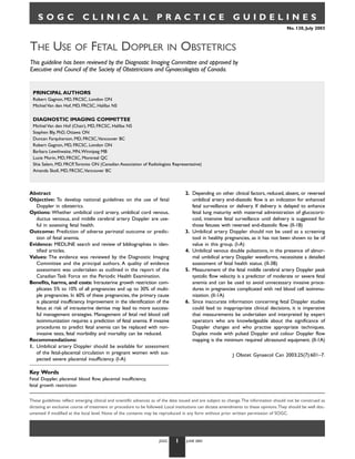
  • 1. SOGC CLINICAL PRACTICE GUIDELINES No. 130, July 2003 THE USE OF FETAL DOPPLER IN OBSTETRICS This guideline has been reviewed by the Diagnostic Imaging Committee and approved by Executive and Council of the Society of Obstetricians and Gynaecologists of Canada. PRINCIPAL AUTHORS Robert Gagnon, MD, FRCSC, London ON Michiel Van den Hof, MD, FRCSC, Halifax NS DIAGNOSTIC IMAGING COMMITTEE Michiel Van den Hof (Chair), MD, FRCSC, Halifax NS Stephen Bly, PhD, Ottawa ON Duncan Farquharson, MD, FRCSC,Vancouver BC Robert Gagnon, MD, FRCSC, London ON Barbara Lewthwaite, MN,Winnipeg MB Lucie Morin, MD, FRCSC, Montreal QC Shia Salem, MD, FRCP,Toronto ON (Canadian Association of Radiologists Representative) Amanda Skoll, MD, FRCSC,Vancouver BC Abstract 2. Depending on other clinical factors, reduced, absent, or reversed Objective: To develop national guidelines on the use of fetal umbilical artery end-diastolic flow is an indication for enhanced Doppler in obstetrics. fetal surveillance or delivery. If delivery is delayed to enhance Options: Whether umbilical cord artery, umbilical cord venous, fetal lung maturity with maternal administration of glucocorti- ductus venosus, and middle cerebral artery Doppler are use- coid, intensive fetal surveillance until delivery is suggested for ful in assessing fetal health. those fetuses with reversed end-diastolic flow. (II-1B) Outcome: Prediction of adverse perinatal outcome or predic- 3. Umbilical artery Doppler should not be used as a screening tion of fetal anemia. tool in healthy pregnancies, as it has not been shown to be of Evidence: MEDLINE search and review of bibliographies in iden- value in this group. (I-A) tified articles. 4. Umbilical venous double pulsations, in the presence of abnor- Values: The evidence was reviewed by the Diagnostic Imaging mal umbilical artery Doppler waveforms, necessitate a detailed Committee and the principal authors. A quality of evidence assessment of fetal health status. (II-3B) assessment was undertaken as outlined in the report of the 5. Measurement of the fetal middle cerebral artery Doppler peak Canadian Task Force on the Periodic Health Examination. systolic flow velocity is a predictor of moderate or severe fetal Benefits, harms, and costs: Intrauterine growth restriction com- anemia and can be used to avoid unnecessary invasive proce- plicates 5% to 10% of all pregnancies and up to 30% of multi- dures in pregnancies complicated with red blood cell isoimmu- ple pregnancies. In 60% of these pregnancies, the primary cause nization. (II-1A) is placental insufficiency. Improvement in the identification of the 6. Since inaccurate information concerning fetal Doppler studies fetus at risk of intrauterine demise may lead to more success- could lead to inappropriate clinical decisions, it is imperative ful management strategies. Management of fetal red blood cell that measurements be undertaken and interpreted by expert isoimmunization requires a prediction of fetal anemia. If invasive operators who are knowledgeable about the significance of procedures to predict fetal anemia can be replaced with non- Doppler changes and who practise appropriate techniques. invasive tests, fetal morbidity and mortality can be reduced. Duplex mode with pulsed Doppler and colour Doppler flow Recommendations: mapping is the minimum required ultrasound equipment. (II-1A) 1. Umbilical artery Doppler should be available for assessment of the fetal-placental circulation in pregnant women with sus- J Obstet Gynaecol Can 2003;25(7):601–7. pected severe placental insufficiency. (I-A) Key Words Fetal Doppler, placental blood flow, placental insufficiency, fetal growth restriction These guidelines reflect emerging clinical and scientific advances as of the date issued and are subject to change.The information should not be construed as dictating an exclusive course of treatment or procedure to be followed. Local institutions can dictate amendments to these opinions.They should be well doc- umented if modified at the local level. None of the contents may be reproduced in any form without prior written permission of SOGC. JOGC 1 JUNE 2003
  • 2. PART I: STANDARD ANTENATAL FETAL SURVEILLANCE tance index or Pourcelot ratio, and the pulsatility index. These indices closely correlate and they can be used interchangeably INTRODUCTION with similar predictive values for perinatal outcome.15,16 Placental insufficiency is the primary cause of intrauterine growth Placental insufficiency can be quantified based on the reduc- restriction in normally formed fetuses and can be identified using tion of end-diastolic Doppler flow velocity into (1) reduced end- umbilical artery Doppler velocimetry.1-4 Umbilical artery diastolic flow velocity, (2) absent end-diastolic flow velocity, and Doppler waveforms provide an estimate of downstream placen- (3) reversed end-diastolic flow velocity. The risk of perinatal tal vascular resistance and placental blood flow.2 There is a strong mortality increases up to 60%, with increasing severity from association between reduced end-diastolic umbilical artery blood reduced to reversed end-diastolic flow velocity.17-32 Therefore, flow velocity and increased vascular resistance in the umbilical- in the presence of umbilical artery reversed end-diastolic flow placental microcirculation.5,6 As well, abnormal umbilical artery velocity, delivery by Caesarean section may be considered if fetal Doppler waveforms have been associated with an increased risk viability is achieved.32 This decision will be influenced by the of fetal acidosis, as measured during cordocentesis,7 and may estimated fetal weight, gestational age, other Doppler parame- improve the performance of the biophysical profile score in pre- ters, and other assessments of fetal health, such as fetal anatom- dicting fetal acidemia and hypercarbia.8 The use of Doppler dur- ical and chromosomal anomalies.33 In cases of prematurity, ing antenatal fetal surveillance has involved assessment of (1) the delivery may be delayed for 48 hours, allowing the maximum umbilical arterial and venous flow velocity waveforms, (2) the fetal benefits of maternal administration of glucocorticoids; fetal cerebral circulation, and (3) the fetal venous circulation, in under such circumstances, continuous fetal heart rate moni- particular the ductus venosus. toring until delivery should be considered.34 ASSESSMENT OF PLACENTAL FUNCTION USING RECOMMENDATION UMBILICAL ARTERY DOPPLER VELOCIMETRY 1. Umbilical artery Doppler should be available for assess- From 7 to 16 days postconception, the yolk sac develops and ment of the fetal-placental circulation in pregnant women early development of the primary chorionic villi takes place. with suspected severe placental insufficiency. (I-A) Thereafter, the chorioallantoic placenta develops in stages con- sisting of invasion of the spiral arteries by endovascular cytotro- At an early gestational age, reduced or absent umbilical phoblast, followed by a second wave of invasion that extends artery end-diastolic flow velocity is an indication for increased into the myometrium. The basic organization of the human fetal surveillance, but not necessarily for immediate deliv- placenta is present by approximately day 20 of pregnancy. Fur- ery.23,35 However, closer to term, severe placental insufficien- ther refinement of this basic structure continues until term, at cy, reflected by absent umbilical artery end-diastolic flow which time there are approximately 50 to 60 primary fetal stem velocity, is an indication for delivery. Fetuses with absent umbil- villi branching into several terminal or tertiary villi. The branch- ical artery end-diastolic flow velocity are more severely growth ing of the stem villi and ensuing development of the non- restricted,2,7 are at higher risk of perinatal morbidity and mor- branching placental microcirculation are responsible for a low tality,19 and require delivery at an earlier gestational age than vascular resistance, the increase in placental blood flow, and the those with end-diastolic flow.21 However, when fetuses are increase in transplacental gas exchange that characterizes human matched for gestational age and birth weight, no differences in placentation. This low umbilical-placental vascular resistance is perinatal outcome are found in the groups with and without also responsible for the elevated end-diastolic blood flow ve- end-diastolic flow velocity.24,36 Although absence of end- locity in the umbilical artery seen during the third trimester in diastolic flow velocity may not affect long-term neurological a normal pregnancy.5,9 A reduction of the branching of the stem outcome, reversal of end-diastolic flow velocity in the umbil- villi and a reduction in the development of the nonbranching ical artery is associated with a wide range of problems at school placental microcirculation result in fewer small arterioles in the age,37 suggesting that it represents intrauterine decompensa- tertiary stem villi, along with a thickened fetal-maternal tion, which may have adverse effects on the developing brain.24 placental interface. This results in abnormally high umbilical- placental vascular resistance,2,10,11 a reduction in umbilical blood RECOMMENDATION flow,10 and chronic fetal hypoxemia.11 2. Depending on other clinical factors, reduced, absent, or With an increase in downstream placental vascular resis- reversed umbilical artery end-diastolic flow is an indica- tance, velocity of the end-diastolic flow in the umbilical cord tion for enhanced fetal surveillance or delivery. If delivery artery is reduced, while the peak-systolic component is not sig- is delayed to enhance fetal lung maturity with maternal nificantly affected.12-14 As a result, several Doppler indices have administration of glucocorticoid, intensive fetal surveil- been used to quantify abnormalities in umbilical artery lance until delivery is suggested for those fetuses with Doppler flow waveforms, including the A/B ratio, the resis- reversed end-diastolic flow. (II-1B) JOGC 2 JULY 2003
  • 3. Randomized clinical trials have demonstrated that the use tion into the fetal abdomen.42 This is particularly important for of umbilical artery velocimetry in high-risk pregnancy (espe- studies obtained in multiple pregnancy, where cord insertion at cially those complicated by hypertension or presumed impaired the umbilicus is relatively easy to obtain to differentiate indi- fetal growth) is associated with a trend to a reduction in peri- vidual fetuses.43 The angle of the fetal Doppler insonation natal deaths (OR 0.71, 95% CI 0.50–1.01).38 The use of should be kept to less than 45˚ for an optimal umbilical artery Doppler ultrasound was also associated with fewer inductions Doppler recording. Because of the potential for variability and of labour (OR 0.83, 95% CI 0.74–0.93) and fewer admissions inaccuracy with fetal Doppler, it is imperative that measure- to hospital (OR 0.56, 95% CI 0.43–0.72), without reports of ments be undertaken by expert operators who are knowledge- adverse effects.38 In high-risk pregnancies complicated with able about the significance of Doppler changes and who practise maternal hypertension, intrauterine growth restriction, or mul- appropriate techniques. Inaccurate information concerning fetal tiple gestation, evidence supports the use of umbilical artery Doppler studies could lead to inappropriate clinical decisions. Doppler studies as part of antenatal assessment.38 As there is no evidence that the use of umbilical artery Doppler has value PART II: SPECIAL CONSIDERATIONS in low-risk pregnancies,39 it should not be used as a screening tool in healthy pregnancies. INTRODUCTION Although the greatest impact on perinatal clinical practice from RECOMMENDATION Doppler research has been the use of umbilical artery Doppler 3. Umbilical artery Doppler should not be used as a screen- assessment of placental function, there have been an increasing ing tool in healthy pregnancies, as it has not been shown number of observational studies that require special considera- to be of value in this group. (I-A) tions. These fetal Doppler studies include assessment of the fetal venous circulation as a marker of severe fetal hypoxia,44-51 pre- FACTORS AFFECTING UMBILICAL diction of fetal hypoxemia using fetal middle cerebral artery,52-60 ARTERY DOPPLER VELOCIMETRY and the prediction of severe fetal anemia using fetal middle Several factors will affect the umbilical artery Doppler wave- cerebral artery Doppler.61-64 form, independent of changes in placental vascular resistance (Table 1). Gestational age-dependent normograms are neces- THE USE OF FETAL VENOUS sary for accurate interpretation of umbilical cord artery DOPPLER VELOCIMETRY velocimetry.14 No correction is necessary for fetal heart rate Blood flow velocity in the fetal systemic venous circulation within the normal range.40,41 In order to reduce methodologi- has a pulsating pattern that reflects changes in central venous cal variability, it is recommended that umbilical artery Doppler pressure, in particular the filling of the atria during ventricular waveforms be measured within 5 cm of the umbilical cord inser- systole and the opening of the atrio-ventricular valves. At the TABLE 1 FACTORS AFFECTING UMBILICAL ARTERY DOPPLER FLOW VELOCITY WAVEFORMS* Gestational age EDFV ratio increases with advancing gestational age15 Fetal heart rate EDFV decreases with decreasing fetal heart rate13,41 Fetal breathing movements Increases variability in the measurements66 Site of measurement EDFV is higher near the umbilical cord insertion into the fetal abdomen than near the placental insertion67 Equipment used: continuous Doppler Continuous Doppler is more a “blind technique” compared with versus pulsed Doppler pulsed Duplex Doppler, allowing 2D real time ultrasound68 User experience Reliability increases with increasing experience69 Radius of the umbilical artery Decreasing radius (vasoconstriction) increases EDFV70 Impedance to pulsatile flow propagation Increasing vascular impedance increases EDFV70 Downstream vascular resistance within the microcirculation Increasing vascular resistance decreases EDFV70–72 Angle of the fetal Doppler insonation Best if less than 45˚73; <15˚ for MCA absolute peak systolic flow velocity62,64 *EDFV = end diastolic flow velocity; MCA = middle cerebral artery. JOGC 3 JULY 2003
  • 4. end of diastole, a reduction in blood velocity occurs due to USE OF DOPPLER TO DETECT FETAL ANEMIA atrial contraction. Blood velocities in the umbilical vein and Several noninvasive methods have been suggested to detect fetal portal circulation are normally continuous and without fluctu- anemia. Umbilical vein maximum velocity and middle cerebral ation.44 Umbilical venous pulsations, particularly double artery peak-systolic flow velocity (MCA-PSV) are the most pulsations, have been associated with perinatal mortality rates promising methods.61,62 A recent systematic review indicated of up to 16% with absent umbilical artery end-diastolic flow that studies evaluating noninvasive techniques to detect fetal velocity, and 60% with reversed umbilical artery end-diastolic anemia were methodologically poor and lacked a standard flow velocity.44,45 However, it is not known if Doppler assess- approach to evaluate the techniques for fetal hemoglobin pre- ment of the fetal umbilical venous circulation improves diction.63 However, since then, it has been shown that the perinatal outcome when compared to assessment of umbilical MCA-PSV is an accurate predictor of severe fetal anemia in artery Doppler velocimetry alone.46-49 pregnancies complicated by red cell alloimmunization.64 Although the correlation between the fetal hemoglobin value RECOMMENDATION and MCA-PSV becomes more accurate as the severity of ane- 4. Umbilical venous double pulsations, in the presence of mia increases, almost 70% of the cordocentesis needed, using abnormal umbilical artery Doppler waveforms, necessi- current standard criteria for assessment of fetal hemoglobin, can tate a detailed assessment of fetal health status. (II-3B) be avoided.62 This approach is likely to decrease the need for cordocentesis and its potential risks. The ductus venosus may play a role in the regulation of venous blood flow between the inferior vena cava and the RECOMMENDATION umbilical vein. Under normoxemic conditions, approximate- 5. Measurement of the fetal middle cerebral artery Doppler ly 40% of the umbilical venous blood flow passes through the peak systolic flow velocity is a predictor of severe fetal ductus venosus.50 During fetal hypoxemia, the proportion of anemia and can be used to avoid unnecessary invasive umbilical venous flow passing through the ductus venosus procedures in pregnancies complicated with red blood increases.52 It is not clear if this increase is the result of an cell isoimmunization. (II-1A) increase in central venous pressure or due to vasodilatation.44 It is reported that a reduction in vascular resistance through In order to accurately measure the MCA Doppler wave- the ductus venosus is responsible for retrograde umbilical forms, pulsed Doppler with colour Doppler flow mapping is venous flow velocity leading to umbilical venous pulsations recommended to visualize the direction of MCA blood flow. during atrial contraction in the presence of fetal hypoxemia.51 Since the MCA-PSV is a measurement of absolute instead of If umbilical venous pulsations are detected in the absence of relative velocity, the angle of the fetal Doppler insonation fetal breathing movements, careful assessment of fetal health should be kept as close as possible to 0˚ for accurate estimate should be considered. Although available in many tertiary cen- of the absolute peak systolic flow velocity. Software-based angle tres, further research is needed on the benefit of umbilical correction cannot be used instead of proper positioning of the venous and ductus venosus Doppler velocimetry before it can transducer since it could lead to erroneous value and interpre- be recommended as a standard of care to evaluate high-risk tation. pregnancies. RECOMMENDATION USE OF MIDDLE CEREBRAL ARTERY 6. Since inaccurate information concerning fetal Doppler VELOCIMETRY TO DETECT FETAL HYPOXIA studies could lead to inappropriate clinical decisions, it The same factors that affect umbilical artery Doppler waveforms is imperative that measurements be undertaken and inter- can also affect fetal cerebral artery Doppler waveforms.52 Fetal preted by expert operators who are knowledgeable about behavioural states can also alter cerebral artery waveforms.53-56 the significance of Doppler changes and who practise Of interest, an increase in pCO2 or a reduction in pO2 will appropriate techniques. Duplex mode with pulsed cause an increase in fetal cerebral arterial Doppler end-diastolic Doppler and colour Doppler flow mapping is the mini- flow velocity, likely related to cerebral vasodilatation.57-59 This mum required ultrasound equipment. (II-1A) phenomenon has been described as the “brain sparing” effect.60 Although an increase in fetal cerebral end-diastolic Doppler flow EVALUATION OF EVIDENCE velocity may reflect chronic fetal hypoxemia, there is no evi- dence that this measurement will provide any additional ben- The quality of evidence and classification of recommendations efit to perinatal outcome beyond the assessment of the umbilical reported in these guidelines has been described using the Eval- circulation alone.23 uation of Evidence criteria outlined in the Report of the Cana- dian Task Force on the Periodic Health Exam (Table 2).74 JOGC 4 JULY 2003
  • 5. TABLE 2 QUALITY OF EVIDENCE ASSESSMENT74 CLASSIFICATION OF RECOMMENDATIONS74 The quality of evidence reported in these guidelines has been Recommendations included in these guidelines have been described using the Evaluation of Evidence criteria outlined in adapted from the ranking method described in the the Report of the Canadian Task Force on the Periodic Health Classification of Recommendations found in the Canadian Exam. Task Force on the Periodic Health Exam. I: Evidence obtained from at least one properly randomized A. There is good evidence to support the recommendation controlled trial. that the condition be specifically considered in a periodic II-1: Evidence from well-designed controlled trials without health examination. randomization. B. There is fair evidence to support the recommendation that II-2: Evidence from well-designed cohort (prospective or the condition be specifically considered in a periodic health retrospective) or case-control studies, preferably from examination. more than one centre or research group. C. There is poor evidence regarding the inclusion or II-3: Evidence obtained from comparisons between times or exclusion of the condition in a periodic health examination, places with or without the intervention. Dramatic results but recommendations may be made on other grounds. in uncontrolled experiments (such as the results of treat- D. There is fair evidence to support the recommendation ment with penicillin in the 1940s) could also be included that the condition not be considered in a periodic health in this category. examination. III: Opinions of respected authorities, based on clinical E. There is good evidence to support the recommendation experience, descriptive studies, or reports of expert that the condition be excluded from consideration in a committees. periodic health examination. REFERENCES 11. Kingdom JC, Burrell SJ, Kaufmann P. Pathology and clinical implications of abnormal umbilical artery Doppler waveforms. Ultrasound Obstet Gynecol 1997;9:271–86. 1. Giles WB,Trudinger BJ, Cook CM. Fetal umbilical artery flow 12. Gagnon R, Challis J, Johnston L, Fraher L. Fetal endocrine responses velocity-time waveforms in twin pregnancies. Br J Obstet Gynaecol to chronic placental embolization in the late-gestation ovine fetus. 1985;92:490–7. Am J Obstet Gynecol 1994;170:929–38. 2. Giles WB,Trudinger BJ, Baird PJ. Fetal umbilical artery flow velocity 13. Mansouri H, Gagnon R, Hunse C. Relationship between fetal heart rate waveforms and placental resistance: pathological correlation. Br J and umbilical blood flow velocity in term human fetuses during labor. Obstet Gynaecol 1985;92:31–8. Am J Obstet Gynecol 1989;160:1007–12. 3. Lackman F, Capewell V, Gagnon R, Richardson B. Fetal umbilical cord 14. Thompson RS,Trudinger BJ, Cook CM. Doppler ultrasound waveforms oxygen values and birth to placental weight ratio in relation to size at in the fetal umbilical artery: quantitative analysis technique. Ultrasound birth. Am J Obstet Gynecol 2001;185:674–82. Med Biol 1985;11:707–18. 4. Lackman F, Capewell V, Richardson B, daSilva O, Gagnon R.The risks 15. Thompson RS,Trudinger BJ, Cook CM. A comparison of Doppler ultra- of spontaneous preterm delivery and perinatal mortality in relation sound waveform indices in the umbilical artery – I. Indices derived from to size at birth according to fetal versus neonatal growth standards. the maximum velocity waveform. Ultrasound Med Biol 1986;12:835–44. Am J Obstet Gynecol 2001;184:946–53. 16. Thompson RS,Trudinger BJ, Cook CM. Doppler ultrasound waveform 5. Adamson SL. Arterial pressure, vascular input impedance, and indices: A/B ratio, pulsatility index and Pourcelot ratio. Br J Obstet resistance as determinants of pulsatile blood flow in the umbilical Gynaecol 1988;95:581–8. artery. Eur J Obstet Gynecol Reprod Biol 1999;84:119–25. 17. Cook CM, Connelly AJ,Trudinger BJ. Doppler assessment of the 6. Thompson RS,Trudinger BJ. Doppler waveform pulsatility index and umbilical circulation. Semin Ultrasound CT MR 1989;10:417–27. resistance, pressure and flow in the umbilical placental circulation: 18. Trudinger B, Cook CM,Thompson R, Giles W, Connelly A. Low-dose an investigation using a mathematical model. Ultrasound Med Biol aspirin improves fetal weight in umbilical placental insufficiency. Lancet 1990;16:449–58. 1988;2:214–5. 7. Yoon BH, Syn HC, Kim SW.The efficacy of Doppler umbilical artery 19. Rochelson B, Schulman H, Farmakides G, Bracero L, Ducey J, Fleischer A, velocimetry in identifying fetal acidosis. A comparison with fetal et al.The significance of absent end-diastolic velocity in umbilical artery biophysical profile. J Ultrasound Med 1992;11:1–6. velocity waveforms. Am J Obstet Gynecol 1987;156:1213–8. 8. Yoon BH, Romero R, Roh CR, Kim SH,Ager JW, Syn HC, et al. 20. Rochelson BL, Schulman H, Fleischer A, Farmakides G, Bracero L, Relationship between the fetal biophysical profile score, umbilical artery Ducey J, et al.The clinical significance of Doppler umbilical artery Doppler velocimetry, and fetal blood acid-base status determined by velocimetry in the small for gestational age fetus. Am J Obstet cordocentesis. Am J Obstet Gynecol 1993;169:1586–94. Gynecol 1987;156:1223–6. 9. Trudinger BJ, Giles WB, Cook CM. Flow velocity waveforms in the 21. Ferrazzi E,Vegni C, Bellotti M, Borboni A, Della PS, Barbera A. Role of maternal uteroplacental and fetal umbilical placental circulations. umbilical Doppler velocimetry in the biophysical assessment of the Am J Obstet Gynecol 1985;152:155–63. growth-retarded fetus. Answers from neonatal morbidity and 10. Hitschold TP. Doppler flow velocity waveforms of the umbilical mortality. J Ultrasound Med 1991;10:309–15. arteries correlate with intravillous blood volume. Am J Obstet 22. Ferrazzi E, Bellotti M,Vegni C, Barbera A, Della PS, Ferro B, et al. Gynecol 1998;179:540–3. Umbilical flow waveforms versus fetal biophysical profile in hyperten- sive pregnancies. Eur J Obstet Gynecol Reprod Biol 1989;33:199–208. JOGC 5 JULY 2003
  • 6. 23. Fong KW, Ohlsson A, Hannah ME, Grisaru S, Kingdom J, Cohen H, et al. 44. Hofstaetter C, Dubiel M, Gudmundsson S.Two types of umbilical Prediction of perinatal outcome in fetuses suspected to have intra- venous pulsations and outcome of high-risk pregnancy. Early Hum uterine growth restriction: Doppler US study of fetal cerebral, Dev 2001;61:111–7. renal, and umbilical arteries. Radiology 1999;213:681–9. 45. Reed KL, Chaffin DG, Anderson CF. Umbilical venous Doppler velocity 24. Schreuder AM, McDonnell M, Gaffney G, Johnson A, Hope PL. pulsations and inferior vena cava pressure elevations in fetal lambs. Outcome at school age following antenatal detection of absent or Obstet Gynecol 1996;87:617–20. reversed end diastolic flow velocity in the umbilical artery. Arch Dis 46. Gudmundsson S. Importance of venous flow assessment for clinical Child Fetal Neonatal Ed 2002;86:F108–14. decision-making. Eur J Obstet Gynecol Reprod Biol 1999;84:173–8. 25. Vossbeck S, de Camargo OK, Grab D, Bode H, Pohlandt F. Neonatal 47. Gudmundsson S, Gunnarsson GO, Hokegard KH, Ingemarsson J, and neurodevelopmental outcome in infants born before 30 weeks of Kjellmer I.Venous Doppler velocimetry in relationship to central gestation with absent or reversed end-diastolic flow velocities in the venous pressure and heart rate during hypoxia in the ovine fetus. umbilical artery. Eur J Pediatr 2001;160:128–34. J Perinat Med 1999;27:81–90. 26. Montenegro N, Santos F,Tavares E, Matias A, Barros H, Leite LP. 48. Hofstaetter C, Gudmundsson S, Dubiel M, Marsal K. Ductus venosus Outcome of 88 pregnancies with absent or reversed end-diastolic velocimetry in high-risk pregnancies. Eur J Obstet Gynecol Reprod blood flow (ARED flow) in the umbilical arteries. Eur J Obstet Biol 1996;70:135–40. Gynecol Reprod Biol 1998;79:43–6. 49. Gudmundsson S,Tulzer G, Huhta JC, Marsal K.Venous Doppler in the 27. Salafia CM, Pezzullo JC, Minior VK, Divon MY. Placental pathology of fetus with absent end-diastolic flow in the umbilical artery. Ultrasound absent and reversed end-diastolic flow in growth-restricted fetuses. Obstet Gynecol 1996;7:262–7. Obstet Gynecol 1997;90:830–6. 50. Kiserud T.The ductus venosus. Semin Perinatol 2001;25:11–20. 28. Karsdorp VH, van Vugt JM, van Geijn HP, Kostense PJ, Arduini D, 51. Reed KL, Chaffin DG,Anderson CF, Newman AT. Umbilical venous Montenegro N, et al. Clinical significance of absent or reversed velocity pulsations are related to atrial contraction pressure end diastolic velocity waveforms in umbilical artery. Lancet waveforms in fetal lambs. Obstet Gynecol 1997;89:953–6. 1994;344:1664–8. 52. Gagnon R, Lamb T, Richardson B. Cerebral circulatory responses of 29. Tannirandorn Y, Phaosavasdi S. Significance of an absent or reversed near-term ovine fetuses during sustained fetal placental embolization. end-diastolic flow velocity in Doppler umbilical artery waveforms. Am J Physiol 1997;273:H2001–8. J Med Assoc Thai 1994;77:81–6. 53. Wladimiroff JW. Behavioural states and cardiovascular dynamics in the 30. Wilson DC, Harper A, McClure G. Absent or reversed end diastolic human fetus; an overview. Early Hum Dev 1994;37:139–49. flow velocity in the umbilical artery and necrotizing enterocolitis. 54. Norwitz ER, Hoyte LP, Jenkins KJ, van der Velde ME, Ratiu P, Arch Dis Child 1991;66:1467. Rodriguez-Thompson D, et al. Separation of conjoined twins with the 31. McParland P, Steel S, Pearce JM.The clinical implications of absent or twin reversed-arterial-perfusion sequence after prenatal planning with reversed end-diastolic frequencies in umbilical artery flow velocity three-dimensional modeling. N Engl J Med 2000;343:399–402. waveforms. Eur J Obstet Gynecol Reprod Biol 1990;37:15–23. 55. van den Wijngaard JA, van Eyck J, Noordam MJ,Wladimiroff JW, 32. Woo JS, Liang ST, Lo RL. Significance of an absent or reversed end van Strik R.The Doppler flow velocity waveform in the fetal internal diastolic flow in Doppler umbilical artery waveforms. J Ultrasound carotid artery with respect to fetal behavioural states. A longitudinal Med 1987;6:291–7. study. Biol Neonate 1988;53:274–8. 33. Trudinger BJ, Cook CM. Umbilical and uterine artery flow velocity 56. Wladimiroff JW,Tonge HM, Stewart PA. Doppler ultrasound assessment waveforms in pregnancy associated with major fetal abnormality. of cerebral blood flow in the human fetus. Br J Obstet Gynaecol Br J Obstet Gynaecol 1985;92:666–70. 1986;93:471–5. 34. du Plessis JM, Hall DR, Norman K, Odendaal HJ. Reversed end diastolic 57. Connors G, Hunse C, Gagnon R, Richardson B, Han V, Rosenberg H. flow velocity in viable fetuses: is there time to wait for the effect of Perinatal assessment of cerebral flow velocity wave forms in the corticosteroids before delivery? Int J Gynaecol Obstet 2001;72:187–8. human fetus and neonate. Pediatr Res 1992;31:649–52. 35. Trudinger BJ, Cook CM, Giles WB, Ng S, Fong E, Connelly A, et al. Fetal 58. Potts P, Connors G, Gillis S, Hunse C, Richardson B.The effect of umbilical artery velocity waveforms and subsequent neonatal outcome. carbon dioxide on Doppler flow velocity waveforms in the human Br J Obstet Gynaecol 1991;98:378–84. fetus. J Dev Physiol 1992;17:119–23. 36. Bekedam DJ,Visser GH, van der Zee AG, Snijders RJ, Poelmann- 59. Gagnon R, Lamb T, Richardson B. Cerebral circulatory responses of Weesjes G. Abnormal velocity waveforms of the umbilical artery in near-term ovine fetuses during sustained fetal placental embolization. growth retarded fetuses: relationship to antepartum late heart rate Am J Physiol 1997;273:H2001–8. decelerations and outcome. Early Hum Dev 1990;24:79–89. 60. Kopecky EA, Ryan ML, Barrett JF, Seaward PG, Ryan G, Koren G, et al. 37. Brar HS, Platt LD. Reverse end-diastolic flow velocity on umbilical Fetal response to maternally administered morphine. Am J Obstet artery velocimetry in high-risk pregnancies: an ominous finding with Gynecol 2000;183:424–30. adverse pregnancy outcome. Am J Obstet Gynecol 1988;159:559–61. 61. Mari G, Deter RL, Carpenter RL, Rahman F, Zimmerman R, Oepkes D, 38. Neilson JP,Alfirevic Z. Doppler ultrasound for fetal assessment in high et al.The use of ultrasonography and Doppler in the prediction of fetal risk pregnancies. Cochrane Database Syst Rev 2000;CD000073. haemolytic anaemia: a multivariate analysis. Br J Obstet Gynaecol 39. Newnham JP, Patterson LL, James IR, Diepeveen DA, Reid SE. An evalu- 1994;101:680–4. ation of the efficacy of Doppler flow velocity waveform analysis as a 62. Mari G, Deter RL, Carpenter RL, Rahman F, Zimmerman R, Moise KJ Jr., screening test in pregnancy. Am J Obstet Gynecol 1990;162:403–10. et al. Noninvasive diagnosis by Doppler ultrasonography of fetal 40. Makikallio K,Tekay A, Jouppila P.Yolk sac and umbilicoplacental hemo- anemia due to maternal re-cell alloimmunization. Collaborative dynamics during early human embryonic development. Ultrasound Group for Doppler Assessment of the Blood Velocity in Anemic Obstet Gynecol 1999;14:175–9. Fetuses. N Engl J Med 2000;342:9–14. 41. Yarlagadda P,Willoughby L, Maulik D. Effect of fetal heart rate on 63. Divakaran TG,Waugh J, Clark TJ, Khan KS,Whittle MJ, Kilby MD. umbilical arterial Doppler indices. J Ultrasound Med 1989;8:215–8. Noninvasive techniques to detect fetal anemia due to red blood cell 42. Mehalek KE, Rosenberg J, Berkowitz GS, Chitkara U, Berkowitz RL. alloimmunization: a systematic review. Obstet Gynecol 2001;98:509–17. Umbilical and uterine artery flow velocity waveforms. Effect of the 64. Mari G, Detti L, Oz U, Zimmerman R, Duerig P, Stefos T. Accurate sampling site on Doppler ratios. J Ultrasound Med 1989;8:171–6. prediction of fetal hemoglobin by Doppler ultrasonography. Obstet 43. Joern H, Rath W. Correlation of Doppler velocimetry findings in twin Gynecol 2002;99:589–93. pregnancies including course of pregnancy and fetal outcome. Fetal 65. Morrow RJ, Adamson SL, Lewin M, Bull SB, Ritchie JW. The influence Diagn Ther 2000;15:160–4. of spontaneous accelerations of fetal heart rate on umbilical artery velocity waveforms. Am J Obstet Gynecol 1989;160:995–7. JOGC 6 JULY 2003
  • 7. 66. Spencer JA, Price J, Lee A. Influence of fetal breathing and movements on variability of umbilical Doppler indices using different numbers of waveforms. J Ultrasound Med 1991;10:37–41. 67. Adamson SL, Morrow RJ, Langille BL, Bull SB, Ritchie JW. Site-dependent effects of increases in placental vascular resistance on the umbilical arterial velocity waveform in fetal sheep. Ultrasound Med Biol 1990;16:19–27. 68. Kurjak A, Dudenhausen JW, Hafner T, Kupesic S, Latin V, Kos M. Intervillous circulation in all three trimesters of normal pregnancy assessed by color Doppler. J Perinat Med 1997;25:373–80. 69. Campbell S,Vyas S, Nicolaides KH. Doppler investigation of the fetal circulation. J Perinat Med 1991;19:21–6. 70. Surat DR, Adamson SL. Downstream determinants of pulsatility of the mean velocity waveform in the umbilical artery as predicted by a computer model. Ultrasound Med Biol 1996;22:707–17. 71. Gagnon R, Johnston L, Murotsuki J. Fetal placental embolization in the late-gestation ovine fetus: alterations in umbilical blood flow and fetal heart rate patterns. Am J Obstet Gynecol 1996;175:63–72. 72. Trudinger BJ, Stevens D, Connelly A, Hales JR, Alexander G, Bradley L, et al. Umbilical artery flow velocity waveforms and placental resistance: the effects of embolization of the umbilical circulation. Am J Obstet Gynecol 1987;157:1443–8. 73. Joern H, Funk A, Goetz M, Kuehlwein H, Klein A, Fendel H. Development of quantitative Doppler indices for uteroplacental and fetal blood flow during the third trimester. Ultrasound Med Biol 1996;22:823–35. 74. Woolf SH, Battista RN, Angerson GM, Logan AG, Eel W. Canadian Task Force on the Periodic Health Exam. Ottawa: Canada Communication Group; 1994. p. xxxvii. JOGC 7 JULY 2003