Msb 206. amino acid catab and fates of cs.2014
•Descargar como PPT, PDF•
8 recomendaciones•4,909 vistas

Metabolism

Recomendados
Más contenido relacionado
La actualidad más candente
La actualidad más candente (20)
Destacado
Destacado (20)
Similar a Msb 206. amino acid catab and fates of cs.2014
Similar a Msb 206. amino acid catab and fates of cs.2014 (20)
Más de Dr. Geoffrey K. K. Maiyoh
Más de Dr. Geoffrey K. K. Maiyoh (20)
Último
Último (20)
Msb 206. amino acid catab and fates of cs.2014
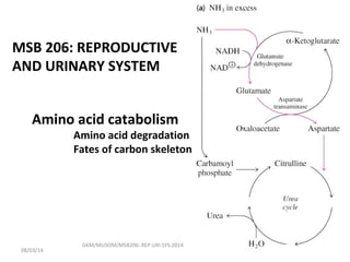
- 1. Amino acid catabolism Amino acid degradation Fates of carbon skeleton GKM/MUSOM/MSB206:.REP.URI.SYS.2014 MSB 206: REPRODUCTIVE AND URINARY SYSTEM 08/03/14
- 2. Amino acids and their R groups • Twenty different amino acid are found in proteins • Most microorganisms and plants can biosynthesize all 20 • Animals (including humans) must obtain some of the amino acids from the diet. • The amino acids that an organism cannot synthesize on its own are referred to as essential amino acids. • Humans require 8 essential amino acids GKM/MUSOM/MSB206:.REP.URI.SYS.2014 08/03/14
- 4. Oxidative degradation of amino acids • Under three different metabolic circumstances in animals: 1. During the normal synthesis and degradation of cellular protein; some amino acids that are released from protein breakdown and are not needed for new protein synthesis undergo oxidative degradation. 2. When a diet is rich in protein and the ingested; amino acids exceed the body’s needs for protein synthesis, the surplus is catabolized; amino acids cannot be stored. 3. During starvation or in uncontrolled diabetes mellitus; when carbohydrates are either unavailable or not properly utilized, cellular proteins are used as fuel. GKM/MUSOM/MSB206:.REP.URI.SYS.201408/03/14
- 5. Removal of the alpha amino group • Every amino acid contains an amino group • The pathways for amino acid degradation therefore include a key 1ST step in which the amino group is separated from the carbon skeleton • It is then shunted into the pathways of amino group metabolism – different based on different organism. GKM/MUSOM/MSB206:.REP.URI.SYS.201408/03/14
- 6. There are multiple transaminase enzymes which vary in substrate specificity. Some show preference for particular amino acids or classes of amino acids as amino group donors, and/or for particular α- keto acid acceptors. H R1 C COO - + R2 C COO - NH3 + O Transaminase H R1 C COO - + R2 C COO - O NH3 + Role of Transaminases (aminotransferases) Catalyze the reversible reaction GKM/MUSOM/MSB206:.REP.URI.SYS.2014 aa aaka ka 08/03/14
- 7. Example of a Transaminase reaction: Aspartate donates its amino group, becoming the α-keto acid oxaloacetate. α-Ketoglutarate accepts the amino group, becoming the amino acid glutamate. aspartate α-ketoglutarate oxaloacetate glutamate Aminotransferase (Transaminase) COO− CH2 CH2 C COO− O COO− CH2 HC COO− NH3 + COO− CH2 CH2 HC COO− NH3 + COO− CH2 C COO− O+ + GKM/MUSOM/MSB206:.REP.URI.SYS.201408/03/14
- 8. In another example, alanine becomes pyruvate as the amino group is transferred to α-ketoglutarate. alanine α-ketoglutarate pyruvate glutamate Aminotransferase (Transaminase) COO− CH2 CH2 C COO− O CH3 HC COO− NH3 + COO− CH2 CH2 HC COO− NH3 + CH3 C COO− O+ + GKM/MUSOM/MSB206:.REP.URI.SYS.201408/03/14
- 9. The Ammonia that is generated following deamination rxn is Transported in the Bloodstream Safely as Glutamate or in combination with Glutamate to form Glutamine • Un-needed glutamine is processed in intestines the, kidneys and liver GKM/MUSOM/MSB206:.REP.URI.SYS.201408/03/14
- 10. Pyruvate – Alanine in SM • In skeletal muscle, excess amino groups are generally transferred to pyruvate to form alanine • Other than Gln and Glu, Alanine is therefore another important molecule in the transport of amino groups to the liver. – See next slide GKM/MUSOM/MSB206:.REP.URI.SYS.201408/03/14
- 11. Glutamate can Donate Ammonia to Pyruvate to Make Alanine • Vigorously working muscles operate nearly anaerobically and rely on glycolysis for energy • Glycolysis yields pyruvate that muscles cannot metabolize aerobically; if not eliminated lactic acid will build up • This pyruvate can be converted to alanine for transport of NH4 into liver GKM/MUSOM/MSB206:.REP.URI.SYS.201408/03/14
- 12. Aminotransferases and pyridoxal phosphate • Aminotransferases are classic examples of enzymes catalyzing bimolecular Ping-Pong reactions • In these rxns, the first substrate reacts and the product must leave the active site before the second substrate can bind. • The incoming amino acid thus binds to the active site, donates its amino group to pyridoxal phosphate, and departs in the form of an –keto acid. • The incoming -keto acid then binds, accepts the amino group from pyridoxamine phosphate, and departs in the form of an amino acid. GKM/MUSOM/MSB206:.REP.URI.SYS.201408/03/14
- 13. The prosthetic group of Transaminase is pyridoxal phosphate (PLP), a derivative of vitamin B6 . pyridoxal phosphate (PLP) N H C O P O− O O OH CH3 C H O − + H2 GKM/MUSOM/MSB206:.REP.URI.SYS.201408/03/14
- 14. In the resting state, the aldehyde group of pyridoxal phosphate is in a Schiff base linkage to the ε-amino group of an enzyme lysine side-chain. N H C O P O− O O O− CH3 HC − + H2 N (CH2)4 Enz H + R H C COO− NH2 Enzyme (Lys)-PLP Schiff base Amino acid GKM/MUSOM/MSB206:.REP.URI.SYS.201408/03/14
- 15. The active site lysine extracts H+ , promoting tautomerization, followed by reprotonation & hydrolysis. N H C O P O− O O O− CH3 HC − + H2 N H C H + R COO− Enz−Lys−NH2 Amino acid-PLP Shiff base (aldimine) The α-amino group of a substrate amino acid displaces the enzyme lysine, to form a Schiff base linkage to PLP. GKM/MUSOM/MSB206:.REP.URI.SYS.201408/03/14
- 16. The amino group remains on what is now pyridoxamine phosphate (PMP). A different α-keto acid reacts with PMP and the process reverses, to complete the reaction. N H C O P O− O O OH CH3 CH2 NH2 H2 R C COO− O − + Enz−Lys−NH2 Pyridoxamine phosphate (PMP) α-keto acidWhat was an amino acid leaves as an α-keto acid. GKM/MUSOM/MSB206:.REP.URI.SYS.201408/03/14
- 17. Several other enzymes that catalyze metabolism or synthesis of amino acids also utilize PLP as prosthetic group, and have mechanisms involving a Schiff base linkage of the amino group to PLP. N H C O P O− O O O− CH3 HC − + H2 N H C H + R COO− Enz−Lys−NH2 Amino acid-PLP Shiff base (aldimine) GKM/MUSOM/MSB206:.REP.URI.SYS.201408/03/14
- 18. Amino acid Carbon skeleton • Amino acids lose their amino groups to form -keto acids - the “carbon skeletons” of amino acids. • The -keto acids undergo oxidation to CO2 and H2O or can be converted by gluconeogenesis into glucose, the fuel for brain. • As in carbohydrate and fatty acid catabolism, the processes of amino acid degradation converge on the central catabolic pathways. • The carbon skeletons of most amino acids find their way to the citric acid cycle. GKM/MUSOM/MSB206:.REP.URI.SYS.201408/03/14
- 19. central catabolic pathways • In cytoplasm (1) • In mitochondria (2, 3 & 4) 25-19 GKM/MUSOM/MSB206:.REP.URI.SYS.201408/03/14 Others e.g; Gluconeogenesis Glycogenesis
- 20. Amino Acid Carbon Skeletons Amino acids are grouped into 2 classes, based on whether or not their carbon skeletons can be converted to glucose: glucogenic ketogenic. GKM/MUSOM/MSB206:.REP.URI.SYS.201408/03/14
- 21. Carbon skeletons of glucogenic amino acids are degraded to: pyruvate, or a 4-C or 5-C intermediate of Krebs Cycle. These are precursors for gluconeogenesis. Glucogenic amino acids are the major carbon source for gluconeogenesis when glucose levels are low. They can also be catabolized for energy, or converted to glycogen or fatty acids for energy storage. GKM/MUSOM/MSB206:.REP.URI.SYS.201408/03/14
- 23. Gluconeogenesis: synthesis of Glucose from none carbohydrates Gluconeogenesis is not just glycolysis in reverse--the enzymes in green print catalyze irreversible reactions GKM/MUSOM/MSB206:.REP.URI.SYS.201408/03/14
- 24. Carbon skeletons of ketogenic amino acids are degraded to: acetyl-CoA, or acetoacetate. Acetyl CoA, & its precursor acetoacetate, cannot yield net production of oxaloacetate, the gluconeogenesis precursor. For every 2-C acetyl residue entering Krebs Cycle, 2C leave as CO2 . Carbon skeletons of ketogenic amino acids can be catabolized for energy in Krebs Cycle, or converted to ketone bodies or fatty acids. They cannot be converted to glucose. GKM/MUSOM/MSB206:.REP.URI.SYS.2014 08/03/14
- 25. The 3-C α-keto acid pyruvate is produced from alanine, cysteine, glycine, serine, & threonine. Alanine deamination via Transaminase directly yields pyruvate. alanine α-ketoglutarate pyruvate glutamate Aminotransferase (Transaminase) COO− CH2 CH2 C COO− O CH3 HC COO− NH3 + COO− CH2 CH2 HC COO− NH3 + CH3 C COO− O+ + GKM/MUSOM/MSB206:.REP.URI.SYS.201408/03/14
- 26. Serine is deaminated to pyruvate via Serine Dehydratase. Glycine, which is also product of threonine catabolism, is converted to serine by a reaction involving tetrahydrofolate (to be discussed later). HO CH2 H C COO− NH3 + C COO− OH2O NH4 + C COO− NH3 + H2C H3C H2O serine aminoacrylate pyruvate Serine Dehydratase GKM/MUSOM/MSB206:.REP.URI.SYS.2014 08/03/14
- 27. The 4-C Krebs Cycle intermediate oxaloacetate is produced from aspartate & asparagine. Aspartate transamination yields oxaloacetate. Aspartate is also converted to fumarate in Urea Cycle. Fumarate is converted to oxaloacetate in Krebs cycle. aspartate α-ketoglutarate oxaloacetate glutamate Aminotransferase (Transaminase) COO− CH2 CH2 C COO− O COO− CH2 HC COO− NH3 + COO− CH2 CH2 HC COO− NH3 + COO− CH2 C COO− O+ + GKM/MUSOM/MSB206:.REP.URI.SYS.201408/03/14
- 28. Asparagine loses the amino group from its R-group by hydrolysis catalyzed by Asparaginase. This yields aspartate, which can be converted to oxaloacetate, e.g., by transamination. C CH2 HC COO− NH3 + OH2N COO− CH2 HC COO− NH3 + H2O NH4 + asparagine aspartate Asparaginase GKM/MUSOM/MSB206:.REP.URI.SYS.201408/03/14
- 29. The 4-C Krebs Cycle intermediate succinyl- CoA is produced from isoleucine, threonine, valine, & methionine. Propionyl-CoA, an intermediate on these pathways, is also a product of β-oxidation of fatty acids with an odd number of C atoms. NB: Isoleucine and Valine are branched-chain amino acids GKM/MUSOM/MSB206:.REP.URI.SYS.2014 08/03/14
- 30. The branched chain amino acids initially share a common pathway. Branched Chain α-Keto Acid Dehydrogenase (BCKDH) is a multi-subunit complex. Genetic deficiency of BCKDH is called Maple Syrup Urine Disease (MSUD). High concentrations of branched chain keto acids in urine give it a characteristic odor. GKM/MUSOM/MSB206:.REP.URI.SYS.201408/03/14
- 31. MSUD • People with MSUD have a mutation that results in a deficiency for one of the 6 proteins that make up this complex. • Therefore, they can't break down leucine, isoleucine, and valine. • They end up with dangerously high levels of these amino acids in their blood, causing the rapid degeneration of brain cells and death if left untreated. GKM/MUSOM/MSB206:.REP.URI.SYS.201408/03/14
- 32. Propionyl-CoA is carboxylated to methylmalonyl-CoA. A racemase yields the L-isomer essential to the subsequent reaction. Methylmalonyl-CoA Mutase catalyzes a molecular rearrangement: the branched C chain of methylmalonyl-CoA is converted to the linear C chain of succinyl-CoA. C CH3 C S-CoA O C CH3 C S-CoA O COO− C C S-CoA O COO− C C COO− C C O H H CoA-S H HH HH H H H H HCO3 − ATP ADP + Pi propionyl-CoA D-methylmalonyl-CoA L-methylmalonyl-CoA succinyl-CoA Propionyl-CoA Methylmalonyl-CoA Methylmalonyl-CoA Carboxylase (Biotin) Racemase Mutase (B12) GKM/MUSOM/MSB206:.REP.URI.SYS.201408/03/14 bk
- 33. The 5-C Krebs Cycle intermediate α-ketoglutarate is produced from arginine, glutamate, glutamine, histidine, & proline. Glutamate deamination via Transaminase directly yields α-ketoglutarate. aspartate α-ketoglutarate oxaloacetate glutamate Aminotransferase (Transaminase) COO− CH2 CH2 C COO− O COO− CH2 HC COO− NH3 + COO− CH2 CH2 HC COO− NH3 + COO− CH2 C COO− O+ + GKM/MUSOM/MSB206:.REP.URI.SYS.201408/03/14
- 34. Glutamate deamination by Glutamate Dehydrogenase also directly yields α-ketoglutarate. − OOC H2 C H2 C C COO− O + NH4 + NAD(P)+ NAD(P)H − OOC H2 C H2 C C COO− NH3 + H glutamate α-ketoglutarate Glutamate Dehydrogenase GKM/MUSOM/MSB206:.REP.URI.SYS.201408/03/14
- 35. Histidine is first converted to glutamate. The last step in this pathway involves the cofactor tetrahydrofolate. Tetrahydrofolate (THF), which has a pteridine ring, is a reduced form of the B vitamin folate. N H H NN HN H2N H H H CH2 HNO C O N H C H COO− C H2 C H2 COO− Tetrahydrofolate (THF) pteridine ρ-aminobenzoate glutamate GKM/MUSOM/MSB206:.REP.URI.SYS.201408/03/14
- 36. In the pathway of histidine degradation, N- formiminoglutamate is converted to glutamate by transfer of the formimino group to THF, yielding N5 - formimino-THF. HC C CH2 H C COO− NH3 +N NH C H − OOC H C CH2 CH2 COO− HN NH C H − OOC H C CH2 CH2 COO− NH3 + THF N 5 -formimino-THF NH4 + H2O H2O histidine N-formimino- glutamate glutamateGKM/MUSOM/MSB206:.REP.URI.SYS.2014 08/03/14
- 37. Aromatic Amino Acids Aromatic amino acids phenylalanine & tyrosine are catabolized to fumarate and acetoacetate. Hydroxylation of phenylalanine to form tyrosine involves the reductant tetrahydrobiopterin. Biopterin, like folate, has a pteridine ring. GKM/MUSOM/MSB206:.REP.URI.SYS.2014 08/03/14
- 38. Overall the reaction is considered a mixed function oxidation, because one O atom of O2 is reduced to water while the other is incorporated into the amino acid product. CH2 CH COO− NH3 + CH2 CH COO− NH3 + HO phenylalanine tyrosine O2 + tetrahydrobiopterin H2O + dihydrobiopterin Phenylalanine Hydroxylase GKM/MUSOM/MSB206:.REP.URI.SYS.2014 08/03/14
- 39. deamination via transaminase) accumulate in blood & urine. Mental retardation results unless treatment begins immediately after birth. Treatment consists of limiting phenylalanine intake to levels barely adequate to support growth. Tyrosine, an essential nutrient for individuals with phenylketonuria, must be supplied in the diet. Transaminase Phenylalanine Phenylpyruvate (Phenylketone) Phenylalanine Deficient in Hydroxylase Phenylketonuria Tyrosine Melanins Multiple Reactions Fumarate + Acetoacetate Genetic deficiency of Phenylalanine Hydroxylase leads to the disease phenylketonuria. Phenylalanine & phenylpyruvate (the product of phenylalanine GKM/MUSOM/MSB206:.REP.URI.SYS.201408/03/14
- 40. High [phenylalanine] inhibits Tyrosine Hydroxylase, on the pathway for synthesis of the pigment melanin from tyrosine. Transaminase Phenylalanine Phenylpyruvate (Phenylketone) Phenylalanine Deficient in Hydroxylase Phenylketonuria Tyrosine Melanins Multiple Reactions Fumarate + Acetoacetate GKM/MUSOM/MSB206:.REP.URI.SYS.2014 Tyrosine is a precursor for synthesis of melanins and of epinephrine and norepinephrine. Individuals with phenylketonuria have light skin & hair color. 08/03/14
- 41. H3C S H2 C H2 C H C COO− NH3+CH2 + O OHOH HH HH Adenine H3C S H2 C H2 C H C COO− NH3+ HS H2 C H2 C H C COO− NH3+ S H2 C H2 C H C COO− NH3+CH2 O OHOH HH HH Adenine methionine homocysteine S-adenosyl- methionine (SAM) S-adenosyl- homocysteine ATP PPi + Pi adenosine H2O acceptor methylated acceptor THF N5 -methyl-THF Methionine → S-Adenosylmethionine by ATP-dependent reaction. GKM/MUSOM/MSB206:.REP.URI.SYS.201408/03/14
- 42. H3C S C H2 C H2 H C COO− NH3+CH2 + O OHOH HH HH Adenine HS C H2 C H2 H C COO− NH3+ S C H2 C H2 H C COO− NH3+CH2 O OHOH HH HH Adenine homocysteine S-adenosyl- methionine (SAM) S-adenosyl- homocysteine adenosine H2O Acceptor (THF) methylated acceptor) SAM is a methyl group donor in synthetic reactions. The resulting S-adenosylhomocysteine is hydrolyzed to homocysteine. Homocysteine may be catabolized via a complex pathway to cysteine & succinyl- CoA. GKM/MUSOM/MSB206:.REP.URI.SYS.2014 08/03/14
- 43. Or methionine may be regenerated from homocysteine by methyl transfer from N5 -methyl-tetrahydrofolate, via a methyltransferase enzyme that uses B12 as prosthetic group. The methyl group is transferred from THF to B12 to homocysteine. Another pathway converts homocysteine to glutathione. H3C S C H2 C H2 H C COO− NH3+ HS C H2 C H2 H C COO− NH3+ methionine homocysteine THF N5 -methyl-THF GKM/MUSOM/MSB206:.REP.URI.SYS.201408/03/14
- 44. The Catabolism of Lysine and Tryptophan are quite complex and will not be discussed in detail GKM/MUSOM/MSB206:.REP.URI.SYS.201408/03/14
- 46. The End Thanks for your attention GKM/MUSOM/MSB206:.REP.URI.SYS.201408/03/14
Notas del editor
- FIGURE 18-15 Summary of amino acid catabolism. Amino acids are grouped according to their major degradative end product. Some amino acids are listed more than once because different parts of their carbon skeletons are degraded to different end products. The figure shows the most important catabolic pathways in vertebrates, but there are minor variations among vertebrate species. Threonine, for instance, is degraded via at least two different pathways (see Figure 18-19, 18-27), and the importance of a given pathway can vary with the organism and its metabolic conditions. The glucogenic and ketogenic amino acids are also delineated in the figure, by color shading. Notice that five of the amino acids are both glucogenic and ketogenic. The amino acids degraded to pyruvate are also potentially ketogenic. Only two amino acids, leucine and lysine, are exclusively ketogenic.