Shoulder Instability Causes and Treatments ExplainedTITLE
•Descargar como PPT, PDF•
66 recomendaciones•7,993 vistas


Recomendados
Recomendados
Más contenido relacionado
La actualidad más candente
La actualidad más candente (20)
Destacado
Destacado (20)
Similar a Shoulder Instability Causes and Treatments ExplainedTITLE
Similar a Shoulder Instability Causes and Treatments ExplainedTITLE (20)
Último
Último (20)
Shoulder Instability Causes and Treatments ExplainedTITLE
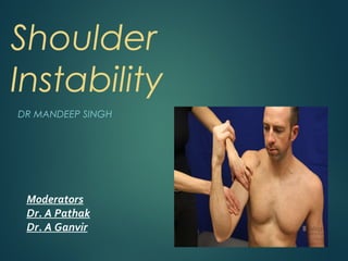
- 1. Shoulder Instability DR MANDEEP SINGH Moderators Dr. A Pathak Dr. A Ganvir
- 2. WHAT IS INSTABILITY? During the use of normal shoulder,humeral head is centered within the glenoid and coracoacromial arch When the shoulder cannot maintain this centered position it is said to be unstable It is not the same as joint laxity allows the shoulder to attain its full range of functional positions while an unstable shoulder prevents normal function of that upper extremity
- 4. Factors contributing to shoulder stability 1. STATIC FACTORS 2. DYNAMIC FACTORS
- 5. STATIC FACTORS Normal glenoid is about 7 degrees retroverted If the retroversion is excessive, it leads to posterior instability of shoulder
- 6. The labrum increases the superoinferior diameter of the glenoid by 75% and the anteroposterior (AP) diameter by 50%
- 7. The bony conformity of the glenoid and humeral head articular surfaces provides some of the stability of the shoulder. Frequently, patients with recurrent dislocations have bony deficits in one or both of these surfaces.
- 8. LIGAMENTS
- 9. • Superior Glenohumeral ligament : Most important check at zero degrees of abduction • Middle Glenohumeral Ligament : Most important check at middle ranges of abduction •Inferior Glenohumeral ligament : Most important check at more than 45 degrees of abduction
- 10. DYNAMIC FACTORS 1. The movement of rotator cuff muscles help to contribute to the negative intra - articular pressure. 2. The rotator cuff muscles themselves make a protective cuff all around the shoulder except inferiorly where shoulder capsule is the weakest.
- 11. Other factors : 1.Muscles around the shoulder - Levator scapulae - Rhomboids -Trapezius 2. Biceps Brachii 3. Proprioceptors
- 12. PATHOLOGICAL ANATOMY LABRAL LESIONS : 1.Bankart lesion 2.Reverse Bankart lesion 3.SLAP Lesion
- 13. BANKART LESION-labral tear at anterior half of glenoid rim
- 15. SLAP Lesion
- 16. CAPSULAR LESIONS: 1. Intra Substance Tear 2. HAGL Lesion 3. Repetitive Micro Trauma 4. Excessive capsular laxity
- 17. HAGL Lesion(Humeral avulsion of the inferior glenohumeral ligament)
- 20. Hill Sach Lesion
- 22. MATSEN’S CLASSIFICATION T Traumatic U Unilateral B Bankart lesion S Surgery is often necessary A Atraumatic M Multidirectional B Bilateral R Rehabilitation is the treatment I If surgery is needed inferior capsular shift is performed
- 23. History Define mechanism Position of arm Point of force Amount of force Electric Shock /Seizure
- 24. CLINICAL EXAMINATION LOOK FEEL MOVE SPECIAL TESTS
- 26. LOOK - Generalized joint laxity - Muscle wasting - Asymmetry - Previous operative scars - Ecchymosis
- 27. FEEL Local temperature Tenderness Any palpable mass Bony defect Muscular weakness Nerve injury
- 28. CLINICAL TESTS The sulcus test.
- 29. Shift and Load Test
- 30. The anterior apprehension test
- 31. The anterior drawer test
- 32. RADIOGRAPHIC EVALUATION A routine AP shoulder radiograph shows overlap of the anterior and the posterior glenoid rims. A true AP radiograph demonstrates superimposition of the anterior and the posterior glenoid rims, producing an excellent view of the glenohumeral joint.
- 33. Normal Shoulder AP view
- 34. Transcapular Y-view of the glenohumeral joint allows assessment of humeral head location in relation to the Glenoid cavity
- 37. Axillary view represents the “gold standard” in radiographic assessment of location of the humeral head relative to the glenoid cavity.
- 39. The stryker notch view
- 41. The west point view
- 42. QUESTIONS TO BE ANSWERED WHILE EVALUATING A PATIENT Is the problem in the glenohumeral joint ? Is the problem one of failure to maintain the humeral head in its centered position ? What mechanical factors are contributing to the instability ? Are these factors amenable to surgical repair or reconstruction ?
- 43. McLaughlin & Cavallaro After acute dislocations, development of recurrence
- 44. Rowe and Sakellarides Frequent dislocations in young athletes Duration of immobilization does not affect recurrence rates
- 45. Burkhart and Debeer; Sugaya et al; Itoi et al Glenoid bone loss more than 20% leads to shoulder instability
- 46. RATIONALE FOR TREATMENT 2 important factors favoring surgical treatment YOUNG AGE HIGH ACTIVITY LEVEL
- 47. EMERGENT MANAGEMENT OF ACUTE DISLOCATIONS
- 48. NON-OPEREATIVE TREATMENT A trial of non-operative treatment is recommended for the following group of patients-a) All patients who sustained a traumatic first time dislocation regardless of age b) Patients > 40 yrs with recurrent instability c) All patients with atraumatic instability
- 49. NON-OPERATIVE TREATMENT PROTOCOL All patients< 30 yrs shoulder immobilized for 3 wks Patients 30-40yrs shoulder immobilized for 1-2 wks Patients >40 yrs the shoulder immobilized for 1 wks Atraumatic instability- immobilization not required Patients with anterior instability-limit ext. rotation to 30 deg. and abd. to < 60 deg. Patients with posterior instability- avoid flex.>60 deg. and int. rotation > 30 deg.
- 50. INDICATIONS FOR OPERATIVE TREATMENT IN INSTABILITY Failure of non operative therapy Young adult with high functional demands Irreducible dislocation Open dislocation
- 51. TREATMENT OPTIONS TYPE OF INSTABILITY PREFERRED SURGERY Traumatic anterior, with Bankart Lesion Open / arthroscopic Bankart repair Traumatic anterior , with no labral lesion, just capsular laxity Open / arthroscopic capsular imbrication AMBRI lesions Lateral capsular shift( modified Neer and Foster ) with closure of rotator interval Recurrent posterior dislocation in association with a reverse Hill-Sachs lesion modified McLaughlin procedure Head defect > 30 – 45 % > 45 % Acute disimpaction / Weber osteotomy Prosthetic replacement Glenoid defect Bristow – Latarjet coracoid transfer Structural bone graft
- 52. OPEN SOFT TISSUE PROCEDURES FOR ANTERIOR INSTABILITY Open Bankart procedure Arthroscopic Bankart procedure Arthroscopic Thermal capsulorraphy Arthroscopic capsular imbrication Putti-Platt procedure Only 3 – 10 % failure rate by various studies 10 – 15 % failure rate by various studies Long term follow up shows high incidence of OA, about 30 %
- 54. Anchor used for repair
- 56. OPEN BONY PROCEDURES FOR ANTERIOR INSTABILITY Bristow procedure Latarjet procedure
- 59. AMBRII Lesions-Idea of management Primary treatment nonoperative Operative management recommended for patients who have continued pain or disability despite an adequate rehabilitation The gold standard is open stabilization
- 60. Capsular shift( modified Neer and Foster )
- 61. POSTERIOR INSTABILITY-A general overview Rare Often missed Often has a component of muscle imbalance Indication for operative treatment is generally continued problems despite rehab.
- 62. Procedures Procedure Description Results Neer’s Capsulorrraphy Posterior capsular tightening Generally unsatisfactory, upto 50 % recurrence Staple capsulorraphy Tightening done with staples Small study group Tieborne and bradley procedure Capsular Imbrication with a horizontal T approach Upto 20 % recurrence Hawkins and Janda procedure Subscapularis advancement and shortening 0 – 5 % recurrence Rockwood Glenloid Plasty with Biceps Tenodesis to the posterior capsule Combined bony and soft tissue procedure Not often done
- 63. ARTHROSCOPIC PROCEDURES FOR POSTERIOR INSTABILITY Posterior capsulolabral reattachment with the help of suture anchors Arthroscopic posterior capsulorrhaphy
- 64. OPEN ANTERIOR PROCEDURES FOR POSTERIOR INSTABILITY McLaughlin procedure Neers modification of McLaughlin procedure
- 65. McLaughlin technique subscapularis
- 67. Some procedures of historic interest Weber osteotomy
- 68. Putty Platt Operation Surgical procedure for stabilizing the glenohumeral joint after recurrent anterior shoulder dislocations. The subscapularis tendon is detached near its insertion on the humerus, the joint opened, and the stump of the tendon on the lesser tuberosity is sutured to the glenoid labrum. Sometimes the procedure is combined with reattachment of the glenoid labrum. Technically an easy procedure Disadvantages: The Putti-Platt procedure is not to be performed on throwers because it can reduce the range of movement in the shoulder. 30 – 35 % incidence of late OA
- 70. ADVANTAGES AND DISADVANTAGES OF ARTHROSCOPIC STABILIZATION ADVANTAGES DISADVANTAGES -Improved cosmesis -Technically demanding -Shorter operative time -Difficult in revision case -Short hospital stay -Difficult in altered anatomy -Decreased morbidity -Cannot address bony defect -Decreased complication -Lower cost
- 71. PHASES OF REHABILITATION Phase I Rest and immobilization. Pain control with nonsteroidal anti-inflammatory drugs and ice applied to the shoulder Phase II Isometric strengthening Isotonic strengthening. Begin exercises with shoulder in adducted, forward- flexed position, progressing to abducted position Phase III Endurance building along with strengthening exercises. Goal: the patient reaches 90% strength in the injured shoulder compared with the uninjured shoulder Phase IV Increase activity to sport- or job-specific activities
- 72. THANK YOU